History of the Procedure
Formalized knowledge of facial reconstruction has been present since the last millennium. The Byzantine physician Oribasius first described procedures for facial reconstruction in the fourth century. The Synagogae Medicae, a 70-volume medical encyclopedia written by Oribasius, describes the principles of basic advancement flaps for brow, nasal, and auricular defects. Interestingly, this ancient writing addresses the importance of flap thickness in relation to its viability and closure without tension in wound healing.
Problem
The prominence and central position of the cheek define its unique characteristics as an anatomic subunit. Zide further divides the cheek into 3 subunits: suborbital, preauricular, and buccomandibular. [1] Cheek wounds can be further classified by depth into superficial, full-thickness, and subcutaneous contour deficits. Reconstruction of the cheek is a challenging problem for a number of reasons. First, cheek contour is paramount to facial aesthetics. No existing reconstructive option can universally recreate the volume loss created by a subcutaneous tissue defect. Second, the dynamic function of the cheek is not easily reproduced. Third, reconstruction can alter or obliterate the lines that divide facial subunits. [2]
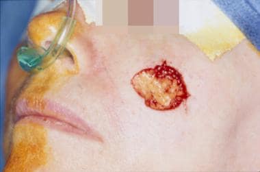
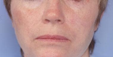
The images below depict a mid-cheek defect before and after reconstruction with M-plasties.
Etiology
Cheek defects occur as a result of 3 main etiologies: neoplasia, burns, and trauma. The cause of the defect contributes significantly in presurgical planning. For example, a skin graft may be considered an acceptable initial alternative to cover the defect from a full-thickness burn. Likewise, a skin graft is a reasonable alternative for patients with a history of radiation for acne as teenagers who are likely to develop multiple skin cancers. However, a traumatic defect is better suited for primary closure or use of a local flap.
Indications
Congenital, traumatic, and Mohs surgery defects all provide indications for cheek reconstruction. A subsequent soft tissue deficit can be corrected with various techniques.
Relevant Anatomy
The cheek is a protuberant structure that extends from the inferior orbital rim superiorly to the mandibular rim inferiorly and from the lateral nasal sidewall and melolabial crease medially to the preauricular area posteriorly.
The external carotid artery (ECA), with contributions from the internal carotid artery (ICA) system, is the predominant arterial blood supply to the skin and muscle of the cheek. The greatest contribution is from the facial artery, which crosses the mandibular border at 2-3 cm anterior to the angle, traverses the face obliquely, and terminates in the angular artery. The transverse facial artery arises from the superficial temporal artery (STA) and follows a path implied by its name across the face. The dorsal nasal artery runs along the path implied by its name and is a terminal branch of the ophthalmic artery, which is a terminal branch of the ICA. Many smaller branches and communications also exist.
The venous drainage system of the cheek predominantly functions via the anterior facial vein, which subsequently communicates with the internal jugular (IJ) vein. However, substantial drainage via the ophthalmic, infraorbital, and deep facial veins communicates with the cavernous sinus. This venous system is valveless, which can lead to bacterial spread from a localized skin infection and subsequent cavernous sinus thrombosis. However, the incidence of such events is low.
Lymphatic drainage in the area is primarily directed to intraparotid, submandibular, and submental lymph nodes.
The nerve supply of the cheek can be divided into sensory and motor systems (see the image below). Sensation to the cheek is carried primarily by the second (maxillary) and third (mandibular) divisions of the trigeminal nerve (cranial nerve V). The facial nerve (cranial nerve VII) provides motor innervation to the muscles of facial expression.
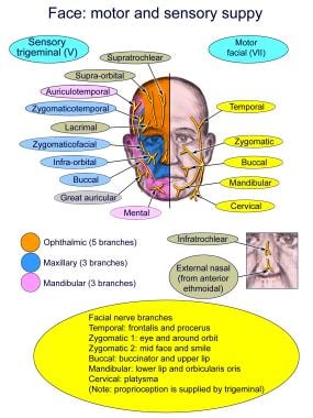
The maxillary division of the trigeminal nerve provides sensation to the cheek via the infraorbital, zygomaticofacial, and zygomaticotemporal branches. The area covered by these nerve branches includes the cornea, lower eyelid, infraorbital region, upper lip, and malar eminence. The mandibular division of the trigeminal nerve provides sensation to the cheek via the mental, buccal, and auriculotemporal branches. The areas innervated by these branches include the lower lip, mandibular border, and temporal regions. See Trigeminal Nerve Anatomy for more information.
The facial nerve exits the temporal bone at the stylomastoid foramen and travels through the parenchyma of the parotid gland, where it branches at the pes anserinus into upper (zygomaticofacial) and lower (cervicofacial) divisions. The upper division forms the temporal and zygomatic branches, which primarily supply the frontalis and orbicularis oculi, zygomaticus major and minor, and levator labii superioris muscles. The lower division forms the buccal, marginal mandibular, and cervical branches, which primarily supply the buccinator, orbicularis oris, and platysma muscles. A great deal of arborization is present between the zygomatic and buccal branches. Indeed, variations in individual anatomy, exact patterns of branching, and patterns of innervation are common. The zygomaticus major, orbicularis oculi, and risorius muscles are of particular clinical relevance because they are innervated via the deep surface of their muscle bellies. See Facial Nerve Anatomy for moreinformation.
The muscles of facial expression, which are anatomically associated with the cheek, include the zygomaticus major and minor, orbicularis oculi, orbicularis oculi, levator labii superioris, platysma, and risorius muscles. A continuous fascial covering known as the superficial musculoaponeurotic system (SMAS) covers each of these muscles. The branches of the facial nerve lie deep to the SMAS as they course more superficially in the anterior face. The more medial and anterior areas of the face have the most superficial facial nerve branches. See Facelift Anatomy for more information.
-
Mid-cheek defect.
-
Mid-cheek defect reconstructed with M-plasties to shorten the length of the final closure.
-
Mid-cheek defect reconstructed with M-plasties. One-month follow-up result.
-
Large medial cheek defect.
-
Large medial cheek defect. Reconstruction with a cervicofacial rotation flap.
-
Eight-month follow-up result. Reconstruction with a cervicofacial rotation flap to correct a large medial cheek defect.
-
Large lateral cheek and tragal defect.
-
Cheek reconstruction with cervicofacial rotation flap, 3-month follow-up result.
-
Diagram of the sensory and motor supply of the face.


