Background
Distraction osteogenesis, also termed callotasis, callus distraction, and osteodistraction, is the surgical technique of generating new bone by progressive stretching of divided segments. Following its introduction, distraction osteogenesis gained immediate interest and has been widely performed in the maxillofacial complex.
History of the Procedure
A crude method of distraction osteogenesis first appeared in the literature in 1905 and was described by Codivilla, who used the technique to elongate a femur.
Ilizarov is the father of modern distraction osteogenesis. In 1951, Ilizarov developed a technique for repairing complex fractures or nonunions of the long bones. While treating a patient with a short amputation stump, Ilizarov performed an osteotomy and applied an external fixator to lengthen the stump with the intention of placing a bone graft. However, by chance, he discovered that the bone grew in the distraction gap, eliminating the need for a bone graft. Later research by Ilizarov demonstrated that the tension-stress effect caused an increase in metabolic activity, an increase in cellular proliferation, and a neovascular ingrowth similar to normal endochondral ossification. Over the ensuing years, Ilizarov perfected the technique in long bones. [1]
Distraction osteogenesis of the mandible is shown in the image below.
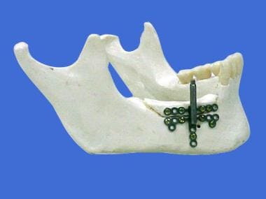 Distraction osteogenesis of the mandible. Alveolar distractor used to increase the height of the alveolar bone. Courtesy of K.L.S. Martin, LP.
In 1992, McCarthy reported a series of 4 young patients (average age, 78 mo) who successfully underwent gradual distraction of the mandible without grafting, transfusion, or intermaxillary fixation. [2] Early in the history of this procedure, distraction osteogenesis of the mandible involved using bulky external distractors. Although these external distractors still have a place in certain applications, a wide variety of intraoral internal distractors are now available; these distractors are small and compact, with increased patient comfort and acceptance.
Distraction osteogenesis is now a feasible treatment option for adults and children with unilateral or bilateral mandibular hypoplasia. It is a treatment option for widening an excessively narrow mandible. Distraction osteogenesis is also a treatment alternative for the management of airway obstruction (eg, obstructive sleep apnea [OSA]), continuity defects of the mandible, reconstruction after a resection, and alveolar crest augmentation.
Problem
Mandibular retrognathia is one of the most common craniofacial deformities; approximately 10% of the population have significant dental overjet. Mandibular retrognathia can be congenital or acquired. Congenital causes include hemifacial microsomia, Treacher Collins syndrome, Pierre Robin syndrome, Goldenhar syndrome, Nager syndrome, and mandibular hypoplasia. Acquired causes of mandibular retrognathia include trauma (ie, condylar fractures occurring at an early age and resulting in bony ankylosis or disrupted growth) and previous surgery for developmental cysts or tumors.
Although less common than retrognathia, the mandible can also exhibit a transverse deficiency, resulting in a severe malocclusion and anterior dental crowding. This condition is difficult to correct with traditional osteotomies, and multiple extractions are often necessary. Distraction osteogenesis of the mandible has become a commonplace procedure with the flexibility to allow the surgeon to address a wide variety of mandibular defects.
Epidemiology
Frequency
Approximately 10% of the population has significant dental overjet. Craniofacial defects amenable to treatment with distraction osteogenesis are not highly prevalent. It is estimated that OSA affects 12 million people in the United States.
Pathophysiology
After a low power osteotomy is performed, distraction osteogenesis begins with the formation of a hematoma between and around the bone segments. The hematoma is converted to a clot, and bone necrosis occurs at the end of the fracture segments. An ingrowth of vasoformative elements and capillaries for the restoration of blood supply forms a soft callus.
Tension is then applied to the soft callus and a dynamic microenvironment is created. Pluripotential mesenchymal cells are activated into fibroblasts and osteoblasts, and type I collagen is laid down parallel to the vector of distraction. Bony trabeculae grow into the fibrous area from the periphery, parallel to the line of tension that occurs during the distraction phase. A bridge of immature bone forms across the distraction gap. A poorly mineralized, radiolucent fibrous interzone is located in the middle of the distraction gap, where the influence of tensional stress is maximal. The interzone functions as the center of fibroblast proliferation and fibrous tissue formation. During the consolidation phase, bony remodeling begins. The regenerate eventually matures into osseous tissue similar to the native bone.
Ilizarov’s study proved that the success of the distraction depended on the rate and rhythm of the force applied on site. The optimal rate of distraction is often reported to be 1 mm/day. Although the majority of authors agree with the distraction rate of 1 mm/day, there are reports suggesting different rates. Chin and Toth applied a distraction rate of up to 3 mm/day and Ramichel et al reported establishing a rate of 2 mm/day. [3, 4] However, distraction of more than 1.5 mm/day may cause delayed ossification or pseudoarthrosis due to local ischemia in the interzone. In their study of 39 patients, Meyer et al reported one failure, defending the approach of setting the rate on a case-by-case basis considering individual variables. [5] A rate of distraction of 0.5 mm or less per day may cause premature consolidation of the bone.
Soft tissue also has the ability to grow linearly along lines of tension. This is referred to as distraction histogenesis. Skin, muscle, nerves, and vascular tissue are generated, not stretched. The advantage is obvious, especially for severe retrognathia, in which the stretched soft tissue envelope can contribute to relapse when a traditional mandibular osteotomy is performed for a large (>10 mm) advancement.
Presentation
Distraction osteogenesis has many advantages over traditional mandibular osteotomies, as delineated below.
Distraction osteogenesis decreases the need for bone grafting for large (>10 mm) mandibular advancements; one can achieve 20 mm or more of advancement without a bone graft and the associated donor site morbidity, scarring, and potential for infection.
The procedure can be performed in infants and children, who would otherwise not be candidates for mandibular osteotomy because of the interference with the developing tooth buds and/or insufficient bone to safely perform a traditional osteotomy. In addition, distraction osteogenesis often has obviated the need for a tracheotomy in newborns and infants with micrognathia and airway obstruction.
This procedure also results in less distortion and loading of the temporomandibular joint than sagittal split osteotomy.
Distraction osteogenesis can be performed in 3 dimensions, that is, advancing, widening, and increasing vertical height of the basal mandibular bone. In addition, the vector and the amount of movement can be tailored to each patient, especially in those with significant facial asymmetry.
Distraction osteogenesis can be used to rotate the anterior portion of the mandible to correct open bites related with mandibular deficiencies.
Greater patient acceptance exists with this procedure, especially with the development of low-profile intraoral devices.
Distraction osteogenesis appears to have a decreased potential for relapse, especially with large advancements; this is because the transported portion experiences a progressively increased movement, instead of being transported in one large block. Due to the process of distraction histogenesis, resistance to advancement by the soft tissue envelope is decreased.
Distraction osteogenesis seems to limit the likelihood of damaging the inferior alveolar nerve.
Widening the mandible, which is difficult to do with traditional osteotomies and presents multiple complications, is possible with distraction osteogenesis.
Bone grafts are not required; hence, there is no donor site morbidity.
Mandibular distraction osteogenesis also has the following limitations [6, 7] :
-
Relative difficulty in performing osteotomies owing to potential damage to lingual mucoperiosteum or vascular plexuses of the floor of the mouth
-
Difficulty in obtaining correct vector of distraction
-
Although rare, relapse and/or resorption of the transport segment
-
Patient discomfort and psychological burden due to the retained distractor device
Indications
Distraction osteogenesis is indicated in the following cases:
-
Severe retrognathia associated with a syndrome (eg, Pierre Robin syndrome, Treacher Collins syndrome, Goldenhar syndrome), especially in infants and children who are not candidates for traditional osteotomies
-
Patients who have unilateral hypoplasia of the mandible (eg, hemifacial microsomia)
-
Nonsyndromic mandibular hypoplasia associated with a dental malocclusion (especially if the advancement exceeds the capabilities of a traditional osteotomy or if the patient is hesitant to undergo a bone graft harvest with the associated morbidity)
-
Mandibular transverse deficiency associated with a dental malocclusion and dental crowding
-
Patients with severe OSA (respiratory disturbance index [RDI] >60) and patients who are obese (body mass index [BMI] >28).
-
Mandibular hypoplasia due to trauma and/or ankylosis of the temporomandibular joint
-
Mandibular continuity defects resulting from excision of tumors and/or aggressive developmental cysts
-
Shortened vertical height of the alveolar bone (Distraction of the alveolar segment can be performed to increase the vertical height in preparation for osteointegrated dental implant placement. In patients who have had a mandibular reconstruction with a free fibular bone graft, the fibular segment can be distracted vertically to facilitate dental implant placement, as depicted in the image below.)
 Distraction osteogenesis of the mandible. Alveolar distractor used to increase the height of the alveolar bone. Courtesy of K.L.S. Martin, LP.
-
Mandibular angle deformity. (The correction includes a rotational component and a distraction component. The rotational component is a hinge mechanism that allows free movement around its center of rotation. The distraction component for angular deformity correction usually consists of a distraction rod with two pivotable connectors at both ends. Type of hinges can be identified based on their location as follows: 1) the opening wedge hinge, 2) the closing wedge hinge, and 3) the translation hinge.)
Relevant Anatomy
The mandible is a U-shaped bone. It is the only mobile bone of the facial skeleton, and, since it houses the lower teeth, its motion is essential for mastication. It is formed by intramembranous ossification. The mandible is composed of 2 hemimandibles joined at the midline by a vertical symphysis. The hemimandibles fuse to form a single bone by age 2 years. Each hemimandible is composed of a horizontal body with a posterior vertical extension termed the ramus.
Placement of the osteotomy depends on the desired vector of distraction. However, certain anatomic structures must be avoided. The osteotomy is best placed either anterior or superior to the mandibular angle; the angle is maintained and the flattening of that portion of the patient's face is prevented. Tooth roots or developing teeth must be avoided when performing the osteotomy or placing the distraction device screw fixation. When the osteotomy is performed, the inferior alveolar nerve must also be avoided. Care must be taken to avoid damaging the lingual nerve when one extends the osteotomy through the lingual aspect of the mandible where the nerve lies in close proximity.
For more information about the relevant anatomy, see Facial Bone Anatomy.
Contraindications
No absolute contraindications to treatment exist. However, relative contraindications are as follows:
-
Patients who are unable or unwilling to comply with the distraction schedule are not ideal candidates for this procedure.
-
Mandibular distraction osteogenesis has been performed on infants as young as 9 days old, [3] but more difficulty is encountered when dealing with small fragile bones in the placement of the distraction device, especially children under 6 years of age.
-
Patients who have inadequate bone structure are not ideal. Adequate bone stock must be available to accept the device and to provide adequate surface area of the osteotomy sites for regeneration. Several authors suggest that distraction may not be a good treatment option if the bone height above the inferior alveolar nerve is less than 6-8 mm or if the transport segment will be shorter than 4-5 mm. [8, 9, 10]
-
In older patients, a decreased number of mesenchymal stem cells may impair bone healing at the distraction site.
-
Patients who have metal allergies are not ideal.
-
Distraction osteogenesis of the mandible. Alveolar distractor used to increase the height of the alveolar bone. Courtesy of K.L.S. Martin, LP.
-
Distraction osteogenesis of the mandible. A reciprocating saw is used to osteotomize inferior, lateral, and superior portions of the mandible. An osteotome is then used to complete the bony cut. Courtesy of Jordan Mastrodonato, MS, Medical Illustrator, Eisenhower Army Medical Center.
-
Distraction osteogenesis of the mandible. Intraoral distraction device in place. Courtesy of K.L.S. Martin, LP.
-
Temporomandibular joint ankylosis following distraction osteogenesis
-
Mandibular distractor used for maxillomandibular advancement for the treatment of OSA. Courtesy of K.L.S. Martin, LP.


