Practice Essentials
Prominent or enlarged blood vessels that exist on the vibratory surface of the vocal fold may eventually cause problems from hemorrhage or mass effect within the lamina propria and may cause dysphonia by disrupting the vibratory pattern and closure of the true vocal folds.
Vascular lesions found within the true vocal fold may threaten the career of a professional vocalist because of recurrent inopportune hemorrhage or scar formation. In the asymptomatic patient, they may create a management dilemma due to the risk of future hemorrhage versus the immediate risks of intervention.
In a retrospective study of 499 vocal performers, Tang et al found that the hemorrhage rate in those with vocal fold varices was 2.68% at 12 months, versus 0.8% in performers without varices. Based on a Cox proportional hazard regression analysis, performers with varices were reported to have a hazard ratio of 10.1 for hemorrhage development compared with the other performers. [1]
An image depicting vascular lesions of the vocal fold can be seen below.
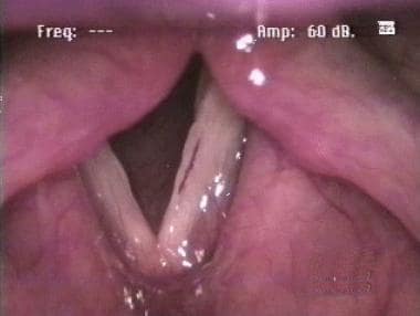 Vocal cords in a performing artist with minor difficulty singing. Videostroboscopy revealed fullness of the entire fold as well as slightly decreased mucosal wave.
Signs and symptoms of vascular lesions of the vocal fold
Vocal fold varices may be entirely asymptomatic, or they may result in dysphonia through hemorrhage and edema, scarring, or mass effect with resultant disruption of the mucosal wave.
Workup in vascular lesions of the vocal fold
Evaluate patients presenting with dysphonia by indirect laryngoscopy and videostroboscopy. Particular attention should be paid to the following:
-
Vocal fold mobility
-
Glottic closure
-
Presence, amplitude, and symmetry of the mucosal wave
Management of vascular lesions of the vocal fold
Medical therapy
Considerations in management include the following:
-
Use of medications with anticoagulant properties should cease if they are not medically necessary
-
Treat any conditions predisposing to trauma and irritation (eg, cough, reflux disease) with the appropriate therapy
-
Voice use should be modified, limiting the frequency, intensity, and duration of voice use and maximizing vocal rest
-
Hard glottal attacks should be avoided and easy-onset patterns used
-
Applicable speech therapy techniques include direct, indirect, and confidential voice therapy
Surgical therapy
Surgical technique begins by identifying the feeding and emptying vessels, which are then photocoagulated sequentially with a carbon dioxide laser or specific photoangiolytic lasers such as the KTP (potassium titanyl phosphate) or 585 nm pulsed dye laser. [2, 3] The primary lesion may then be excised via a microflap approach or photocoagulated, depending upon its size. The goal of surgical excision is preservation of the mucosal cover with minimal disruption of the underlying tissue.
Direct surgical excision of the vascular abnormalities is another treatment option. This method uses cold steel phonomicrosurgical techniques and instruments.
Problem
No widely accepted system of nomenclature is available for vascular lesions. Microvascular lesions of the true vocal folds are known as varices, capillary ectasias, papillary ectasias, capillary and venous lakes, and spider telangiectasias. The anatomic variations based on these terms are subtle, and treatment approaches are similar regardless of the type. A varix is a prominent, dilated, and commonly tortuous vein found on the surface of the vocal fold. Ectasias are distinguished by a coalescent hemangiomatous appearance.
Epidemiology
Frequency
Vocal fold varices occur most commonly in female professional vocalists, although they are not rare in males. Postma et al found a prevalence of 3.5% among their patient population, with 14.5% of those cases occurring in female professional voice users. [4] Prevalence in the general population is unknown.
Etiology
Formation of varices is related to vocal use, abuse, and trauma. [5] Most patients presenting with symptoms stemming from these lesions are professional voice users. Repeated trauma may lead to new blood vessel formation and weakening of the vessel walls.
Pathophysiology
The immediate cause of vocal fold varices is unknown, although they may originate from shearing stress along the lateral fold near the termination and reversal point of the mucosal wave. A hormonal cause has been postulated because of the prevalence in female singers, but this has not been proven. In addition, physiologic and histologic changes associated with menses may increase the risk of variceal hemorrhage. However, hormonally directed therapy has not been successful in treatment of these lesions. The predilection may be attributable to the unique anatomy and associated mechanics of voice production in female vocalists.
Presentation
The clinical presentation of vocal vascular lesions is highly variable. Vocal fold varices may be entirely asymptomatic, or they may result in dysphonia through hemorrhage and edema, scarring, or mass effect with resultant disruption of the mucosal wave. Dysphonia may be severe with an acute, dramatic onset. This presentation typically appears after episodic vocal abuse or straining. Other presentations can be subtle, with patients having an apparently normal voice while being easily fatigued or incurring loss of normal vocal range. Patients with recurrent hemorrhage may relate a history of episodes of hoarseness followed by resolution.
Patients may have completely normal sounding voices with a pronounced varix on the surface of the true vocal fold. Patients presenting immediately after an acute bleed may have extensive ecchymosis and hemosiderosis of the involved true vocal fold. Acute hemorrhage may resolve without event, or it may transform into a hemorrhagic polyp, cyst, or scar, which then causes dysphonia through a mass effect or vibratory margin effects.
In women, the appearance of the lesion may depend on the stage of the menstrual cycle; therefore, periodic examinations may be required to accurately establish severity.
Indications
Indications for surgical intervention in a patient with a vascular lesion of the vocal fold include enlargement of the lesion, recurrent hemorrhage, development of a mass in conjunction with the varix, unacceptable dysphonia, and uncertainty as to the diagnosis.
Relevant Anatomy
Vascular lesions appear on the superior surface of the vocal fold or, less commonly, along the vibratory margin. Because of the large numbers of vascular arcades found in the vocal fold, they are not critical to the blood supply of the tissue. Varices may manifest as abnormally dilated capillary arcades running in the anterior-to-posterior direction or as clusters of capillaries. Another formation is a dot, which represents the tip of a vascular loop rising superficially from the underlying mucosa. Finally, venous lakes may form that are so large as to appear as a chronic area of hemorrhage. Hochman et al postulated that vascular lesions are more likely to form on the superior lateral surface of the vocal fold because of the shearing forces generated by the termination of the mucosal wave at that point. [6]
-
Vocal cords in a performing artist with minor difficulty singing. Videostroboscopy revealed fullness of the entire fold as well as slightly decreased mucosal wave.
-
Hemorrhagic polyp on the surface of the true vocal fold in a professional singer. Note the fullness along the medial edge of the true vocal fold in the area of the lesion.
-
Untreated or recurrent hemorrhage can evolve into a large hemorrhagic polyp. Conservative therapy has little chance of success at this point, and these lesions can lead to scarring if untreated.


