Practice Essentials
Osteoradionecrosis (ORN) is a condition of nonvital bone in a site of radiation injury. [1] ORN can be spontaneous, but it most commonly results from tissue injury. The absence of reserve reparative capacity is a result of the prior radiation injury. Even apparently innocuous forms of trauma, such as denture-related injury, ulcers, or tooth extraction, can overwhelm the reparative capacity of the radiation-injured bone. Traditionally, three grades of disease (I, II, III) are recognized. Grade I ORN is the most common presentation; exposed alveolar bone is observed. Grade II designates ORN that does not respond to hyperbaric oxygen (HBO) therapy and requires sequestrectomy/saucerization. Grade III is demonstrated by full-thickness involvement and/or pathologic fracture. Therefore, patients can demonstrate grade I or grade III ORN at initial presentation. ORN is assessed through imaging studies.
ORN of the mandible is depicted in the images below.
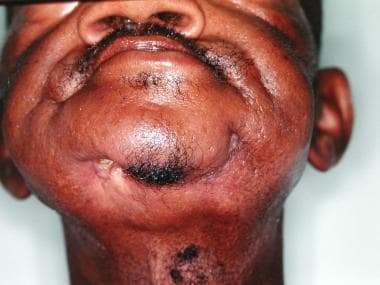 This patient developed osteoradionecrosis (ORN) following radical radiotherapy. His primary tumor was located in the floor of mouth. An orocutaneous fistula is demonstrated here. A pathologic fracture was evident on examination. Biopsies were negative for carcinoma.
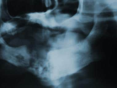 This is the panoramic radiograph of the patient seen in the image above. Bone necrosis and pathologic fracture are evident.
Signs and symptoms of osteoradionecrosis of the mandible
These include the following:
-
Pain
-
Swelling
-
Trismus
-
Exposed bone
-
Pathologic fracture
-
Malocclusion
-
Oral cutaneous fistula formation
Workup in osteoradionecrosis of the mandible
Plain radiography of the mandible, or Panorex, depicts areas of local decalcification, osteolysis or sclerosis.
Computed tomography (CT) scanning and magnetic resonance imaging (MRI) may allow early diagnosis of ORN and better delineate the extent of disease.
Management of osteoradionecrosis of the mandible
Prior to beginning radiation therapy, all patients should undergo a thorough dental evaluation, including full mouth radiographs, dental and periodontal diagnosis, and prognosis for each tooth.
To prevent radiation caries, patients should begin daily fluoride treatment with 1% neutral sodium fluoride gel in prefabricated trays for 5 minutes each day. This practice should continue for life.
Medical therapy in the treatment of ORN is primarily supportive, involving nutritional support along with superficial debridement and oral saline irrigation for local wounds.
HBO transiently elevates tissue oxygen tension and stimulates fibroblastic proliferation and oxygen-dependent collagen synthesis. This allows for angiogenesis in the radiated bed. This does not totally resolve the radiation injury, however, and some degree of tissue hypoxia persists.
Surgical treatments include transoral sequestrectomy with primary wound closure. Patients may also undergo transcutaneous mandibular resection with wound closure and mandibular fixation with an external fixator or maxillomandibular fixation.
Epidemiology
Frequency
ORN is rare in patients who receive less than 60 gray (Gy) radiation therapy. Patients with ORN who receive less than 60 Gy and more than 50 Gy have been reported, but these cases are extremely rare. The overall incidence of ORN has decreased since the latter part of the 20th century. In general, studies from prior to the 1970s showed an ORN incidence from 5.4-11.8%. Studies in the early years of the 21st century, however, placed the incidence closer to 3.0%. [2] The true frequency of ORN is impossible to determine because no mechanism exists for reporting the disease. Incidence is increased in patients who receive combined chemotherapy-radiation. The Radiation Therapy Oncology Group (RTOG) requires their members to report radiation toxicity including ORN; however, the disease is probably under-reported.
More valuable than an understanding of frequency is an appreciation for the decrease in reparative capacity in tissue exposed to more than 60 Gy of radiation.
Etiology
ORN can be either spontaneous or the result of an insult. Spontaneous ORN occurs when, in the process of otherwise normal turnover of bone, the degradative function exceeds new bone production. ORN develops following injury when the reparative capacity of bone within an irradiated field is insufficient to overcome an insult. Bone injury can occur through direct trauma (eg, tooth extraction [84%], related cancer surgery or biopsy [12%], denture irritation [1%]) or by exposure of the irradiated bone to the hostile environment of the oral cavity secondary to overlying soft tissue necrosis. [3] The cumulative progressive endarteritis caused by radiotherapy results in insufficient blood supply (tissue oxygen delivery) to effect normal wound healing.
A study by Chronopoulos et al indicated that risk factors for grade III osteoradionecrosis include active smoking, excessive alcohol use, diabetes mellitus, and dental treatment and/or local pathologic conditions. The study involved 115 patients (153 lesions). [4]
A study by Huang et al found that in head and neck cancer patients, the presence of sialadenitis was associated with a 2.55-fold increase in the risk for osteonecrosis of the jaw (ONJ), with the likelihood of ONJ development being particularly high in oral cancer patients with both sialadenitis and radiation exposure (odds ratio = 15.9). [5]
The images below depict a patient who developed ORN following tooth extractions.
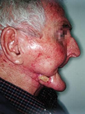
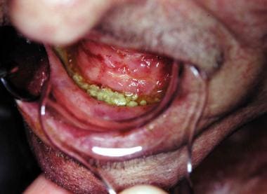 This patient developed ORN following tooth extractions. Sequential debridement was attempted prior to patient referral.
 The patient seen in the image above developed a pathologic fracture at the mandibular angle. He underwent resection of the area of the fracture. At the time of surgery, surgeons thought the patient had bleeding bone, but further ORN is evident.
A retrospective study by Kubota et al indicated that in patients with head and neck squamous cell carcinoma, independent risk factors for ORN of the jaw include oropharyngeal or oral cancer, as well as administration of 60 Gy of radiation to more than 14% of the jaw’s volume (V60 >14%). The investigators found the 3-year cumulative incidence rate of jaw ORN to be 8.6% in patients with V60 of greater than 14%, compared with 2.5% in those with V60 of 14% or less. In addition, patients with a primary oropharyngeal or oral cancer site had a 3-year cumulative incidence rate of 9.3%, compared with 1.4% in those with other cancers. [6]
In a retrospective study of ORN development in association with postoperative radiation therapy in patients with oral cavity cancer, Möring et al found that for active smokers at diagnosis who received 60 Gy or more to a volume of over 40%, the 1-year incidence of ORN was 29%. This was in contrast to an estimated cumulative 1-year incidence of 8.4% for the entire cohort. Forty one of the 46 cases of ORN in the study were in the mandible. [7]
A study by Caparrotti et al found ORN of the mandible to be relatively uncommon in patients with squamous cell carcinoma of the oropharynx who underwent curative-intent treatment with intensity-modulated radiotherapy (IMRT). Even so, it was reported that ORN continued to develop more than 5 years posttreatment in these patients. The study also found that the ORN rate may be reduced by minimizing the mandibular volumes receiving more than 50 Gy or over 60 Gy during treatment. In addition, the data indicated that smoking and bisphosphonate use are modifiable risk factors for ORN. [8]
Pathophysiology
ORN was first described by Marx in 1983 as hypovascularity, hypocellularity, and local tissue hypoxia. [9, 10] Prior to this, many other theories existed regarding the etiology of ORN. The report by Marx, clinical experience, and subsequent research support this now widely accepted theory.
The irradiated mandible, periosteum, and overlying soft tissue undergo hyperemia, inflammation, and endarteritis. These conditions ultimately lead to thrombosis, cellular death, progressive hypovascularity, and fibrosis. The radiated bed is hypocellular and devoid of fibroblasts, osteoblasts, and undifferentiated osteocompetent cells.
Mandibular ORN develops most commonly after local trauma, such as dental extractions, biopsies, related cancer surgery, and periodontal procedures, but it may also occur spontaneously.
Presentation
Clinical signs and symptoms include the following:
-
Pain
-
Swelling
-
Trismus
-
Exposed bone
-
Pathologic fracture
-
Malocclusion
-
Oral cutaneous fistula formation
On physical examination, missing hair follicles, surface texture changes, and color changes are common findings that assist clinicians in assessment of the area of radiation injury.
Relevant Anatomy
In a histologic study of irradiated osteoradionecrotic mandibles, several characteristic changes were noted. The inferior alveolar artery (the predominant arterial blood supply to the body of the mandible) and periosteal arteries had significant intimal fibrosis and thrombosis. Normal marrow was replaced by dense fibrous tissue with loss of osteocytes. Finally, the study noted buccal cortical necrosis with sequestrum formation and periosteal fibrosis with a tendency to detach from the cortex. [11] In the elderly, the inferior alveolar artery’s flow to the mandible diminishes and the periosteum and muscle attachments predominate as the primary blood supply. The thrombosis of the inferior alveolar artery and surgical disruption of this soft tissue blood supply may contribute to the development of osteoradionecrosis (ORN).
-
This patient developed osteoradionecrosis (ORN) following radical radiotherapy. His primary tumor was located in the floor of mouth. An orocutaneous fistula is demonstrated here. A pathologic fracture was evident on examination. Biopsies were negative for carcinoma.
-
This is the panoramic radiograph of the patient seen in the image above. Bone necrosis and pathologic fracture are evident.
-
This patient developed ORN following tooth extractions. Sequential debridement was attempted prior to patient referral.
-
The patient seen in the image above developed a pathologic fracture at the mandibular angle. He underwent resection of the area of the fracture. At the time of surgery, surgeons thought the patient had bleeding bone, but further ORN is evident.
-
An absence of healing is evident in this radiograph following extraction of a tooth within a field of radiation therapy.
-
Osteoradionecrosis developed in the patient seen in the image above. Osteolysis is clearly evident.
-
Pathologic fracture has developed in this case of osteoradionecrosis (ORN). This constitutes, by definition, stage III disease. This is the same patient seen in the 2 images above.


