Practice Essentials
The natural history of cervicofacial lymphangioma is the presence of a mass in the head and neck area. Lymphatic malformation or lymphangioma is a rare neoplasm or congenital rest that presents in the head and neck region in more than 70% of cases.
Cervicofacial lymphangioma is noted before birth, at birth, or in the first few months of life in less than 50% of patients with this condition. The initial appearance of cystic hygroma or lymphangioma in adulthood is less common. The growth rate of this neoplasm is variable. Slow progression followed by pseudoregression eventually gives rise to reappearance. Rapid growth or engorgement with lymph or blood is often associated with direct infection, trauma, or a secondary respiratory or skin infection. Spontaneous regression has been reported in as many as 15% of patients with cervicofacial lymphangioma. Malignant change has not been reported.
Whether patients with this neoplasm present in childhood or adulthood, the identification of a mass requires a diagnosis and management plan. Management may be complex and difficult because enlargement often results in functional and cosmetic problems. Attempts at a classification have been created to differentiate between the capillary, cystic, and cavernous forms of cervicofacial lymphangioma; however, the histologic appearance is not always uniform, which suggests a variable clinical and pathologic appearance of essentially the same disease. Determining location, persistence, growth patterns, and functional and cosmetic problems associated with lymphangioma usually requires a full diagnostic evaluation. Proactive therapy chosen on the basis of a careful clinical assessment follows the diagnosis.
An image depicting a lymphatic malformation can be seen below.
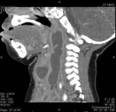 Reconstruction of CT in sagittal dimension. This patient was intubated because of respiratory distress caused by the large lymphatic malformation.
Signs and symptoms of cervicofacial lymphangioma
Lymphangioma in the head and neck generally involves a swelling or mass that is soft to palpation and well circumscribed or diffuse. It may have defined or ill-defined borders and is often associated with a bluish discoloration.
Symptoms and findings include the following:
-
Diplopia
-
Proptosis
-
Respiratory distress
-
Dysphagia
-
Dysphonia
Orofacial manifestations include mandibular or maxillary deformation, rotation, and malocclusion (ie, crossbite).
Workup in cervicofacial lymphangioma
Imaging studies include the following:
-
Barium swallow and/or upper and lower gastrointestinal (GI) series
-
Chest radiography
-
Computed tomography (CT) scanning
-
Magnetic resonance imaging (MRI)
-
Ultrasonography
-
Arteriography
-
Screening ultrasonography and MRI
-
Lymphoscintigraphy
-
Nuclear magnetic imaging (NMR) and MRI
Depending on the lesion’s anatomic location, other diagnostic studies may include the following:
-
Full neuro-ophthalmic examination for orbital or central lesions
-
Laryngoscopy, bronchoscopy, and/or esophagoscopy for lesions of the upper aerodigestive system
-
Abdominal endoscopy and/or colonoscopy for lower GI lesions
Tissue diagnosis, as a direct and final correlate to imaging studies, is the best and most consistent confirmation of lymphangioma.
Management of cervicofacial lymphangioma
Medical therapy
Carbon dioxide laser therapy has been effective in managing upper airway lesions and superficial mucosal microcystic lesions.
Intralesional sclerotherapy with group A Streptococcus pyogenes of human origin (OK-432) has had some success in controlling lymphangiomas.
Surgical therapy
The primary intention in the surgical treatment of lymphangiomas is to accomplish total resection. However, because of lesion size, lesion location, and a myriad of previously mentioned variables, total resection is not always possible.
A combined sequential approach is recommended for mixed lesions as well as extensive lesions that involve both the mucosa and soft tissues.
The particular surgical procedure relates to the location of the lymphangioma and the structures involved.
Epidemiology
Frequency
Cervicofacial lymphangioma is uncommon, representing fewer than 6% of benign tumors of childhood. Incidence has been reported to be less than 2.8 per 1000 population. No sex preponderance or side predilection has been reported.
Age
A retrospective study by Cho et al of 40 patients with cervicofacial lymphatic malformations found that 73% of cases presented with symptoms prior to age 2 years. [1]
Etiology
The origin of lymphangiomas is controversial. Theories include lymphangiomas as true neoplasms, hamartomas, or congenital dysplasias of the lymphatics. To determine the role of angiogenesis in the pathogenesis of lymphangioma, Sidle et al (2005) examined patients' specimens for expression of angiogenic inducer vascular endothelial growth factor (VEGF) and angiogenic inhibitor pigment epithelium-derived factor (PEDF) using immunohistochemical analysis. [2] Staining patterns of VEGF and PEDF were evaluated. Histological evidence of increased angiogenesis, including microvascular density, stromal fibrosis, and inflammation, were graded in each group and correlated with recurrence.
Lymphangioma specimens demonstrated histological evidence of increased angiogenic activity including multiple areas of increased VEGF staining combined with little PEDF staining. Recurrent specimens had increased histological evidence of angiogenesis as well as increased VEGF and decreased PEDF activity compared with nonrecurrent lesions.
They concluded that lymphangiomas exhibit tumorlike pathogenesis owing to the high expression of angiogenic inducers compared with the low expression of inhibitors. Recurrence may be influenced by this imbalance of angiogenic mediators. Further research with antiangiogenic therapy using agents such as PEDF analogues or anti-VEGF receptor antibodies is indicated.
However, most physicians favor the theory that dysplastic lymphatic tissue is sequestered in a target tissue space or organ during fetal development. Sabin (1909) proposed that lymphatics are derived from primitive veins by budding endothelial cells. [3] Because jugular sacs are the largest formations, the high incidence in the neck correlates well with clinical studies and experience. Incomplete canalization of the lymphatics causes obstruction of lymph flow and cyst or cavern development. These formations are analogous to blood-containing cavernous hemangiomas. Target organs and tissues include the retroperitoneum, chest wall, lung, mesentery, pancreas, scrotum, tongue, floor of the mouth, larynx, neck, lip, mediastinum, parotid gland, adrenal gland, axilla, diaphragm, gallbladder, spleen, skull base, colon, breast, subdural space, and pelvis.
A recent case series (2013) of 141 patients concluded that airway involvement of lymphatic malformations predominantly affects the oral cavity and oropharynx and no cases of glottis, subglottic, or tracheal involvement have been seen. [4]
Pathophysiology
Macroscopically, lymphangiomas are large cavernous, cystic, or complex multilocular masses that extend into tissue fascial planes. Patients usually present with a painless enlarging mass. Cystic types infiltrate into the surrounding tissue by fingerlike multiloculated extensions. Within these multiloculated honeycombed cysts is a clear-to-cloudy, sometimes blood-tinged, fluid with the consistency of uncooked egg whites. Cyst walls are indistinct; penetration and decompression of the cyst is a common occurrence during surgical excision. In many cases, cervical fetal lymphangi oma may be diagnosed before birth by means of ultrasonography. Before 30 weeks' gestation, cervical lesion is almost always associated with chromosomal abnormalities and congenital cardiac malformations usually in the septated type. A high rate of spontaneous abortion is encountered in this type.
The different types of cervicofacial lymphangioma are classified as follows:
-
Lymphangioma circumscriptum is a simplex superficial red macular or vesicular lesion of mucous membranes or skin.
-
Lymphangioma capillary type is a simplex lesion of dilated capillarylike channels.
-
Lymphangioma cavernosa is a simplex lesion of dilated lymphatic channels with deep extension and without cyst formation.
-
Lymphangioma cystica (ie, cystic hygroma) is composed of large lymphatic cysts that expand into adjacent soft tissue planes and are well defined, circumscribed, or lobulated.
-
Lymphangioma complex is composed of multiloculated poorly defined cysts extending to more than one anatomic area, tissue plane, or organ system.
Presentation
Children or, less commonly, adults usually present with a mass in the head and neck area. Approximately 90% of cases of cervicofacial lymphangioma become clinically apparent by the patient's third year of life. Common head and neck sites in childhood are the cervical area, floor of the mouth, and the tongue. In the aforementioned study by Cho et al, cervicofacial anomalies were found primarily on the left side and in the V2-V3 nerve area. [1] Orofacial manifestations include mandibular or maxillary deformation, rotation, and malocclusion (ie, crossbite). In a study by Orvidas et al, the most common location involved was the submandibular region, followed by the parotid and cheek. [5]
-
Obtain a careful history.
-
Perform a complete head and neck examination as well as a general examination.
-
Lymphangioma in the head and neck generally involves a swelling or mass that is soft to palpation and well circumscribed or diffuse. It may have defined or ill-defined borders and is often associated with a bluish discoloration.
-
Regional venous congestion and the absence of pain are common.
-
Common additional symptoms and findings include diplopia, proptosis, respiratory distress, dysphagia, and dysphonia.
-
Children with massive head and neck and chest lymphangiomas have higher mortality and morbidity. The involvement of the airway is an important prognostic factor. [6]
Differential diagnosis consideration
Differentiation from other cervical congenital masses or malignancy is necessary in children and adults. The presence of a mass in the larynx or trachea is usually associated with airway compromise and may require differentiation from a subglottic hemangioma. The presence of a mass in an anatomic location generates a differential diagnosis, particularly when combined with diagnostic studies. The cystic nature of the process and the bluish hue often associated with this growth frequently resemble characteristics of a hemangioma. Lesions in the floor of the mouth, particularly when bulging in the submental or submaxillary region, may simulate a plunging ranula. Other differential considerations in the head and neck/axillary region include a branchial cleft cyst, a thyroglossal duct cyst, an axillary lymphocele, a varix, a lipoma, a schwannoma, a parotid cyst, malignant tumors, or other growths capable of arising in this anatomic area.
Kura and Rane (2011) documented a case of cervicofacial actinomycosis mimicking lymphangioma circumscriptum. It presented as "lumpy jaw" with draining sinuses that discharged the characteristic "sulfur granules." It responded well to penicillin. [7]
Location of different classifications of cervicofacial lymphangioma
Cystic lymphatic malformation is more common in the neck, while the cavernous type usually appears in the lips, tongue, cheek, and floor of the mouth. The anterior two thirds of the tongue is the most common intraoral site, producing macroglossia. Enlargement of this congenital rest in the upper aerodigestive tract can result in airway distress, dysphagia, aspiration, and difficulty breastfeeding in a child. Cystic hygroma may be a solitary or complex opaque neck mass that spreads along multiple tissue planes.
Cavernous lymphangioma may be well circumscribed or diffuse. Extension into the skin produces a bluish discoloration. The diffuse form tends to infiltrate into the surrounding tissue planes, around blood vessels and nerves with poorly defined borders. An attempt at staging has been reported. This system stages lymphangioma from early T1 to late T4 disease and is based on cosmetic changes, functional deficits, age of patient at diagnosis, and the number of sites involved.
Indications
Indications for therapy in the neonate with cervical lymphangioma are complex. Severe cosmetic changes do not necessitate immediate intervention unless functional impairment is occurring. Failure to thrive because of ineffective breastfeeding in a child and aspiration and airway compromise during the first 3 months of life are clear indications for immediate intervention. Rapid growth at any age is another indication for immediate intervention. Assess persistence or recurrence of disease in older age groups according to location, functional loss, and cosmetic impairment. Lymphangioma is best treated by means of surgical resection. However, surgical resection may be associated with a significant neurologic deficit and high rate of recurrence of the lymphangioma, which is reported to approach 81% in the cystic type arising above the hyoid bone.
Contraindications
The principle contraindication to surgery is the circumstance in which further intervention will result in serious functional or cosmetic consequences rendering the quality of life below the standards of a reasonable functional life. This problem often arises in clinical situations in which previous therapy has resulted in significant complications that are often neurologic. Multiple recurrences usually indicate that the probability of cure from additional surgery is low. These recurrences are usually associated with lymphangiomas diagnosed in patients younger than 1 year or with lymphangiomas involving the lip, hypopharynx, larynx, tongue, and floor of the mouth.
-
Reconstruction of CT in sagittal dimension. This patient was intubated because of respiratory distress caused by the large lymphatic malformation.


