Continually Updated Clinical Reference
 
 
  All Sources     eMedicine     Medscape     Drug Reference     MEDLINE
 
eMedicine - Skin Cancer: Merkel Cell Carcinoma : Article by

Quick Find
Authors & Editors
Introduction
Workup
Treatment
Outcome and Prognosis
Future and Controversies
Multimedia
References


Patient Education
Click here for patient education.


Author: Michael J Pitman, MD, Assistant Professor, New York Medical College; Director, The Voice and Swallowing Institute; Director, Division of Laryngology, Department of Otolaryngology, New York Eye and Ear Infirmary

Michael J Pitman is a member of the following medical societies: Alpha Omega Alpha, American Academy of Otolaryngology-Head and Neck Surgery, and Voice Foundation

Coauthor(s): James M Pearson, MD, Staff Physician, Department of Otolaryngology - Head and Neck Surgery, New York Eye and Ear Infirmary; James D Williams, MD, FACS, Consulting Staff, Department of Ear, Nose and Throat, Granville Medical Center

Editors: M Abraham Kuriakose, MD, DDS, FRCS, Chairman, Head and Neck Institute, Amrita Institute of Medical Sciences; Francisco Talavera, PharmD, PhD, Senior Pharmacy Editor, eMedicine; M Sherif Said, MD, PhD, Assistant Professor of Pathology, Director of Head and Neck Pathology, Department of Pathology, University of Colorado Health Sciences Center; Christopher L Slack, MD, Otolaryngology-Facial Plastic Surgery, Private Practice, Associated Coastal ENT; Medical Director, Treasure Coast Sleep Disorders; Arlen D Meyers, MD, MBA, Professor, Department of Otolaryngology-Head and Neck Surgery, University of Colorado School of Medicine

Author and Editor Disclosure

Synonyms and related keywords: Merkel cell carcinoma of the skin, MCC, trabecular carcinoma of the skin, neuroendocrine carcinoma of the skin, cutaneous neoplasm, ectodermal dysplasia, Bowen disease, squamous cell carcinoma, basal cell carcinoma, solar keratoses, lentigo maligna, B cell lymphoma, radiation exposure, keratoacanthoma, amelanotic melanoma, epidermal cysts, lymphoma, metastatic carcinoma of the skin, skin cancer, Merkel cell, skin tumor, metastatic oat cell carcinoma, lymphadenopathy, Mohs surgery, neck dissection

Merkel cell carcinoma (MCC) is an uncommon and aggressive cutaneous neoplasm that lacks distinguishing clinical features. More than half of MCCs occur in the head and neck of elderly people in areas of actinically damaged skin. The most common site of occurrence is the periorbital region. MCC has a propensity to recur and to cause local and distant metastases. Distant metastases indicate a condition that is nearly always fatal.

The diagnosis is based on a combination of light microscopy, electron microscopy, and immunohistochemistry. Current treatment consists of wide local excision with adjuvant irradiation. Neck dissection used for clinically positive nodes, and chemotherapy is given for advanced disease.

If the prognosis of patients with MCC is to be improved, early diagnoses are needed, and further understanding of the roles of neck dissection, radiation therapy, and chemotherapy must be attained.

History of the Procedure

Freidrich Sigmund Merkel, a German histopathologist, first described the Merkel cell in 1875. He fixed and stained the skin of geese and ducks and demonstrated touch cells in the snouts of pigs. These clear-staining cells at the dermoepidermal junction were near myelinated nerve fibers. Merkel postulated that these cells acted as mechanoreceptors in all animals.

Cyril Toker first described MCC in 1972. On the basis of the histologic characteristics of the tumor, he named it trabecular cell carcinoma of the skin.

Subsequent studies involving immunohistochemistry and electron microscopy revealed that these tumors originate from the Merkel cell.

Problem

MCC is a deadly disease with a poor likelihood for survival. Local recurrence occurs in 44% of patients; multiple local recurrences occur in 15%. These tumors appear as rapidly growing, painless nodules in elderly Caucasian individuals or in young adults with ectodermal dysplasia syndromes. The mean age at presentation is 68 years, and no sex bias is observed. MCCs usually appear as indurated plaques or violaceous (red or deep purple) solitary and dome-shaped nodules. The surface is typically shiny, with telangiectasias and possibly ulceration. Most tumors measure 0.7-1.2 cm in diameter.

MCCs usually occur in sun-damaged skin. They are often found near other lesions of actinically damaged skin, including skin involved with Bowen disease, squamous cell carcinoma, basal cell carcinoma, solar keratoses, or lentigo maligna. MCC has also been linked to previous radiation exposure and B-cell lymphoma.

Approximately 53% of MCCs occur in the head and neck; 35% occur in the extremities. In the head and neck, 46% of tumors occur in the periorbital region; 29%, on the cheek; 18%, on the eyelid; and 17%, on the forehead. Other sites in the head and neck include the lips (9%), ears (7%), nose and neck (5.4%), and scalp (4%) (see Image 1).

Tumors have also been reported in areas not exposed to sun, such as the nasal cavity, buccal mucosa, gingiva, hard palate, and postauricular skin. 

About 3% of patients with MCC have tumors at several sites. Approximately 11-15% of patients present with clinically positive nodes.

The nonspecific characteristics of MCC lead to a lengthy differential diagnosis that includes basal cell carcinoma, squamous cell carcinoma, keratoacanthoma, amelanotic melanoma, epidermal cysts, lymphoma, and metastatic carcinoma of the skin. As a result, MCC is rarely diagnosed until biopsy is performed.

Frequency

Since MCCs were first described in 1972, more than 600 cases have been reported in the literature; 321 of these cases have involved the head and neck.

The reported annual incidence of MCC is 0.2-0.45 case per 100,000 population. MCC is 100 times rarer than melanoma.

Recent evidence suggests that the incidence of MCC is increasing. In an analysis of the Surveillance, Epidemiology and End Results (SEER) database, Hodgson (2005) reported that the incidence of MCC has increased 3-fold between 1986 and 2001.

Etiology

The Merkel cell is found in the skin of fish, amphibians, reptilians, avians, and mammals. It is an ovoid or round cell in the basal layer of the epidermis, lying parallel to the surface. The cell has scant cytoplasm and a round or oval nucleus with fine, evenly dispersed chromatin. The cells cluster in areas of sensory perception, such as fingertips, the tip of the nose, and tactile hair follicles.

Ultrastructural evaluation of the Merkel cell reveals desmosomal connections with surrounding keratinocytes; intracytoplasmic aggregates of intermediate filaments; and numerous, membrane-bound, dense core granules located in short, spinous, cytoplasmic processes that synapse with adjacent terminal nerve endings.

Immunohistochemical studies of the Merkel cell have demonstrated the presence of neuron-specific enolase (NSE), an amine precursor uptake and decarboxylation (APUD) cell marker. Studies have also shown staining for cytokeratins 8, 18, and 19.

The origin of the Merkel cell is still controversial. The cell has both epithelial and neuroendocrine elements. This finding has led some to hypothesize that the cell is derived from an epidermal stem cell in the basal layer of the epidermis that is capable of differentiation along either lineage. An alternative hypothesis, one stimulated by the presence of calcitonin and other hormones, suggests that the cell may be of neural crest origin.

The exact function of the Merkel cell has yet to be delineated, but most believe that it acts to modulate mechanoreception.

Clinical

MCC commonly appears as a painless mass on or just under the skin surface. Appropriate clinical diagnosis is often delayed because of a lack of symptoms. The tumor may take on an erythematous or violaceous appearance. Bleeding and superficial ulceration are late findings suggestive of advanced disease. Regional lymph node metastasis is common, even with tumors smaller than 2 cm.

In a representative case that demonstrates common findings, a patient was an 89-year-old Caucasian woman with a 6-month history of an enlarging painless mass involving the right side of her nose. No pain or bleeding was associated with this mass. Her medical history included no previous cutaneous malignancies or sun exposure.

Physical examination revealed a smooth violaceous discolored mass measuring 2 X 3 cm involving the right nasal ala. The mass deeply invaded the full thickness of the nasal skin, with evidence of right nasal obstruction (see Image 2). The rest of her facial skin contained no additional lesions. The bilateral intraparotid and jugulodigastric nodes were normally sized.

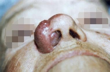
The patient underwent right-sided partial rhinectomy, with at least 5-mm margins from the visible borders of the tumor. Frozen sections revealed that all margins were free of disease. Reconstruction was accomplished immediately with a nasolabial flap. The patient's postoperative treatment included radiation therapy of 45 Gy for 5 weeks. The patient was free from recurrence at 2 years after surgery, when she died from causes unrelated to this mass.


Lab Studies

Baseline laboratory studies should include a CBC count, a chemistry profile, and liver function tests.

Imaging Studies

  • Because of the difficulties in distinguishing metastatic oat cell carcinoma from MCC, chest radiography should be performed.
  • Obtain CT scans of the chest, abdomen, and pelvis to rule out metastases.
  • Some have suggested that obtaining octreotide scanning may also be helpful for ruling out metastases.
  • The role of [18F]2-fluoro-D-2-deoxyglucose (FDG) positron emission tomography (PET) remains undefined.

    • Recent reports indicate that FDG-PET may aid in the staging and following up MCCs.
    • Yao et al (2005) reported that pretreatment FDG-PET scans revealed metastatic disease in subcentimeter lymph nodes that were not appreciated on initial CT images. Likewise, posttreatment FDG-PET scans revealed responses to therapy, with the level of FDG uptake correlating with complete responses to treatment and with residual disease.

Histologic Findings

Diagnosing MCC is difficult and requires a high degree of suspicion. Light microscopy, electron microscopy, and immunohistochemistry may be needed to confirm the diagnosis.

Light microscopy

On occasion, MCCs may be diagnosed with histology alone, but confirmation with immunohistochemistry and/or electron microscopy is always encouraged. Diagnosis by means of light microscopy alone is difficult because the appearance of MCCs is similar to that of many other undifferentiated small-cell neoplasms, especially other APUD tumors, such as metastatic oat cell carcinoma. In fact, 66% of MCCs are misdiagnosed when studied with light microscopy alone.

These points emphasize the need to consider MCC when small cell tumors are being diagnosed with light microscopy. Light microscopy reveals round-to-polygonal neoplastic cells that are compactly arranged. Rare fusiform cells are also observed. A ball-in-mitt arrangement of cells is often described, where 1 or 2 crescentic tumor cells wrap around a round tumor cell. A grenz zone usually separates the tumor from the epidermis. As a result, the epidermis is commonly spared because the MCC alternatively extends into the subcutaneous tissues, vessels, and lymphatics.

Gould et al (1985) described a widely accepted histologic classification of MCC. The classification details 3 specific cellular patterns.

The most common pattern is the intermediate cell type. These tumors display a large nest of cells without organoid architecture or recognizable palisading. A distinct disassociation exists between cells. Areas of focal necrosis and lymphocytic invasion are typical. Cytoplasm is moderate, nuclei are vesicular, and mitoses are abundant.

The trabecular cell type is observed in 25% of MCCs. It is believed to be the original tumor Toker described (1972). In this class, the cells are arranged in organoid clusters with interconnected trabeculae separated by strands of connective tissue. Clusters may show glandlike organization. Individual cells are compactly arranged round-to-polygonal cells. Cytoplasm is abundant, and the nuclei are round, centrally located, and vesicular. Pleomorphism and mitotic activity is mild to moderate.

The final and least common class is the small cell variation. This pattern consists of solid sheets and clusters of cells separated by abundant stroma, with large areas of necrosis. The cells are small with scant cytoplasm and hyperchromatic nuclei. Pleomorphism and mitoses are common.

Electron microscopy

Because of the difficulty in diagnosing MCC with light microscopy, electron microscopy plays an important role in the diagnosis of MCC. The ultrastructure of the tumor is similar to that of the normal Merkel cell. The cells are round to ovoid and intimately apposed to adjacent tumor cells, with desmosomal junctions to surrounding keratinocytes. One of the most consistent findings is the aggregation of intermediate filaments in a paranuclear location. Other characteristic findings are membrane-bound, dense core granules. The granules are usually concentrated in the periphery or in dendritelike processes.

Immunohistochemistry

Immunohistochemistry is often used to confirm MCC. Merkel cell tumors stain positively for NSE, as would any APUD cell tumor. They also demonstrate perinuclear staining with antikeratin antibodies to low-molecular-weight cytokeratins 8, 18, and 19. These 2 markers are the most constant immunohistochemical markers and are often said to be present in 100% of MCCs. A third marker, neurofilament protein, is used to distinguish MCC form oat cell carcinoma. Neurofilament protein is seen in nearly all MCCs but few oat cell carcinomas.

Other markers present with variable frequency are chromogranin, synaptophysin, vasoactive intestinal peptide (VIP), calcitonin, bombesin, corticotropic hormone (ACTH), met-encephalon, gastrin, and somatostatin.

Finally, the absence of certain markers also helps in the diagnosis of MCC by ruling out other tumors. S-100 is seen in melanoma, whereas leukocyte common antigen is present in lymphoma. Neither of these is found in MCC.

Staging

A widely used staging system is one Yiengpruksawan et al (1991) suggested, as follows.

  • Stage I - Absence of lymphadenopathy
    • Stage IA - Tumors <2 cm
    • Stage IB - Tumors >2 cm
  • Stage II - Positive regional lymphadenopathy
  • Stage III - Evidence of distant metastases


Surgical therapy

As with most tumors, treatment is based on the stage. The Yiengpruksawan system is used here (Yiengpruksawan, 1991).

Stage I

Aggressive wide local excision is the treatment of choice for the primary tumor. A 2.5- to 3-cm margin is recommended because it is thought to decrease the risk of recurrence. All margins should be confirmed with frozen sections.

The role of elective neck dissection in the absence of clinically positive nodes is controversial. Because of the low incidence of MCC, most reports are based on retrospective or anecdotal studies. Lymph node metastases develop in approximately 55% of patients; therefore, some authors recommend prophylactic neck dissection in all patients. Others recommend neck dissection after the tumor reaches 2 cm in diameter.

Silva et al (1984) recommend neck dissection for tumors with 10 or more mitoses per high-power field, for cases of lymphatic invasion, or for tumors composed of small cells. Cotlar et al (1986) recommend lymphadenectomy for tumors present longer than 6 weeks. Tumors in the midline present the problem of bilateral drainage. Goepfert et al (1984) and Hitchcock et al (1988) recommend that patients with such lesions undergo a bilateral neck dissection.

Recent studies have been conducted to investigate the efficacy of sentinel node biopsy to determine whether neck dissection is necessary. These studies have been small, but their results indicate that a negative sentinel node may obviate neck dissection.

When one contemplates neck dissection, realize that no indications are universally accepted and that the effect of regional lymphadenopathy on overall survival is uncertain.

MCC is a radiosensitive tumor, and radiotherapy is currently used as an adjuvant. Numerous authors have recommended postexcision irradiation of the primary site and lymph node drainage. Goepfert et al (1984) offer these specific indications for radiotherapy: (1) primary tumor >1.5 cm, (2) margins are <2 mm, or (3) evidence of lymphatic invasion.

In 1 of the largest series to date, adjuvant radiation did not substantially reduce local or regional recurrence.

The recommended dosing schedule is 45-50 Gy for 5 weeks; this is increased to 56-65 Gy for tumors with positive margins. These doses are similar to those used to treat squamous cell carcinoma. Therapy for recurrent localized disease or extensive unresectable stage I disease is similar to that given for stage II disease because lymphadenopathy is more likely to occur in this situation than in primary disease.

Stage II

A widely accepted practice is for patients with regional node metastases or local or regional recurrence to undergo excision of the primary lesion and lymph node dissection. Adjuvant radiation therapy to the primary site and regional nodes is generally recommended in addition to neck dissection. Chemotherapy is also currently advocated for stage II disease. Because of the morphologic and immunohistochemical similarities of MCC to small cell lung cancer, these regimens have been used as treatment models.

Pharmacologic agents most commonly used are doxorubicin and cyclophosphamide. Other agents are cisplatin, vincristine, etoposide, methotrexate, bleomycin, and 5-fluorouracil. However, reports of these treatments to date have consisted of small studies and anecdotal evidence. MCCs often respond to chemotherapy; however, as with small cell carcinoma, remission is brief. No chemotherapeutic protocol has notably increased survival rates.

Stage III

The development of distant metastases portends a poor prognosis, with a mean life expectancy of 5 months. As in stage II disease, many types of chemotherapeutic agents have been used with brief success in treating stage III disease, with no increase in the survival rate. The role of radiation therapy in disseminated disease is to achieve palliation.

Follow-up

After treatment, monitor patients closely. Recommended follow-up is every month for 6 months, every 3 months for the next 2 years, and every 6 months thereafter.


MCC is a deadly disease with a poor outlook for survival. Local recurrence occurs in 44% of patients; multiple local recurrences occur in 15%. These recurrences usually happen within 5 months after the primary lesion is treated. About 15% of patients have palpable nodes at the time of diagnosis. Lymph node metastases eventually develop in 55% of patients, and distant metastases develop in 34%. Most metastases occur before the eighth month after diagnosis.

The areas where metastases are most likely to occur are the liver, bone, brain, and lung. The presence of distant metastases is the only factor that is consistently predictive of the outcome. The mean time to death after the discovery of distant metastases is 5 months. Mortality rates for patients with distant metastases are 75-100%. In patients without distant metastases, mortality rates are 4%.


Recent advances in the treatment of MCC that are still under investigation are Mohs surgery for excision of the primary lesion and lymphoscintigraphy, intraoperative mapping of lymph nodes, and biopsy of sentinel lymph node to treat and stage occult neck disease.

O'Connor et al (1997) compared the efficacy of Mohs surgery to that of wide local excision. Following up 86 patients, they determined that Mohs surgery fared well compared with the standard treatment of wide local excision.

When weighing the choice between wide local excision and Mohs surgery, one must consider the advantage of Mohs surgery because it allows for histologic control of margins of the tumor, minimizing the extent of excision. In the head and neck, conservation of tissue is imperative to preserve vital structures. Considering this information, many authors now advocate Mohs excision in lieu of wide local excision.

In MCC, the prognosis and the treatment of the disease is largely based on the presence or absence of metastases. To permit appropriate treatment and staging, the clinician must determine the state of the regional lymphatics in the clinically negative neck. At present, prophylactic neck dissection is advocated, but the procedure causes morbidity. In addition, because drainage patterns in the head and neck are notoriously ambiguous and difficult to predict, which lymph node basins should be dissected is often unclear.

For the treatment of melanomas of the head and neck, preoperative lymphoscintigraphy and intraoperative lymphatic mapping have been used to successfully identify draining lymph node basins and localize the sentinel lymph node for biopsy. Preliminary studies have demonstrated that, in MCC, the status of the sentinel node is predictive of the status of the remaining lymph node basin.

Recent work in this area by Schmalbach et al (2005) supports these preliminary findings. Regional failure in the setting of negative findings on sentinel lymph node biopsy was observed in 1 (13%) of 8 patients. This rate of regional recurrence compares favorably with mean rates of regional recurrence reported in the literature. If data large studies confirm these findings, sentinel lymph node biopsy may provide an accurate and less morbid alternative to neck dissection for the treatment and staging of regional occult neck disease in MCC.


Media file 1:  Common distribution of Merkel cell carcinoma in the head and neck.
Click to see larger pictureClick to see detailView Full Size Image
Media type:  Image

Media file 2:  Merkel cell carcinoma affecting the right nasal ala.
Click to see larger pictureClick to see detailView Full Size Image
Media type:  Photo


  • Agelli M, Clegg LX. Epidemiology of primary Merkel cell carcinoma in the United States. J Am Acad Dermatol. Nov 2003;49(5):832-41. [Medline].
  • Ames SE, Krag DN, Brady MS. Radiolocalization of the sentinel lymph node in Merkel cell carcinoma: a clinical analysis of seven cases. J Surg Oncol. Apr 1998;67(4):251-4. [Medline].
  • Boyer JD, Zitelli JA, Brodland DG, D'Angelo G. Local control of primary Merkel cell carcinoma: review of 45 cases treated with Mohs micrographic surgery with and without adjuvant radiation. J Am Acad Dermatol. Dec 2002;47(6):885-92. [Medline].
  • Cotlar AM, Gates JO, Gibbs FA Jr. Merkel cell carcinoma: combined surgery and radiation therapy. Am Surg. Mar 1986;52(3):159-64. [Medline].
  • Crown J, Lipzstein R, Cohen S, et al. Chemotherapy of metastatic Merkel cell cancer. Cancer Invest. 1991;9(2):129-32. [Medline].
  • Ecker HA Jr, Abt AB, Graham WP 3d, Herceg S. Trabecular or Merkel-cell carcinoma of the skin. Plast Reconstr Surg. Oct 1982;70(4):485-9. [Medline].
  • Fenig E, Lurie H, Klein B, Sulkes A. The treatment of advanced Merkel cell carcinoma. A multimodality chemotherapy and radiation therapy treatment approach. J Dermatol Surg Oncol. Sep 1993;19(9):860-4. [Medline].
  • Feun LG, Savaraj N, Legha SS, et al. Chemotherapy for metastatic Merkel cell carcinoma. Review of the M.D. Anderson Hospital's experience. Cancer. Aug 15 1988;62(4):683-5. [Medline].
  • Goepfert H, Remmler D, Silva E, Wheeler B. Merkel cell carcinoma (endocrine carcinoma of the skin) of the head and neck. Arch Otolaryngol. Nov 1984;110(11):707-12. [Medline].
  • Gomez LG, DiMaio S, Silva EG, Mackay B. Association between neuroendocrine (Merkel cell) carcinoma and squamous carcinoma of the skin. Am J Surg Pathol. Mar 1983;7(2):171-7. [Medline].
  • Gould VE, Moll R, Moll I, et al. Neuroendocrine (Merkel) cells of the skin: hyperplasias, dysplasias, and neoplasms. Lab Invest. Apr 1985;52(4):334-53. [Medline].
  • Haag ML, Glass LF, Fenske NA. Merkel cell carcinoma. Diagnosis and treatment. Dermatol Surg. Aug 1995;21(8):669-83. [Medline].
  • Heenan PJ, Cole JM, Spagnolo DV. Primary cutaneous neuroendocrine carcinoma (Merkel cell tumor). An adnexal epithelial neoplasm. Am J Dermatopathol. Feb 1990;12(1):7-16. [Medline].
  • Hill AD, Brady MS, Coit DG. Intraoperative lymphatic mapping and sentinel lymph node biopsy for Merkel cell carcinoma. Br J Surg. Apr 1999;86(4):518-21. [Medline].
  • Hitchcock CL, Bland KI, Laney RG 3d, et al. Neuroendocrine (Merkel cell) carcinoma of the skin. Its natural history, diagnosis, and treatment. Ann Surg. Feb 1988;207(2):201-7. [Medline].
  • Hodgson NC. Merkel cell carcinoma: changing incidence trends. J Surg Oncol. Jan 1 2005;89(1):1-4. [Medline].
  • Koljonen VS. Merkel cell carcinoma. World J Surg Oncol. Feb 8 2006;4(1):7. [Medline].
  • Kuhajda FP, Olson JL, Mann RB. Merkel cell (small cell) carcinoma of the skin: immunohistochemical and ultrastructural demonstration of distinctive perinuclear cytokeratin aggregates and a possible association with B cell neoplasms. Histochem J. May 1986;18(5):239-44. [Medline].
  • Kwekkeboom DJ, Hoff AM, Lamberts SW, et al. Somatostatin analogue scintigraphy. A simple and sensitive method for the in vivo visualization of Merkel cell tumors and their metastases. Arch Dermatol. Jun 1992;128(6):818-21. [Medline].
  • Lawenda BD, Thiringer JK, Foss RD, Johnstone PA. Merkel cell carcinoma arising in the head and neck: optimizing therapy. Am J Clin Oncol. Feb 2001;24(1):35-42. [Medline].
  • Mendenhall WM, Mendenhall CM, Mendenhall NP. Merkel cell carcinoma. Laryngoscope. May 2004;114(5):906-10. [Medline].
  • Merkel F. Stzellen und Tastkorperchen bei den Haustieren und beim Menschen. Arkiv Mikroskopische Anatomie Entwicklungsmechanik. 1875;11:636-52.
  • Messina JL, Reintgen DS, Cruse CW, et al. Selective lymphadenectomy in patients with Merkel cell (cutaneous neuroendocrine) carcinoma. Ann Surg Oncol. Jul-Aug 1997;4(5):389-95. [Medline].
  • O'Brien CJ, Uren RF, Thompson JF, et al. Prediction of potential metastatic sites in cutaneous head and neck melanoma using lymphoscintigraphy. Am J Surg. Nov 1995;170(5):461-6. [Medline].
  • O'Connor WJ, Brodland DG. Merkel cell carcinoma. Dermatol Surg. Mar 1996;22(3):262-7. [Medline].
  • O'Connor WJ, Roenigk RK, Brodland DG. Merkel cell carcinoma. Comparison of Mohs micrographic surgery and wide excision in eighty-six patients. Dermatol Surg. Oct 1997;23(10):929-33. [Medline].
  • Pan D, Narayan D, Ariyan S. Merkel cell carcinoma: five case reports using sentinel lymph node biopsy and a review of 110 new cases. Plast Reconstr Surg. Oct 2002;110(5):1259-65. [Medline].
  • Pilotti S, Rilke F, Bartoli C, Grisotti A. Clinicopathologic correlations of cutaneous neuroendocrine Merkel cell carcinoma. J Clin Oncol. Dec 1988;6(12):1863-73. [Medline].
  • Pitale M, Sessions RB, Husain S. An analysis of prognostic factors in cutaneous neuroendocrine carcinoma. Laryngoscope. Mar 1992;102(3):244-9. [Medline].
  • Rice RD Jr, Chonkich GD, Thompson KS, Chase DR. Merkel cell tumor of the head and neck. Five new cases with literature review. Arch Otolaryngol Head Neck Surg. Jul 1993;119(7):782-6. [Medline].
  • Schmalbach CE, Lowe L, Teknos TN, et al. Reliability of sentinel lymph node biopsy for regional staging of head and neck Merkel cell carcinoma. Arch Otolaryngol Head Neck Surg. Jul 2005;131(7):610-4. [Medline].
  • Sharma D, Flora G, Grunberg SM. Chemotherapy of metastatic Merkel cell carcinoma: case report and review of the literature. Am J Clin Oncol. Apr 1991;14(2):166-9. [Medline].
  • Silva EG, Mackay B, Goepfert H, et al. Endocrine carcinoma of the skin (Merkel cell carcinoma). Pathol Annu. 1984;19 Pt 2:1-30. [Medline].
  • Stein JM, Hrabovsky S, Schuller DE, Siegle RJ. Mohs micrographic surgery and the otolaryngologist. Am J Otolaryngol. Nov-Dec 2004;25(6):385-93. [Medline].
  • Straka JA, Straka MB. A review of Merkel cell carcinoma with emphasis on lymph node disease in the absence of a primary site. Am J Otolaryngol. Jan-Feb 1997;18(1):55-65. [Medline].
  • Suarez C, Rodrigo JP, Ferlito A, et al. Merkel cell carcinoma of the head and neck. Oral Oncol. Sep 2004;40(8):773-9. [Medline].
  • Tennvall J, Biorklund A, Johansson L, Akerman M. Merkel cell carcinoma: management of primary, recurrent and metastatic disease. A clinicopathological study of 17 patients. Eur J Surg Oncol. Feb 1989;15(1):1-9. [Medline].
  • Toker C. Trabecular carcinoma of the skin. Arch Dermatol. Jan 1972;105(1):107-10. [Medline].
  • Tuneu A, Pujol RM, Moreno A, et al. Postirradiation Merkel cell carcinoma. J Am Acad Dermatol. Mar 1989;20(3):505-7. [Medline].
  • Wanebo HJ, Harpole D, Teates CD. Radionuclide lymphoscintigraphy with technetium 99m antimony sulfide colloid to identify lymphatic drainage of cutaneous melanoma at ambiguous sites in the head and neck and trunk. Cancer. Mar 15 1985;55(6):1403-13. [Medline].
  • Wasserberg N, Schachter J, Fenig E, et al. Applicability of the sentinel node technique to Merkel cell carcinoma. Dermatol Surg. Feb 2000;26(2):138-41. [Medline].
  • Wells KE, Cruse CW, Daniels S, et al. The use of lymphoscintigraphy in melanoma of the head and neck. Plast Reconstr Surg. Apr 1994;93(4):757-61. [Medline].
  • Wilder RB, Harari PM, Graham AR, et al. Merkel cell carcinoma. Improved locoregional control with postoperative radiation therapy. Cancer. Sep 1 1991;68(5):1004-8. [Medline].
  • Yao M, Smith RB, Hoffman HT, et al. Merkel cell carcinoma: two case reports focusing on the role of fluorodeoxyglucose positron emission tomography imaging in staging and surveillance. Am J Clin Oncol. Apr 2005;28(2):205-10. [Medline].
  • Yiengpruksawan A, Coit DG, Thaler HT, et al. Merkel cell carcinoma. Prognosis and management. Arch Surg. Dec 1991;126(12):1514-9. [Medline].
  • Zeitouni NC, Cheney RT, Delacure MD. Lymphoscintigraphy, sentinel lymph node biopsy, and Mohs micrographic surgery in the treatment of Merkel cell carcinoma. Dermatol Surg. Jan 2000;26(1):12-8. [Medline].

Skin Cancer: Merkel Cell Carcinoma excerpt

Article Last Updated: Apr 30, 2007