Overview
Soft tissue volume replacement with injectable materials has been a challenging problem for facial plastic and reconstructive surgeons. The quest for a safe, long-lasting, and predictable material still continues today.
Early investigators used materials based on availability and ease of application. Paraffin wax, petrolatum, vegetable oils, lanolin, silicone oil, and beeswax have been used for facial augmentation, but long-term follow-up revealed problems (eg, foreign body granulomas).
Although autogenous fat transplants have been used for decades, the field of minimally invasive soft tissue augmentation expanded with the introduction of injectable bovine collagen in 1981.
The ideal material for soft tissue replacement/augmentation has not yet been identified, whether injectable or surgically implanted; permanent or temporary; biological or synthetic; allograft, homograft, or xenograft. Therefore, materials must be judged on relative merits and disadvantages. This article reviews injectable soft tissue fillers.
The images below depict soft tissue implants.
 Patient immediately following bovine collagen injection (1 cc) for lip augmentation
Indications
Soft tissue implants are used for the following purposes:
-
To reconstruct surgically or traumatically created tissue voids
-
To restore bulk to aging tissues in order to correct soft tissue folds or rhytides
-
To augment tissue for cosmetic enhancement
Anatomy
Traditionally, skin is divided into 3 layers, epidermis, basement membrane, and dermis. The epidermis is mainly composed of cells, while the dermis contains extracellular matrix, nerves, vasculature, and adnexal structures.
Injectable soft tissue fillers are generally injected into the dermal layer. The dermal layer allows for host incorporation of the material because of the presence of fibroblasts in the extracellular matrix and vasculature. Some materials are preferably placed in the subcutaneous layer because of implant nodularity and palpability.
Injectable Materials
Synthetic Materials
Silicone
Silicone injection into facial tissues was popularized in the 1960s and 1970s with the introduction of medical grade silicone (MDX 4-4011) by Dow Corning. Microdroplets of silicone are dispersed within the dermal tissues. Fibrosis around these droplets localizes the material, and it is seemingly well tolerated in small amounts in the face.
Silicone oil has many advocates among those who used it prior to Food and Drug Administration (FDA) withdrawal of approval. However, silicone, although chemically well tolerated, becomes encapsulated as a foreign body by a chronic inflammatory reaction. Giant cells surround the material and cannot process any ingested material, establishing a low-grade inflammatory nidus. Fibrous tissue surrounds and encapsulates the silicone; this capsule is avascular and is a potential site of infection. A number of late infections, granulomas, and palpable masses have been reported following silicone use. Currently, silicone oil is approved by the FDA for ophthalmic use only but is being used off label for facial soft tissue augmentation.
Bioplastique
Bioplastique (Uroplasty BV, Netherlands) is a biphasic material. It consists of solid silicone particles (ranging in size from 100-400 µm) suspended in a polyvinylpyrrolidone (CHNO) carrier.
Once injected, the material elicits a low-grade inflammatory reaction. The carrier is removed by the body and excreted by the kidneys. Collagen encapsulates and localizes the silicone, and animal studies have shown no evidence of foreign body migration. Deposition of collagen progresses, replacing the organic component of the material in a ratio slightly higher than 1:1. Therefore, overcorrection is not advised.
Case series have reported no major complications with Bioplastique other than overcorrection. Subcutaneous placement is recommended to avoid palpable nodularity. Bioplastique is not yet approved by the FDA.
Arteplast
Arteplast was introduced in Europe in 1991. Arteplast is an injectable material composed of microspheres of polymethylmethacrylate (PMMA) suspended in a gelatin solution. Gelatin is resorbed and replaced by native collagen. A rat model has shown that collagen deposition is detected at 3 weeks, and the density of collagen increases thereafter.
The decrease in space between microspheres is believed to be the result of collagen deposition and consolidation, leading to loss of volume correction. PMMA beads average 20-40 µm in size and are theoretically subject to phagocytosis.
Artecoll/Bellafill
Artecoll was introduced in 1995 and was intended to replace Arteplast. Artecoll consists of smooth PMMA spheres (30-40 µm in size) suspended in bovine collagen (ie, 25% PMMA, 75% collagen, by weight).
Human studies indicated a need for subdermal placement and slight overcorrection. Follow-up over 1-2 years showed that 90% of patients were satisfied with treatment.
An improved form of PMMA, ArteFill, which was of greater purity, was approved in 2006 by the FDA for correction of moderate nasolabial folds (with use in the lips specifically contraindicated). In 2014, the product's name was changed to Bellafill, and in 2015, it was approved by the FDA for the treatment of acne scars. [1, 2]
Radiesse
Radiesse (Bioform, Inc, San Mateo, Calif) is a suspension of calcium hydroxylapatite (CaHA) particles in glycerin and saline. Once injected just below the dermis, the glycerin carrier is absorbed, and a mild fibrous response is deposited around the CaHA particles, stabilizing the implants and adding further bulk. The clinical effect of Radiesse is estimated to be between 12 and 18 months. Caution is advised when treating the lips, as a high risk of subcutaneous nodules has been noted when treating this area. Radiesse is currently approved by the FDA for correction of nasolabial folds.
Sculptra
Sculptra (poly-L-lactic acid, Dermik Laboratories, Berwyn, Pa) is a preparation of poly-L-lactic acid particles that are injected into the superficial subdermis, where a fibrous response is elicited. Sculptra is injected in 4-6 sessions while a volume response is created. Persistence is reported to last up to 2 years. Sculptra is currently FDA approved for the treatment of HIV protease inhibitor–induced lipodystrophy.
Biological Materials
Injectable bovine collagen
The FDA approved injectable bovine collagen (ie, Zyderm) in 1981. Zyderm is a suspension of collagen fibrils from chemically processed bovine skin. Zyplast contains the same material, but collagen fibers are cross-linked with glutaraldehyde.
Human and bovine collagens differ in terminal telopeptide units. These telopeptides must be enzymatically removed during the Zyderm manufacturing process to reduce immunogenicity.
Zyderm and Zyplast (Collagen Corp, Palo Alto, CA) require skin testing because of a 3% reactivity risk. Studies show an average of 4-6 months of histologic persistence for Zyderm; most users of bovine collagen describe 3-4 months of clinical effect from Zyplast injection, while Zyderm results are shorter lived. CosmoDerm and CosmoPlast were introduced to limit allergenic potential and are analogs of Zyderm and Zyplast, respectively. Collagen fibers for these are produced by human fibroblasts in vitro, rather than derived from bovine skin. Longevity of clinical effect is similar.
Zyderm I and I, Zyplast, CosmoDerm I and II, and CosmoPlast have been withdrawn from the US market, with the lag time needed for skin testing having contributed to reduced popularity for bovine collagen. [3]
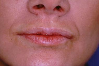
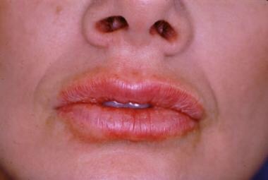
A patient can be seen prior to bovine collagen injection for lip augmentation in the first image below and immediately following injection in the second image below.
Patient immediately following bovine collagen injection (1 cc) for lip augmentation
Injectable porcine collagen
Porcine collagen cross-linked in a 3-dimensional structure has been available as Evolence. This material is best injected into the deep dermis or at the dermal-subdermal border, and its clinical effects appear to last about 12 months. Although clinical results were generally quite good, [4] the manufacturer has withdrawn this material from sale in the United States.
Hyaluronan derivatives
Hyaluronic acid (HA) is a polysaccharide that plays an integral role in stabilizing the extracellular matrix. It does not elicit any humoral or cell-mediated immune response and therefore does not require pretreatment skin tests.
Restylane (Medicis Aesthetics, Scottsdale, Ariz) is a gel formed by cross-linking bacteria fermented HA via carbon bridges and particulated into beads at 200 µm in size. The same manufacturer also supplies Perlane, which is chemically identical to Restylane but has a larger particle size. Perlane is injected subdermally and lasts approximately 9-12 months, while Restylane is injected into the mid-dermis and persists for 5-7 months.
Hylaform (Inamed Aesthetics, Santa Barbara, Calif) is an insoluble derivative formed by treating animal-derived HA with vinyl sulfone, while Captique is bacteria-derived HA cross-linked in a similar way.
Juvéderm is a spectrum of materials composed of nonanimal source HA cross-linked in a way similar to Restylane, but to a much greater degree. It has been approved for facial soft tissue augmentation by the FDA, and seems to behave similarly to Restylane and Perlane.
In clinical trials, Restylane and Juvéderm have been shown to persist clinically in nasolabial folds as long as 6 months, while Hylaform and Hylaform Plus persist only about 3 months (Hylaform and Hylaform Plus are no longer sold in the United States). FDA-approved modifications of Restylane (Perlane) and Juvéderm Ultra (Juvéderm Ultra Plus) are thicker materials with larger HA particles designed to be injected subdermally. [5] These fillers work particularly well for treatment of nasolabial folds and last 12 months or more. [6, 7]
Belotero is a recent addition to the US market and is also an HA derivative. Belotero appears to yield a smoother correction, especially soon after treatment, but is otherwise very similar to Restylane and Juvéderm. [8]
Other HA materials include Elevess and Prevelle. Published studies with these materials are lacking, although they are attractive fillers because they contain lidocaine, which aids in providing maximal patient comfort.
Fibrel
Fibrel (Mentor Corp, Goleta, Ga) is a mixture of plasma, porcine-derived gelatin, and e-aminocaproic acid. The mixture is injected intradermally. The resultant tissue reaction leads to fibrinogen deposition, followed by fibrinolysis and collagen formation.
Fibrel is FDA-approved for treatment of depressed scars and rhytides. Studies describe increasing collagen content of the wound site over the first 6 months, followed by partial reduction of collagen by 9 months. Overcorrection is recommended. Fibrel is currently not available commercially.
Homologous Materials
Dermalogen
Homologous collagen dispersion (Dermalogen, Collagenesis Inc, Beverly, Mass) is a suspension of processed dermis obtained from the tissue banks accredited by the American Association of Tissue Banks (AATB).
A mechanical process homogenizes the decellularized material to produce a suspension of type I collagen, traces of types III and VI collagen, elastin, fibronectin, chondroitin sulfate, and other proteoglycans.
Dermalogen is well tolerated and elicits a low-grade inflammatory response. It is injected into the superficial to mid-dermal level. Dermalogen appears to persist between 3-6 months. Dermologen is currently not clinically available.
Cymetra
Acellular dermal graft (AlloDerm Acellular Tissue, LifeCell Corp, Branchburg, NJ) is prepared from donated human skin that is processed into an acellular matrix of dermal proteins and proteoglycans.
The FDA classifies it as banked human tissue when used for damaged or insufficient soft tissue replacement. To some degree, the persistence of sheet AlloDerm placed through an incision appears to be permanent (ie, 20-50% at >1 year).
An injectable form, Cymetra allows injection through a 26-gauge needle and promotes deep dermal fibrosis. A series of injections is necessary in order to build adequate augmentation.
Autologous Materials
Autologous fat injections may offer dual benefits, as most patients desire reduction of some area of excess adipose tissue. Tissue harvested via liposuction can be used for volume augmentation. This has been well described by Coleman. However, this technique appears to be highly operator-dependent. Most workers have been unable to duplicate Coleman's results and instead describe only a 10-20% residual effect after the first 12 months.
Isolagen (Isolagen Technologies Inc, Paramus, NJ) refers to cultured autologous fibroblasts. Following a skin biopsy, autologous fibroblasts are expanded in vitro. A suspension of these fibroblasts is injected into the dermis, where, theoretically, they produce collagen.
West and Alster treated 11 patients with Isolagen, noting persistent correction in nasolabial folds as long as 6 months following injection. [9] Longer follow-up is necessary. Isolagen is currently sold as LaViv (Fibrocell Science, Inc.)
Surgically Implanted Materials
Synthetic Materials
Expanded polytetrafluoroethylene (e-PTFE) is by far the most commonly used synthetic implantable material for soft tissue augmentation. [10] Currently, it is marketed as Gore-Tex subcutaneous augmentation material (SAM) or Advanta implants. These materials have been used extensively for nasolabial fold correction, lip augmentation, and prevention of Frey syndrome following parotidectomy. E-PTFE is very well tolerated and glides easily through tissue, making the necessary surgical cavity only slightly larger than the implant itself. The material is microporous and displays minimal tissue ingrowth. To promote tissue stabilization of the implant, Advanta implants have a dual porosity, with a softer, larger pore center and a smaller pore periphery that allows limited soft tissue ingrowth.
Biological Materials
Homologous
Acellular dermal graft (AlloDerm, LifeCell Corp, Branchburg, NJ) is derived from skin obtained from tissue banks. The skin is de-epithelialized, and the dermis is then decellularized, leaving a matrix of dermal protein. AlloDerm can be used for correction of nasolabial folds, for correction of atrophic lips, and for reconstruction of large-volume deficits following tumor extirpation. Long-term data is still pending, but AlloDerm does seem to persist indefinitely to some variable degree.
Autologous
Many autologous materials have been suggested (eg, fascia lata, dermal-fat grafts). Fascia is generally too thin to provide significant bulk. Dermal-fat grafts can be used successfully, especially if skin excision is already planned; careful and thorough removal of all epidermis is necessary to prevent epidermal inclusion cyst development.
Complications
Although infection is always a possibility with use of any of the materials described above, some materials have unique complications. Silicone can cause foreign body reactions and rejection of the implant for up to 20 years following implantation. Synthetic implants of any kind can produce nodularity if placed in an improper plane. Zyderm and Zyplast cause allergic responses in up to 3% of those injected. Sculptra has been associated with subcutaneous nodules in over 50% of patients in one study, although this is now believed to be a technical issue related to inadequate hydration and mixing of the filler after reconstitution.
A retrospective study by Kern et al indicated that patients who undergo injectable filler treatments performed by qualified clinicians only rarely suffer serious adverse events. Out of 7659 individuals in the study who underwent filler injections, as administered by board-certified dermatologists, the investigators identified only four such events, including three instances of delayed-onset skin nodule formation and one instance of vascular compromise followed by cutaneous necrosis. A total of 18,013 mL of filler was administered to the patient group, with hyaluronic acid derivatives making up 74.1% of this volume, and poly-L-lactic acid and calcium hydroxylapatite making up 19.19% and 6.71%, respectively. [11]
Future and Controversies
The search for a natural, safe, easy to administer, and permanent soft tissue filler continues. Great strides have been made in the last 25 years. As the understanding of tissue physiology continues to improve, the ideal soft tissue implant may eventually be produced.
-
Patient prior to bovine collagen injection for lip augmentation.
-
Patient immediately following bovine collagen injection (1 cc) for lip augmentation


