Overview
Background
Electronystagmography (ENG) is a study used to clinically evaluate patients with dizziness, vertigo, or balance dysfunction. ENG provides an objective assessment of the oculomotor and vestibular systems. [1, 2]
The vestibular system monitors the position and movements of the head to stabilize retinal images. This information is integrated with the visual system and spinal afferents in the brain stem to produce the vestibuloocular reflex. [3]
Essentially, the standard ENG test battery consists of the following 3 parts:
-
Oculomotor evaluation
-
Positioning/positional testing
-
Caloric stimulation of the vestibular system
Indications
Although ENG is the most widely used clinical laboratory test to assess vestibular function, normal ENG test results do not necessarily mean that a patient has typical vestibular function. ENG abnormalities can be useful in the diagnosis and localization of site of a lesion. However, many abnormalities are nonlocalizing; therefore, the clinical history and otologic examination of the patient are vital in formulating a diagnosis and treatment plan for a patient who presents with dizziness or vertigo.
Although ENG testing cannot be used to determine the specific site of lesion, the information acquired can be integrated with history, symptoms, and other test results to aid in diagnosis. Comparing results obtained from various subtests of an ENG evaluation assists in determining whether a disorder is central or peripheral. In peripheral vestibular disorders, the side of lesion can be inferred from the results of caloric stimulation and, to some degree, from positional findings. An ENG evaluation can also be useful in ruling out potential causes of dizziness.
In a large study of patients tested with ENG in a wide range of clinical settings, Stockwell [4] found abnormal test results in approximately 39% of patients tested, and only about 29% of test results revealed the site of lesion. Peripheral vestibular abnormalities were found in approximately 23%, whereas central abnormalities were found in only approximately 5% of patients tested.
Periprocedural Care
Patient Education & Consent
Inform patients that ENG may cause dizziness, nausea, or both. Patients should be advised to limit food intake prior to examination and arrange for transportation after the examination, which usually takes 1-1.5 hours.
For many parts of the test, mental tasking is necessary to prevent central suppression of responses. If the examiner does not speak the patient's language or the patient is hearing impaired, an interpreter may be necessary to assist in instructing, explaining, and mental tasking.
Pre-Procedure Planning
Outer and middle ear status should be confirmed prior to caloric assessment. Presence of drainage in the outer ear canal may affect the results obtained with air caloric stimulation because moisture changes the calibrated temperature, thus limiting interpretation. Pressure equalization tubes or perforation of the tympanic membrane precludes the use of water irrigation.
Large unilateral perforations limit the interpretation of air caloric irrigations. Large perforations can increase stimulation with cool air above calibrated expectation and can exhibit a cooling effect for warm air as moisture of the middle ear mucosa is evaporated. Excessive cerumen must be removed prior to any caloric stimulation. Middle ear fluid limits the effective stimulation of the vestibular system with air and water.
Many medications can affect test results. With physician approval, patients should discontinue all medications, unless contraindicated, for 24-72 hours prior to testing. Any medications taken should be clearly noted on the test results. Alcohol consumption can affect ENG test results for 72 hours after ingestion; results are unpredictable because alcohol can be an agonist or antagonist.
Equipment
ENG equipment varies from basic to advanced. Traditional systems use a strip recorder for acquisition of responses. Although some traditional systems may still be in use, most systems currently provide computerized stimulus generation, response acquisition, and interpretation.
Stimulus Generation
Oculomotor stimulus generation was initially accomplished with manually controlled objects. Gaze and saccadic testing used dots placed on the wall and/or ceiling at specified places (eg, 10 degrees, 20 degrees, and 30 degrees in each direction), optokinetic testing was conducted with a rotating drum with stripes of contrasting colors, and smooth pursuit testing was conducted with a swinging pendulum or metronome.
Although these systems could provide gross evaluation of the various oculomotor subsystems, fine analysis of latency and accuracy of eye movements in response to the stimulus was not possible. Systems with computer-generated stimuli locked to the acquisition of the responses allow for more sophisticated analysis (eg, latency, amplitude). Computerized systems may use light-emitting diodes on a light bar anchored to the wall or in a self-contained oculomotor stimulator or computer-generated stimuli projected on a screen or video monitor.
Response Acquisition
For response acquisition, the clinician can choose to use only 1 channel to evaluate horizontal eye movements. However, a minimum of 2 channels is recommended; this facilitates evaluation of both horizontal and vertical eye movements. A 3-channel recording system further increases the power of analysis by allowing horizontal recording of each eye and vertical recording of one. Finally, some newer systems provide acquisition of both horizontal and vertical movements of each eye.
Electrodes
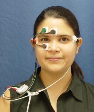
Traditional ENG includes the use of electro-oculography to objectively measure eye movements. This recording is possible because of the corneal-retinal potential difference; the cornea is electropositive relative to the retina. With a fixed recording site, voltage differences can be recorded for eye movements. Electrodes are placed around the patient’s eyes to record the corneal-retinal potential differences. By placing electrodes on both a horizontal and vertical axis around the eyes, tracings are produced for eye movements on both axes (see the image below).
Infrared Video
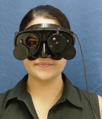
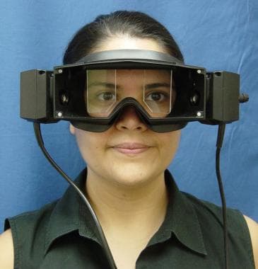
Increasingly, clinicians are using infrared technology to record eye movements for ENG. These systems are commonly referred to as videonystagmography (VNG). Horizontal and vertical tracings of eye movements are produced by the camera tracking the pupil of the eye. Since the camera uses infrared technology, these tracings can be made with the patient in complete darkness, thereby eliminating any visual fixation points. The systems may use a camera for one or both eyes, as shown in the images below.
VNG offers many advantages that make it a preferred method over electrodes. [5] Probably the most important of these advantages is that all eye movements are captured on video and can be viewed by the clinician during and after testing. In addition to capturing horizontal and vertical eye tracings, the clinician is able to visually assess for torsional eye movement by observing the striations of the iris. This is especially important for the diagnosis of benign paroxysmal positional vertigo.
According to a study of 100 patients suffering from vestibular disorders of peripheral, central and mixed origin, VNG is equally effective as ENG for assessing vertigo and distinguishing peripheral and central vestibular lesions. [6]
Patient Preparation
If the patient has back or neck injuries, consider positional testing (head hanging) and the Dix-Hallpike maneuver to avoid further complications.
The clinician may want to screen for vertebrobasilar insufficiency prior to any positional or positioning test that may constrict the vertebrobasilar artery (ie, head-hanging or Dix-Hallpike maneuvers). This may include having the patient engage in mental tasking (eg, counting, reciting multiplication tables) while gradually tilting the head back and then holding. Change in cognitive status or reports of lightheadedness may be significant. This screening method is especially important for older patients.
Technique
Approach Considerations
For the oculomotor portion of the evaluation, patients must have adequate vision to follow targets. Although the oculomotor portion cannot be completed without adequate vision, the rest of the ENG and VNG test battery can be conducted in patients with limited or absent vision.
It is also important to note that oculomotor stimulation with lights may cause seizures.
Oculomotor Evaluation
The visual system provides information about whether the environment is stationary or mobile. Stabilization of visual information is accomplished by foveation. The visual system is subserved by the motor system, the premotor system, and the command (control) eye movement system, which controls saccadic eye movements, pursuit, and vergence.
The oculomotor portion of ENG assesses eye movement function for the various command eye movement systems in the absence of vestibular stimulation. For oculomotor evaluation, the patient is presented with various visual stimuli while his or her eye movements are recorded.
Saccades (Calibration) Test
The saccade test, also called the calibration test, evaluates the saccadic eye movement system. [7] This system is responsible for rapid eye movements and refixation of the target on the fovea.
For saccadic testing, one may place dots on the wall or ceiling at specified distances from each other (usually center and 10, 20, and 30 degrees off center) and then instruct the patient to look back and forth between the dots, keeping the head fixed.
More current technology allows the examiner to control (via computer) the presentation of pinpoints of light on a light bar, on a projection screen, or in an oculomotor stimulator. This allows randomized presentation of the stimuli and more specific assessment of eye movements. With older technology, a clinician could only visually assess the tracings of eye movements to look for gross deviations from normal. With computer-generated stimuli and analysis, the clinician can assess latency and velocity of eye movements.
Saccadic test results are influenced by patient cooperation and visual acuity. Various drugs can also affect performance. Accuracy, latency, and velocity should all be taken into consideration when interpreting saccades.
The following accuracy issues may be noted.
Also known as calibration overshoot, hypermetric saccades occur when a patient has difficulty measuring the distance required for the muscular act necessary for following the target. In these cases, the clinician may note an overshoot of the target followed by a correction. Patients who consistently exhibit hypermetric saccades may have ocular dysmetria, which is suggestive of a central nervous system (CNS) lesion at the level of the cerebellum.
Ocular flutter is evidenced as a type of spiky overshoot; the patient overshoots the target several times with a short duration between overshoots. This is also suggestive of CNS pathology.
With hypometric saccades, the patient undershoots the target. Occasionally undershooting a target is normal; the undershoot must be reproducible and must occur frequently to be considered abnormal. If extreme, hypometric saccades are suggestive of basal ganglia pathology.
Multistep saccades occurs when a patient undershoots the target and then attempts to correct with multiple small saccades. This pattern is suggestive of CNS pathology.
Also called glissade, postsaccadic drift is seen as a drifting of eye movement after the saccade. This pattern is consistent with cerebellar pathology.
Pulsion is seen in vertical saccades in patients with posterior inferior or superior cerebellar artery syndrome. The pattern includes a pulling to the left or right of the eyes when completing vertical saccades.
Disorders in latency are primarily due to prolongation of saccades. Saccades characterized by short latency are usually due to an artifact or the patient anticipating the position of the target. If prolongation of saccades is evident, the examiner should rule out inattention or uncooperative behavior. Prolongations of more than 400 ms in attentive and cooperative patients may be suggestive of CNS pathology.
Asymmetrical latencies can occur in patients with lesions in the occipital or parietal cortex. For these patients, saccades in one direction may be normal with a prolongation of saccades in the opposite direction.
The following velocity issues may be noted.
If saccadic slowing is observed, the examiner should first rule out drowsiness or drug effects. In the absence of these, saccadic slowing is consistent with various CNS or ocular disorders, including oculomotor weakness, degenerative conditions, basal ganglia pathology, and cerebellar disorders.
As with saccades that evidence an abnormally short latency, abnormally fast saccades are usually an artifact and may be due to technical difficulties. However, in some cases, abnormally fast saccades may suggest CNS or ocular pathology (ie, ocular flutter). [8]
Asymmetry in saccadic velocity can be observed as an asymmetry between the eyes or between directions. When asymmetry is evident, the clinician may suspect ocular nerve or muscle pathology (ie, lesions, palsies). CNS pathology may also be suspected. A lesion in the medial longitudinal fasciculus that causes internuclear ophthalmoplegia may evidence as asymmetrical saccadic velocity.
Gaze Testing
Gaze testing is conducted to evaluate for the presence of nystagmus in the absence of vestibular stimulation. In the ENG test battery, essentially 3 types of information are obtained: presence or absence of spontaneous nystagmus (no task or center gaze); presence, absence, or exacerbation of nystagmus with addition of off-center gaze tasks to stress the system; and fixation suppression of spontaneous nystagmus.
For gaze testing, the patient is instructed to look straight ahead and then fixate on a target 30 degrees to the right, left, up, and down. Fixation is maintained for approximately 30 seconds in center gaze and 10 seconds in eccentric gaze. The test can be conducted with the same targets placed on the wall or ceiling for calibration or with computer-generated stimuli as mentioned above.
For evaluation of spontaneous nystagmus, eliminating any possibility of suppression is important. Fixation suppression can be eliminated by having the patient's eyes open in a dark room with Frenzel lenses or dark goggles used for infrared assessment. With electrode-based systems, spontaneous nystagmus may be evaluated with the patient’s eyes closed. In addition, the patient may be asked to participate in a number of mental tasks (eg, answering questions, counting by multiples of 2).
Square-wave jerks are the most common abnormality found with eyes closed (absence of visual fixation). Caution must be exercised in the interpretation of square-wave jerks. Many healthy patients exhibit this pattern with their eyes closed. Furthermore, the frequency of square-wave jerks increases with age. In young patients, square-wave jerks may be considered atypical if they occur more frequently than once per second or with visual fixation. In such cases, square-wave jerks are suggestive of a cerebellar disorder. [9]
Spontaneous nystagmus may indicate either central or peripheral pathology. The presence of nystagmus with visual fixation is always diagnostically significant.
Peripheral indicators include horizontal or horizontal rotary nystagmus, nystagmus suppressed by visual fixation, non–direction-changing nystagmus, and nystagmus exacerbated by gazing in the direction of the fast phase.
Central indicators include vertical nystagmus, nystagmus not suppressed by fixation, and direction-changing nystagmus.
By Alexander's law, nystagmus evident with visual fixation always beats in the same direction and increases when the patient gazes in the direction of the fast phase. Nystagmus decreases or disappears when the patient gazes in the direction opposite to the fast phase. This pattern is often seen in peripheral vestibular disorders and occasionally in central disorders.
Unilateral gaze-paretic nystagmus only occurs with eccentric gaze in one direction. Elicited nystagmus beats in the direction of the gaze. This is consistent with CNS pathology.
With bilateral gaze-paretic nystagmus, when the patient gazes to the right, nystagmus is elicited that beats to the right; when the patient gazes to the left, left-beating nystagmus occurs. This pattern suggests CNS pathology.
Bruns nystagmus is a combination of unilateral gaze-paretic nystagmus and vestibular nystagmus, which is evidenced as nystagmus in both directions of a gaze that is asymmetrical. Bruns nystagmus is associated with extra-axial mass lesions on the side of the gaze-paretic nystagmus.
Congenital nystagmus often has a spiky appearance and increases with lateral gaze. Congenital nystagmus may decrease in velocity or completely disappear in the absence of visual fixation.
Rebound nystagmus is characterized by a burst of nystagmus that lasts approximately 5 seconds and begins when the eyes return to center gaze. When this is present, the clinician may suspect brainstem or cerebellar lesions.
For peripheral lesions, nystagmus that is evident with eyes closed or in the dark should be suppressed by visual fixation. If visual fixation does not suppress nystagmus, CNS pathology is possible.
Smooth Pursuit Tracking
The smooth pursuit system is responsible for following targets within the visual field. Tracking can be evaluated horizontally and vertically. As a rule, vertical tracking is not as smooth as horizontal, even in healthy subjects. Care should be taken in interpreting smooth pursuit test results in geriatric and pediatric patients. Tracking is also affected by attention and patient cooperation.
In smooth pursuit tracking, the patient is instructed to follow a sinusoidal moving target with his or her eyes only. The target can be a pendulum, metronome, or computer-generated stimulus.
Tracking test results should resemble a smooth sinusoid. Breakup of movement may indicate CNS pathology. Nystagmus may be seen in tracking test results when also observed in spontaneous (center gaze) recordings.
Optokinetic Testing
For optokinetic testing, the patient tracks multiple stimuli. These may take the form of stripes on a rotating drum or a stream of lighted dots across a light bar or the field of an oculomotor stimulator. Some recent devices allow more natural stimulation, such as a full-field array of moving stars or trees. Stimuli are moved at a rate of 300, 400, or 600 per second in each direction. Some clinics use both slow and fast speeds; others test at one intermediate speed only.
Eye movements that are generated by moving fields resemble nystagmus. The clinician primarily evaluates symmetry of the response. If responses are not symmetrical, CNS pathology may be suspected. Some patients are unable to appropriately complete this task in either direction.
Positioning and Positional Testing
Dix-Hallpike Maneuver
The Dix-Hallpike maneuver is conducted specifically to assess the presence or absence of nystagmus associated with BPPV. [10, 11] When test results are classically positive, canalith repositioning and vestibular rehabilitation therapy may be indicated. [12]
The Dix-Hallpike maneuver is performed by turning a patient's head to the right or left and then briskly assisting him or her to a supine position with the head hanging to the right or left. The patient is left in this position for a brief period (at least 20 seconds) while eye movements are observed. Finally, the patient is returned to a sitting position. If nystagmus is observed, the test is repeated to evaluate fatigability of the response. Because one of the typical signs of BPPV is fatigability, the Dix-Hallpike maneuver should be completed before any other positional testing.
Patients with posterior canal BPPV present with a geotropic rotary nystagmus. [13] Because the rotary component is not acquired with traditional electrode systems, the clinician must use infrared technology or Frenzel lenses to observe the direction of rotation. Response can be suppressed with visual fixation. Keeping the eyes open in a room with enough light for the examiner to observe eye movements may not allow accurate representation of the response.
If rotary nystagmus is observed, the results must have the following 4 characteristics to be considered classically positive:
-
Delayed onset, in which nystagmus begins a few seconds after the patient is positioned
-
Transient burst of nystagmus, in which nystagmus lasts less than a minute
-
Subjective report of vertigo
-
Fatigability, in which the response decreases with repetition
In addition to these 4 characteristics, the clinician may observe a reversal of the nystagmus. When the patient resumes the sitting position, the nystagmus may begin beating in the opposite direction.
When a classically positive response is observed during the Dix-Hallpike maneuver, a peripheral lesion on the side that is down when the nystagmus occurs may be indicated.
Positional Tests
The examiner places the patient in each position and evaluates him or her for a minimum of 20-30 seconds. Mental tasking is used to keep the patient from suppressing nystagmus. Visual suppression must also be avoided by the use of infrared goggles or with the patient’s eyes closed with electrodes. Some standard positions used include the following:
-
Head hanging
-
Supine
-
Supine, head right
-
Supine, head left
-
Lateral right
-
Lateral left
Many clinics do not assess in the lateral right and left positions unless nystagmus is observed in the supine position with the head to the right or left. The lateral right and left positions are used to rule out neck rotation as a cause for nystagmus.
Nonstandard positions include any position in which the patient reports dizziness (eg, dizziness when bending to tie shoes).
If no nystagmus is observed in any position, results are considered normal. For results to be considered abnormal, the nystagmus observed in positional testing should exceed 6 degrees per second, change direction in any 1 position, persist in at least 3 different positions, or be intermittent in all positions. Lesser degrees of nystagmus are of questionable pathologic significance.
If spontaneous nystagmus is observed, the nystagmus observed during positional testing must show an increase in velocity to be considered a significant positional finding.
Peripheral indicators include the following:
-
Direction-fixed nystagmus
-
Direction of nystagmus changing in different positions in a geotropic pattern (also consider a horizontal canal variant of BPPV in this case)
-
Latency of onset
-
Fatigability
Central indicators include the following:
-
The direction of nystagmus changing in different positions in an ageotropic pattern
-
The direction of nystagmus changing in a single position (a strong indicator for CNS pathology)
-
Immediate onset of nystagmus
-
Nonfatigability
Caloric Stimulation
Caloric stimulation of the vestibular system offers an assessment of the lateral semicircular canal. [14] This is a valuable tool because it allows for the objective measurement of function from each labyrinth individually. This test is considered nonphysiologic since isolating and stimulating one labyrinth at a time in nature is physiologically impossible. The equivalent speed of stimulation for this test is a very low frequency. Although this is a limiting factor in diagnosis, other tests of vestibular function at higher frequencies are currently not readily available in most clinical settings. [15, 16]
Vestibular stimulators include water, air, and closed-loop cuff; water and air are commonly used. Water caloric irrigations provide a strong stimulus but cannot be used with patients with pressure equalization tubes or perforation of the tympanic membrane. Use of water as a stimulus should also be avoided in patients whose immune systems are compromised.
For caloric stimulation, the patient is placed in a reclining position with his or her head at a 30-degree angle. This position orients the lateral semicircular canals in the most vertical plane. Before recording responses to caloric stimulation, spontaneous nystagmus is evaluated in this position. Caloric stimulation can be accomplished with an alternating binaural bithermal, simultaneous binaural bithermal, or monothermal protocol. Simultaneous binaural bithermal protocols are not traditionally used clinically; thus, alternating binaural bithermal and monothermal protocols are discussed here.
In patients with responsive vestibular systems, caloric irrigation produces nystagmus in a predictable manner:
-
Cool irrigations: The fast phase of nystagmus beats in the direction opposite to the stimulated ear (ie, cool irrigation in the right ear causes left-beating nystagmus)
-
Warm irrigations: Nystagmus beats in the direction of the stimulated ear (ie, warm stimulation of the right ear produces right-beating nystagmus)
For a comparison of responses between ears, the test must be performed in exactly the same manner for each ear. Careful otoscopic examination allows the stimulus to be directed appropriately and at an equivalent depth in each ear canal. Furthermore, when using electrodes, recalibration is recommended prior to each irrigation to account for variations in corneal-retinal potential. [17]
Alternating Binaural Bithermal Caloric Stimulation
Alternating binaural bithermal caloric stimulation, which is probably the most commonly used protocol for caloric stimulation, includes the following conditions:
-
Right ear cool (RC)
-
Left ear cool (LC)
-
Left ear warm (LW)
-
Right ear warm (RW)
This test order elicits nystagmus responses of opposing directions with each change in temperature or side (eg, left beating, then right beating). However, evidence shows that no physiological order effect is present after caloric irrigations. [17] Thus, for practical reasons, some practitioners recommend beginning with warm irrigation of the suspect ear, since that test result is the most useful for the assessment should the patient decline further testing at that point.
During each stimulation, the cool or warm stimulus is delivered for a preset time that is determined by the type of stimulus and normative data. The patient is instructed to engage in a mental task during delivery of the stimulus. After the stimulus is removed, recording continues while the patient is mentally tasked.
The patient is given a break of 3-5 minutes between the RC and LC calorics and between the LW and RW. A break of 8-10 minutes is given between the LC and LW conditions.
When adequate data have been recorded, fixation suppression testing is conducted.
Monothermal Caloric Irrigations
Some clinics use monothermal caloric irrigations as a screening test for patients with normal oculomotor, positional, and positioning test results. For the monothermal screening test, only warm caloric irrigations are conducted. The warm stimulus has a stimulatory effect on the labyrinth and thus may increase the likelihood of revealing residual function. [18]
If the responses from each ear are equivalent and robust, testing is terminated. If a difference in responses exists between the 2 ears or the responses are less than 20 degrees per second, bithermal caloric testing is conducted.
Ice Water Caloric Stimulation
Ice water caloric stimulation is used to determine whether residual vestibular function exists in patients who have no caloric response with standard stimuli.
To administer this test, water and ice are placed in a cup. In the standard position for caloric testing, a basin is placed beneath the patient's ear, and the patient's head is turned to the side. With a syringe, 2 mL of water is placed into the external ear canal and retained for 20 seconds. At the end of 20 seconds, the water is drained into the basin, and the head is returned to the normal position for caloric testing. Recordings are obtained to determine the presence or absence of nystagmus.
Interpretation
In the normal vestibular system, adequate, equivalent responses should be obtained from each ear. A normal caloric response does not rule out a vestibular pathology, however, since this test only measures a response from part of the labyrinth at a very low frequency of stimulation.
Slow phase velocity is determined for each recording for use in the following calculations.
Unilateral weakness (UW) is used to evaluate symmetry, as calculated by the following equation:
%UW = [((RC + RW) – (LC + LW))/(RC + RW + LC + LW)] × 100
A negative number indicates a right unilateral weakness, and a positive number indicates a left unilateral weakness. Unilateral weakness is indicative of a peripheral vestibular lesion that involves the nerve or end-organ on the side of the weakness. In many clinics, a UW greater than 25% is significant.
Average caloric responses of 6 degrees per second or less are consistent with a bilateral weakness. Borderline bilateral weakness is noted when the average responses are between 7-9 degrees per second. Abnormally weak bilateral responses may be due to bilateral peripheral vestibular pathology or central interruption of the vestibuloocular reflex. When a borderline bilateral weakness or bilateral weakness is observed, drug effects should be excluded.
If the patient has spontaneous nystagmus, directional preponderance (DP) is evident, as calculated by the following equation:
%DP = [((LC + RW) – (RC + LW))/(RC + RW + LC + LW)] × 100
In general, a directional preponderance greater than 20-30% is considered significant.
Fixation Suppression
After each caloric stimulus, the patient is instructed to fixate on a light or other stationary target. Fixation should normally eliminate or greatly reduce the induced nystagmus. If visual fixation does not inhibit nystagmus, central pathology at the level of the brain stem is indicated. The clinician must ensure that fixation suppression testing is completed as soon as the peak nystagmus response has been recorded. Fixation suppression testing is not accurate if the clinician waits to introduce the stimulus until the response has naturally decayed.
Compute the fixation index (FI):
FI = EO/EC,
where EO = nystagmus with eyes open (visual fixation) and EC = nystagmus with eyes closed (without visual fixation). An FI of 0.60 or greater is considered significant.
Special Applications
Pressure Fistula Test
Some patients have vertigo caused by a perilymph fistula. Fistula testing can be conducted with ENG to determine which patients are likely to benefit from perilymph fistula repair surgery. [19]
For the pressure fistula test, the patient is placed in an upright position. While the patient is involved in mental tasks, recordings are conducted with the patient’s eyes open in the dark (wearing infrared goggles) or with eyes closed with electrodes. The presence or absence of spontaneous nystagmus is noted. Next, a probe from an immittance bridge is placed in the ear canal and a seal is obtained. Pressure is then varied from 0-200 mm H2 O and held for approximately 15 seconds. Pressure is then decreased to -400 mm H2 O and held for 15 seconds. The patient is questioned for subjective symptoms.
The presence of a fistula is confirmed if the pressure causes nystagmus.
Tullio Phenomenon
This is not as routinely used as pressure fistula testing, but the Tullio phenomenon is an option for patients on whom a hermetic seal cannot be obtained.
Follow the procedure for a pressure fistula test, but instead of varying the air pressure, present a 500-Hz tone at 95 dB for no longer than 3 seconds. Monitor eye movements and ask the patient about his or her subjective experience.
Results of Tullio phenomenon testing are positive if nystagmus is observed.
Head-Shake Nystagmus Test
The head-shake nystagmus test is most useful in the assessment of vestibular disorders that produce asymmetries in vestibular function. [20, 21, 22, 23]
For this test, the patient is placed in an upright sitting position with his or her head tilted forward 30 degrees. The examiner rotates the head back and forth (45 degrees to either side), completing 30 full cycles at a frequency of about 2 cycles per second. If nystagmus is observed following head rotation, eye movements should be recorded for at least 1 minute.
Nystagmus produced after the head rotation is considered significant if at least 5 consecutive beats of at least 2 degrees per second are observed. Head-shake nystagmus is described by the following classifications:
-
Monophasic, in which the nystagmus produced does not change directions
-
Biphasic, in which the nystagmus produced initially beats in one direction and then fatigues and reverses directions
-
Paretic, in which the initial nystagmus observed beats away from the side of lesion
-
Reversed, in which the initial nystagmus observed beats toward the side of lesion
-
Cross-coupled, in which a strong vertical nystagmus is produced by head shaking on the horizontal axis
A positive finding of head-shake nystagmus is highly suggestive of an underlying vestibular pathology. A negative head-shake nystagmus test result does not rule out a vestibular pathology. Head-shake nystagmus usually beats away from the side of a peripheral lesion; however, because this is not the rule, head-shake nystagmus should be used in conjunction with other vestibular tests in determining the side of the lesion.
Cross-coupled head-shake nystagmus is suggestive of CNS pathology.
Vibration-Induced Nystagmus
Nystagmus may be induced in patients by stimulating the head and/or neck with vibrations. Vibration-induced nystagmus may be useful in the diagnostic confirmation of unilateral lesions.
For vibration-induced nystagmus, the patient is placed in an upright sitting position and a vibrator is applied to the mastoid. Eye movements are recorded in the dark while a vibratory stimulus of 100 Hz is presented to the mastoid. Recordings should be obtained separately for the right and left mastoid. Vibratory stimulation may likewise be presented to the sternocleidomastoid muscles to induce vibration-induced nystagmus. The vibration-induced nystagmus test should be considered with caution if the patient has any history of a detached retina.
A positive finding of vibration-induced nystagmus occurs when nystagmus appears during vibratory stimulation and disappears when the stimulation terminates. The nystagmus may have several components but is predominantly horizontal. Healthy subjects have reportedly displayed up to 1.5 degrees per second of vibration-induced nystagmus with mastoid stimulation. [24]
In patients with a unilateral lesion, the vibration-induced nystagmus most often beats in the direction of the nonaffected side. The exception to this is Meniere disease, in which the vibration-induced nystagmus may beat in either direction. [25]
Questions & Answers
Overview
What is electronystagmography (ENG)?
What is the standard electronystagmography (ENG) test battery?
When is electronystagmography (ENG) indicated?
What is included in patient education about electronystagmography (ENG)?
What is included in pre-procedure planning for electronystagmography (ENG)?
What equipment is needed to perform electronystagmography (ENG)?
What is stimulus generation in electronystagmography (ENG)?
What is response acquisition in electronystagmography (ENG)?
What is the role of electrodes in electronystagmography (ENG)?
What is the role of infrared video in electronystagmography (ENG)?
What is included in patient preparation for electronystagmography (ENG)?
What is required for electronystagmography (ENG) oculomotor evaluation?
What is assessed with electronystagmography (ENG) oculomotor evaluation?
What is the saccade test in electronystagmography (ENG)?
What is Gaze testing in electronystagmography (ENG)?
What is smooth pursuit tracking in electronystagmography (ENG)?
What is optokinetic testing in electronystagmography (ENG)?
What is the Dix-Hallpike maneuver in electronystagmography (ENG)?
Which findings from electronystagmography (ENG) are characteristics of rotary nystagmus?
How is positional testing in electronystagmography (ENG) performed?
What is the role of caloric stimulation in electronystagmography (ENG)?
How is caloric stimulation electronystagmography (ENG) performed?
What is alternating binaural bithermal caloric stimulation electronystagmography (ENG)?
What are monothermal caloric irrigations in electronystagmography (ENG)?
What is ice water caloric stimulation electronystagmography (ENG)?
How are caloric stimulation electronystagmography (ENG) results interpreted?
What is fixation suppression in caloric stimulation electronystagmography (ENG)?
What is a pressure fistula test in electronystagmography (ENG)?
What is Tullio phenomenon in electronystagmography (ENG)?
What is a head-shake nystagmus test in electronystagmography (ENG)?
How is head-shake nystagmus classified in electronystagmography (ENG)?
What does a positive finding of head-shake nystagmus on electronystagmography (ENG) suggest?
What is a vibration-induced nystagmus test in electronystagmography (ENG)?
-
Electrode montage for electronystagmography testing.
-
Binocular camera recording for videonystagmography.
-
Binocular camera recording for videonystagmography.