Continually Updated Clinical Reference
 
 
  All Sources     eMedicine     Medscape     Drug Reference     MEDLINE
 
eMedicine - Reflux Laryngitis : Article by

GERD Resource Center

View all GERD Articles

GERD Multimedia Library


Quick Find
Authors & Editors
Introduction
Clinical
Differentials
Workup
Treatment
Medication
Follow-up
Miscellaneous
Multimedia
References

Related Articles
Acute Laryngitis

Chronic Laryngitis, Infectious or Allergic

Functional Voice Disorders

Laryngeal Manifestations of Stroke

Laryngeal Stenosis

Malignant Tumors of the Larynx

Postcricoid Area, Malignant Tumors


Patient Education
Heartburn/GERD/Reflux Center

Gastroesophageal Reflux Disease FAQs

Reflux Disease (GERD) Overview

Reflux Disease (GERD) Causes

Reflux Disease (GERD) Symptoms

Reflux Disease (GERD) Treatment

Laryngitis Overview

Laryngitis Causes

Laryngitis Symptoms

Laryngitis Treatment


Author: Ann L Edmunds, MD, PharmD, Consulting Staff, ENT Physicians; Consulting Staff, Department of Otolaryngology, Boys Town National Research Hospital

Ann L Edmunds is a member of the following medical societies: Alpha Omega Alpha, American Academy of Otolaryngic Allergy, American Academy of Otolaryngology-Head and Neck Surgery, American College of Surgeons, and Nebraska Medical Association

Coauthor(s): Pamela A Mudd, BS, MD, Resident Physician, Department of Otolaryngology, University of Colorado Health Science Center; Jeegar Jailwala, MD, Consulting Staff, Carolina Gastroenterology Consultants, PLLC; Reza Shaker, MD, Chief, Professor, Department of Internal Medicine, Division of Gastroenterology and Hepatology, Medical College of Wisconsin; Bardia Amirlak, MD, Fellow, Department of Plastic and Reconstructive Surgery, Case Western School of Medicine

Editors: Clark A Rosen, MD, Director, University of Pittsburgh Voice Center; Associate Professor, Department of Otolaryngology and Communication Science and Disorders, University of Pittsburgh School of Medicine; Francisco Talavera, PharmD, PhD, Senior Pharmacy Editor, eMedicine; Robert M Kellman, MD, Professor and Chair, Department of Otolaryngology and Communication Sciences, State University of New York Upstate Medical University; Christopher L Slack, MD, Otolaryngology-Facial Plastic Surgery, Private Practice, Associated Coastal ENT; Medical Director, Treasure Coast Sleep Disorders; Arlen D Meyers, MD, MBA, Professor, Department of Otolaryngology-Head and Neck Surgery, University of Colorado School of Medicine

Author and Editor Disclosure

Synonyms and related keywords: refluxed gastric acid, supraesophageal gastroesophageal reflux disease, supraesophageal GERD, atypical gastroesophageal reflux disease, atypical GERD, laryngopharyngeal reflux, LPR, extraesophageal complications of gastroesophageal reflux disease, extraesophageal complications of GERD, chronic laryngeal disorder, pharyngolaryngeal manifestations ofgastroesophagealrefluxdisease, pharyngolaryngeal manifestations of GERD, reflux-induced supraesophageal lesions, aerodigestive tract disorders, empiric antireflux treatment, abnormal esophageal acid exposure, recurrent hoarseness, persistent hoarseness, persistent throat clearing, halitosis, hypersialorrhea, globus, regurgitation, heartburn, dysphagia, posterior laryngitis, contact ulcer of larynx, pachyderma laryngis, vocal cord granuloma, subglottic stenosis, recurrent mouth ulcers, idiopathic pulmonary fibrosis, recurrent noninfectious pulmonary infiltrates, recurrent sinusitis, refractory sinusitis, dental erosions, Sandifer syndrome, deglutitive dysfunction of upper aerodigestive tract structures, neurallymediated chronic cough,refluxlaryngitis

Background

Since the late 1960s, gastroesophageal acid reflux has been implicated in the pathogenesis of several extraesophageal disorders, including laryngitis (Cherry, 1968). Although the cause-effect relationship has been strengthened by more recent evidence, the body of evidence on causation, diagnosis, and treatment of these increasingly diagnosed disorders is still evolving.

Various symptoms, functional and structural abnormalities that involve the larynx, and other contiguous structures positioned proximal to the esophagus constitute the spectrum of these disorders. Patients presenting with extraesophageal reflux–related signs and symptoms may account for up to 10% of an otolaryngologist's practice (Koufman, 1991). A large amount of gastroesophageal reflux (GERD)–associated and laryngopharyngeal reflux (LPR)–associated processes are treated primarily by otolaryngologists. This list includes the following:

  • Chronic laryngitis
  • Hoarseness
  • Globus sensation
  • Chronic cough or throat clearing
  • Dysphagia
  • Halitosis
  • Chronic rhinosinusitis
  • Laryngeal malacia
  • Laryngeal stenosis
  • Laryngeal carcinoma

Various terms such as LPR, supraesophageal GERD, atypical GERD, and extraesophageal complications of GERD have been used to describe this group of symptoms and signs. Although addressed by various terms, these basically represent supraesophageal complications due to reflux of gastric acid content through the esophageal/pharyngeal/laryngeal/pulmonary axis. Although these symptoms were previously thought to constitute the spectrum of GERD, laryngopharyngeal reflux (LPR) is today thought to be a distinct entity and should be managed differently (Ford, 2005).

Failing to recognize LPR is dangerous, while overdiagnosis of LPR can lead to unnecessary costs and missed diagnosis. Inflamed laryngeal tissue affected by LPR is more easily damaged from intubation, has a high risk of progressing to contact granulomas, and may evolve to symptomatic subglottic stenosis (Maronian, 2001). In a recent report, LPR symptoms were found to be more prevalent in patients with esophageal adenocarcinoma than were typical GERD symptoms, and they often represented the only sign of disease (Reavis, 2004). On the other hand, increased awareness may lead to overdiagnosis of the condition because typical LPR symptoms are nonspecific and can occur in processes such as infection, vocal abuse, allergy, smoking, inhaled irritants, and alcohol abuse (Ford, 2005). Caution must also be taken to rule out serious processes that may present with similar symptoms, such as laryngeal cancer, before proceeding with conservative management.

Laryngopharyngeal reflux is the term used in this article to discuss the pathogenesis of reflux laryngitis.

Pathophysiology

Major factors that have led clinicians to associate chronic supraesophageal disorders with reflux of gastric acid include the frequent lack of an etiology for some chronic laryngeal symptoms and findings, the recurrent or persistent nature of these disorders, and the benefit of empiric antireflux treatment as reported by multiple observational studies. However, the cause-effect relationship has been difficult to establish for several reasons, including the following:

  • GERD is a prevalent disorder, but only a small proportion of these patients have supraesophageal problems.
  • Although a significant subset of these patients may have abnormal esophageal acid exposure, in most patients, esophageal symptoms or esophagitis is absent.
  • Ascertaining whether supraesophageal disorders result from neurally mediated cough and chronic throat clearing or from direct injury from mucosal contact with substances in the refluxate has been difficult. However, most believe that the mucosa of the pharyngolarynx is not designed to handle the direct injury of acid or pepsin found in the refluxate.
  • Response to agents that inhibit gastric acid secretion has not been as clear as response for esophageal signs and symptoms of reflux disease.

Unfortunately, a direct relationship between refluxed gastric acid and most of these suspected supraesophageal complications has been difficult to conclusively establish to date. This dilemma is further complicated by the fact that patients with suspected pharyngeal and laryngeal complications of reflux disease frequently lack the characteristic features of GERD, including its symptom of heartburn, and some patients may have suspected reflux-induced supraesophageal and esophageal peptic injuries, which are independent of each other.

Two hypotheses exist about how gastric acid precipitates extraesophageal pathologic response. The first purports direct acid-pepsin injury to the larynx and surrounding tissues. The second hypothesis suggests that acid in the distal esophagus stimulates vagal-mediated reflexes that result in bronchoconstriction and chronic throat clearing and coughing, eventually leading to mucosal lesions. These 2 mechanisms may act in combination to produce the pathologic changes seen in LPR (Burton, 2005).

The following 4 physiological barriers protect the upper aerodigestive tract from reflux injury:

  • The lower esophageal sphincter
  • Esophageal motor function with acid clearance
  • Esophageal mucosal tissue resistance
  • The upper esophageal sphincter

The delicate ciliated epithelium of the respiratory tract is sensitive to damage when these mechanisms fail. Dysfunction in the cilia leads to mucus stasis. The accumulation of mucus produces sensations that provoke chronic throat clearing. Direct irritation of the upper airway by gastric refluxate can cause laryngospasm, producing symptoms of chronic coughing and choking.

The combination of direct injury by refluxate and symptoms such as chronic laryngospasm and throat clearing can lead to vocal cord edema, contact ulcers, and granulomas that cause other LPR-associated symptoms such as hoarseness, globus pharyngeus, and sore throat.

Evidence suggests that in both healthy and patient populations the refluxed gastric acid may come into contact with structures as high as the pharynx. Furthermore, several signs of laryngeal irritation, which are generally considered to be signs of LPR, were found to be present in a high percentage of asymptomatic individuals on laryngoscopic examination (Milstein, 2005).

These findings suggest the existence of interindividual variability in terms of mucosal resistance to acid exposure, both in the esophagus and pharyngolarynx. Currently, the understanding of the pharyngolaryngeal defense mechanisms against refluxed acid is limited, and the natural history of the disease is unknown. This problem is further magnified by the fact that pharyngolaryngeal lesions may have multiple etiologies with similar appearance and presentation.

More recent investigation into defense mechanisms against refluxed acid in the larynx and surrounding tissues suggests a possible mechanism of increased susceptibility in some patient populations. Defense mechanisms in the epithelium of the esophagus and larynx are known to differ. Active bicarbonate production is pumped into the extracellular space in the esophagus but not into the larynx. Recent investigations suggest that laryngeal tissues are protected from reflux damage by a carbonic anhydrase in the mucosa of the posterior larynx. The carbonic anhydrase enzyme catalyzes hydration of carbon dioxide to produce bicarbonate, which neutralizes the acid in refluxate. Carbonic anhydrase isoenzyme III, expressed at high levels in normal laryngeal epithelium, was shown to be absent in 64% of biopsy specimens from laryngeal tissues of LPR patients (Gill, 2005).

Frequency

United States

GERD is one of the most common disorders; US population surveys, for example, suggest that as many as 50% of adults (or 60 million people) have symptoms of heartburn at least once a month. More than one quarter of adult Americans use antacids 3 or more times per month. Although nearly half of the US population experiences occasional heartburn, only 4-7% report daily symptoms. This group of patients most likely represents those with significant esophageal complications of reflux disease.

The true incidence of GERD might be underestimated because of the relatively low proportion of individuals who seek medical attention for reflux symptoms. One report found that only 5% of patients with symptoms of heartburn and regurgitation had visited a physician because of this problem within the preceding year. An estimated 4-10% of chronic nonspecific laryngeal disorders in otolaryngology clinics are associated with reflux disease.

A recent retrospective review showed a significant increase in US ambulatory care visits for GERD, from a rate of 1.7 per 100 to 4.7 per 100 over 12 years. Otolaryngologists appeared to have an increasingly prominent role in management of this disease (Altman, 2005).

Mortality/Morbidity

Symptoms of laryngopharyngeal reflux are more prevalent in patients with esophageal adenocarcinoma (EAC) than typical GERD symptoms and may represent the only sign of disease. Chronic cough is an independent risk factor associated with the presence of EAC (Reavis, 2004). Therefore, LPR symptoms should be assessed in the screening for esophageal cancers and Barrett esophagus. LPR may be a significant risk factor for the development of EAC.

Chronic LPR is a risk factor for symptomatic subglottic stenosis, laryngeal malacia, laryngeal stenosis, and laryngeal carcinoma.

Race

No particular racial predilection reported.

Sex

A slightly higher prevalence in males than females may exist (55% vs 45%).

Age

The percentage of patients with GERD who are older than 44 years appears to be slowly growing.


History

The most common symptoms used by ENT physicians to diagnose GERD-related laryngitis or laryngopharyngeal reflux (LPR) included globus, throat clearing, cough, and hoarseness; sore throat and dysphagia were considered less useful (Ahmed, 2006).

The typical symptoms of LPR, as listed above, can be caused by chronic irritation of the vocal cords due to overuse, smoking, alcohol, infection, and allergies and other environmental irritants.

Furthermore, history alone is often insufficient to elicit clues that suggest acid reflux as a cause of these symptoms. Most patients with suspected laryngeal complications of GERD may have no esophageal symptoms.

Most ENT physicians reported that they relied significantly more on symptoms, rather than on laryngoscopic signs, in diagnosing LPR.

Belfasky et al (2002) published the self-administered 9-item reflux symptom index (RSI) to assist clinicians in documenting the presence and degree of LPR symptoms, both before and after treatment (see Image 1).

  • Supraesophageal
    • Globus (This was the primary symptom in 4% of visits and was unrelated to the severity of reflux symptoms in a large cohort of more than 4000 general otolaryngology clinic patients. Globus was not associated with any specific psychometric parameters in 88 gastroenterology clinic patients.)
    • A history of persistent throat clearing
    • Chronic cough
    • Halitosis
    • Recurrent or persistent hoarseness, especially in the morning
  • Esophageal symptoms associated with LPR
    • Regurgitation: A history of regurgitation, particularly at nighttime, associated with cough or with symptoms suggesting aspiration is the most significant clue to the possibility of supraesophageal complications of GERD. Unfortunately, this symptom complex occurs in a minority of patients.
    • Heartburn: The presence of heartburn symptoms can be a significant indication, but it should be defined appropriately in terms of the specific location and description of these symptoms. In a study of a large number of patients with suspected otolaryngologic complications of GERD, only 43% had classic symptoms of heartburn, regurgitation, or dysphagia. Heartburn was reported by 72% of 50 patients with idiopathic hoarseness and normal laryngoscopic findings who were refractory to speech therapy. Overall, GERD was demonstrable in 40% of patients by either esophagraphy or a 24-hour ambulatory pH study.
    • Other classic GERD-related history findings: These include patient symptoms related to the intake of tomato-based and/or spicy foods. These types of questions are important screening tools. Patients should also be asked about the frequency of using over-the-counter antacids. Often, patients deny having heartburn but then respond positively to questioning regarding problems with specific food types and/or frequent use of antacids.

Physical

Visualization of the larynx and vocal cords for signs of LPR requires a laryngoscopic examination. The most useful signs of GERD-related laryngitis or LPR were reported to be erythema, edema, presence of posterior commissure bar, and cobblestoning, while pseudosulcus vocalis; ulcers; and ventricular obliteration, nodules, polyps, and leukoplakia were reported to be less useful (Ahmed, 2006).

Pseudosulcus vocalis (see below) was shown to be reported in as many as 90% of LPR cases. In a separate study, pseudosulcus was show to have a 70% sensitivity and 77% specificity in patients with LPR. This further supports that the presence of pseudosulcus vocalis is suggestive of LPR (Belfasky, 2002).

Belfasky et al (2002) developed an 8-item clinical severity scale to document LPR findings during fiberoptic laryngoscopy, which are quantified as the reflux finding score (RFS) (see Image 2). The following 8 items are assessed to aid in the diagnosis of LPR:

  1. Pseudosulcus vocalis
  2. Ventricular obliteration
  3. Erythema/hyperemia
  4. Vocal fold edema
  5. Diffuse laryngeal edema
  6. Posterior commissure hypertrophy
  7. Granuloma/granulation
  8. Thick endolaryngeal mucus

  • Posterior laryngitis: The classic laryngeal physical findings of LPR reported in the otolaryngology literature are edema and erythema of the posterior commissure.
  • Pseudosulcus vocalis: The medial edge of the vocal cord appears to have a linear indentation due to diffuse infraglottic edema.
  • Vocal cord granuloma
  • Subglottic stenosis - Subglottic stenosis is a significant complication associated with chronic pharyngeal acid exposure.
  • Contact ulcer of larynx
  • Additional signs related to laryngopharyngeal reflux
    • Recurrent or refractory sinusitis: A recent study on the long-term outcome of adult patients who underwent functional endoscopic sinus surgery indicated that GERD predicted poor symptom relief.
    • Dental erosions: Patients have a smooth, glazed, dished-out appearance of the dentin on the lingual surfaces of the teeth. The deleterious effect of regurgitated gastric acid on the teeth has been suggested in reports dating back to the early 1970s. These include association of dental erosions with hiatal hernia, chronic vomiting, rumination, alcoholic gastritis, and regurgitation, as well as anorexia nervosa and bulimia. Dental erosions are defined as the loss of tooth substance by a chemical process that does not involve bacteria. Dental erosions are hard dished-out areas with a smooth and glistening base as opposed to the soft, dark, and jagged-edge lesions of dental caries. The prevalence of dental erosions in patients with GERD was reportedly 20-55%, in contrast to 2-18% in the general population.
    • Sandifer syndrome: The unique neck posture in Sandifer syndrome is a clue to acid reflux disease in infants or young children. This posture is an anatomic defense mechanism against repetitive acid reflux.

Causes

  • Retrograde reflux of gastric acid or other contents (ie, pepsin) or both into the supraesophageal aerodigestive tract with mucosal injury from direct contact
  • Damage to cilia from refluxate that leads to mucous stasis and chronic throat clearing and cough, with consequent symptoms of laryngeal inflammation and irritation
  • Gastroesophageal reflux that leads to neurally mediated chronic cough and throat clearing with consequent symptoms with or without tissue injury
  • A defect in carbonic anhydrase isoenzyme III
  • Deglutitive pharyngolaryngeal abnormalities that lead to abnormal laryngeal exposure to contents transported in antegrade direction (possible role of defective airway protective defense mechanisms)


Acute Laryngitis
Chronic Laryngitis, Infectious or Allergic
Functional Voice Disorders
Laryngeal Manifestations of Stroke
Laryngeal Stenosis
Malignant Tumors of the Larynx
Postcricoid Area, Malignant Tumors

Other Problems to be Considered

Deglutitive dysfunction
Primary disorder of esophageal musculature or neural supply
Degenerative disorders
Myasthenia gravis
Peripheral disorder due to diabetes
Vagal injury
Esophageal, pharyngeal, thyroid, or laryngeal neoplasm


Imaging Studies

  • Barium esophagography
    • A demonstration of a structural abnormality on barium contrast esophagraphy may supply useful clues to the presence of GERD (eg, the presence of hiatal hernia or distal esophageal narrowing or stricture). The former finding may be a clue; the latter is evidence of damage secondary to GERD.
    • Although reflux of gastric barium into the esophagus during fluoroscopy is not specific for diagnosing reflux disease, spontaneous frequent barium reflux to the aortic arch correlates well, in the authors' experience, with patients who have extensive esophageal acid exposure revealed by pH monitoring. Overall barium esophagography has a sensitivity of only 33% in diagnosing reflux (Burton, 2005).
    • Pharyngeal reflux was noted in 25% of patients with globus who were evaluated with esophagraphy. However, the diagnostic yield of this technique in regard to pharyngolaryngeal complications of reflux disease is unacceptably low. Furthermore, the technique does not reveal intermittent reflux or provide assessment of the status of the fine mucosa.

Procedures

  • Laryngoscopy
    • The laryngoscopic examination is the primary procedure for diagnosing laryngopharyngeal reflux (LPR). As stated above, several signs of posterior laryngeal irritation are usually seen, with edema and erythema being the most useful for diagnosis. Pseudosulcus vocalis is also thought to be somewhat specific for LPR.
    • Several signs suggestive of LPR have been shown to be present in a high percentage of asymptomatic individuals, raising questions about the diagnostic specificity of the laryngoscopic examination. Furthermore, pseudosulcus vocalis was found in up to 37% of asymptomatic individuals. One criticism of the following study was the inclusion of subclinical reflux disease (Milstein, 2005).
    • Laryngeal examination with the more commonly used flexible laryngoscopy is more sensitive but less specific than rigid laryngoscopy in revealing laryngeal tissue irritation in suspected LPR (Milstein, 2005).
  • Endoscopic examination of the esophagus
    • Demonstrating signs of esophageal inflammation at endoscopic examination does not incriminate GERD as the possible etiology in a supraesophageal disorder. However, it does help to build a possible scenario for the role of acid reflux and alerts the clinician to a possible explanation for the patient's problems. Unfortunately, the presence of esophagitis revealed during endoscopic examination is not a constant finding in patients with suspected supraesophageal complications of GERD and has rarely been documented in patients with reflux laryngitis. Prior reports have shown that less than 30% of patients with extraesophageal manifestations of reflux show endoscopic evidence of esophagitis.
    • Although the absence of physical damage to the esophagus in most patients with suspected supraesophageal complications of GERD appears at first glance to be a paradox, most investigators in this field have come to accept this fact based on the assumption that the magnitude of acid reflux that reaches the pharynx may be adequate for causing pharyngolaryngeal lesions but inadequate for inducing esophageal damage. This is possibly caused by differences in tolerance to acid exposure between esophageal and pharyngeal mucosa.
    • Often, patients with suspected supraesophageal complications of GERD have been treated with antacids at doses acceptable for healing esophagitis but inadequate for treating the suspected supraesophageal complications. In these situations, the macroscopic peptic lesion of the esophageal lining may have disappeared. Subtle distal esophageal scars or pitting above the gastroesophageal junction is a hallmark of gastroesophageal reflux. Obviously, the presence of Barrett columnar lining, with or without associated esophageal inflammation, indicates the presence of acid reflux disease.
  • Ambulatory 24-hour pharyngoesophageal pH monitoring
    • Ambulatory pharyngoesophageal pH monitoring was once considered the criterion standard for diagnosing reflux. However, this diagnostic modality is less sensitive in those with extraesophageal manifestations of GERD such as reflux laryngitis. Studies have shown that the distal proximal and hypopharyngeal pH monitoring are only 70%, 50%, and 40% sensitive in detecting reflux (Ahmed, 2006). Furthermore, recent data suggest that abnormal findings of pH monitoring do not predict response to therapy.
    • In esophageal pH monitoring, a distal pH probe is located 5 cm above the lower esophageal sphincter (LES) by tradition, and the proximal pH probe is usually placed 20 cm above the LES, just below the upper esophageal sphincter. A third pH probe is placed in the pharynx to simultaneously record changes associated with acid escape into the pharynx. The pH readings are recorded for 24 hours while the patient indicates onset and end of meals, sleep, and symptoms events. Information provided by this test includes the frequency, duration, and site of reflux events.
    • The reference range of total acid exposure when the pH probe is positioned beneath the upper esophageal sphincter is approximately 0-1% over a 24-hour period. However, a significantly greater percentage of distal reflux episodes reached the proximal esophagus in a group of patients with laryngitis compared with control groups, and the number of pharyngeal reflux episodes and acid exposure time were also significantly greater in the laryngitis group.
    • Overall proximal esophageal and pharyngeal reflux events are short-duration events. Given the sensitivity of the supraesophageal regions to acid exposure, comparable acid exposure time to the esophagus may not be necessary for the development of lesions. Consequently, the number of reflux events may be a better diagnostic clue than acid exposure time.
    • The most meaningful computed values are those that indicate the total time of esophageal exposure to a pH level of less than 4.0 and a differentiation of total acid exposure in the upright versus the supine position. LPR is confirmed if the total time of exposure is greater than 1% over 24 hours.
    • The reference range of values for the proximal probe varies between various centers and currently lacks standardization. Such lack of standardization remains a significant concern with regard to the diagnostic accuracy of this modality.

Histologic Findings

Posterior laryngitis is characterized by hyperplasia of the squamous epithelium with a chronic inflammatory infiltrate in the submucosa. Disease progression leads to the epithelium becoming atrophic and ulcerated with deposits of fibrin, granulation tissue, and fibrosis in the submucosa.


Medical Care

Because of reservations regarding specificity of the laryngoscopic examination, many physicians have opted to begin a trial of empiric therapy.

Four categories of drugs are used in treating laryngopharyngeal reflux (LPR): proton pump inhibitors (PPIs), H2-receptor agonists, prokinetic agents, and mucosal cryoprotectants.

PPIs are the mainstay of treatment. PPIs are the most effective drugs in treating GERD that involves the esophagus. Acid reflux events are decreased by greater than 80%, and healing of esophagitis is reported in 80-90% of patients. The response to medical therapy in patients with suspected supraesophageal complications of GERD is not as efficacious as that noted in esophageal complications of GERD. Although PPIs appear to be effective, higher doses for a longer duration are necessary as compared with esophageal GERD disease.

Based on these clinical experiences, a similar approach for the treatment of suspected supraesophageal complications of GERD was recommended by the Working Party at the First Multidisciplinary Symposium on Supraesophageal Complications of Reflux Disease. The recommendation calls for a double standard dose of PPI therapy initially for patients with suspected supraesophageal complications of GERD and a duration of therapy for at least 3, and possibly 6, months.

At the completion of this initial trial, assessment of the patient's symptoms and the response to therapy should be critically evaluated. Before medical therapy can be considered unsuccessful, adequate esophageal and gastric acid suppression should be documented. Recently, a noncontrolled study reported the results of PPI therapy in 16 patients with persistent posterior laryngitis for whom H2 receptor therapy was unsuccessful. Omeprazole treatment ranged from 6-24 weeks with a dosage of 40 mg of omeprazole at nighttime. (This dose was increased to 40 mg bid for 6 wk in 4 patients with continuing symptoms.) At the conclusion of the study, both the laryngoscopy scores and the esophageal symptom indices improved significantly. However, symptoms recurred after the discontinuation of acid suppressant therapy, suggesting that acid reflux was indeed the underling etiology.

In cases of unsuccessful medical therapy, consideration needs to be given to nonacidic refluxate. Multichannel intraluminal impedance testing may be indicated to look for nonacid, as well as gaseous, events as a possible cause.

The importance of long-term treatment for laryngeal complications of reflux disease is stressed because the injury to the epithelium is a chemical burn and takes weeks to months to resolve. For most patients, an 8-week course of antisecretory treatment, used for esophageal reflux injury, is inadequate. Recurrence of symptoms is common in patients who require PPI therapy for initial treatment.

Table 2 shows key features of the 7 studies that evaluated efficacy of antireflux medical treatment. These studies were published from 1991-1997 and reported on 346 adult patients with otherwise unexplained posterior laryngitis suspected to be caused by GERD who received antireflux medical treatment in an uncontrolled nonblinded clinical trial.

Table 2. Outcomes Reported by Trials of Antireflux Medical Treatment of Reflux Laryngitis

Author n Pharmacologic Intervention Treatment Duration
wk
Symptom Improvement Laryngoscopic Improvement Follow-up
wk
Repeat Treatment
Laryngeal Esophageal
Koufman 33 Ranitidine 300-600 mg/d or
Famotidine 80 mg/d
24 85% 85% 44 50%
Kamel 16 Omeprazole 40 mg/d 6-24 79% 96% 56% 6 Majority
Hanson 182 Step-wise treatment
Famotidine 20 mg/d,
Omeprazole 20-40 mg/d
6-12 96% 96% >6-12 79%
Shaw 68 Omeprazole 20 mg bid 12 Significantly improved 40% Significantly improved None
Wo 21 Omeprazole 40 mg/d 8 40% 48% 50% 8 38%
Metz 10 Omeprazole 20 mg bid 4 60% 100%
Hanson 16 Omeprazole 20 mg/d 6-9 Significantly improved acoustic parameters

Intervention generally consists of standard antireflux nonpharmacologic measures and acid suppression with a variable dose of omeprazole and in one trial with H2 receptor antagonists (H2RA). The duration of intervention varied from 6-24 weeks, and postintervention follow-up varied considerably.

Outcome was generally assessed by change in symptom score and laryngoscopic severity score. One study reported only acoustic parameters as the outcome, and another study evaluated only patient-reported overall improvement. Except for one study, others performed ambulatory pH monitoring in only selected patients with refractory symptoms. Most studies reported recurrent symptoms off treatment after an initial favorable response maintained while on treatment.

However, at present, difficulties exist with the interpretation of trials using PPIs for treatment of patients with suspected supraesophageal complications of GERD. This is because studies contain small groups of patients, treatment duration is very short, and no control groups have been included. Future studies using PPIs in patients with suspected supraesophageal complications of GERD require properly designed controlled protocols to fully evaluate treatment efficacy. H2RA and prokinetic medications have not, for the most part, found an effective role in treating patients with suspected supraesophageal GERD complications. Because the efficacy of diagnostic testing is not 100% in substantiating the role of acid reflux in supraesophageal disorders, at times a therapeutic trial may be the only recourse. In this situation, attempts at maximal acid suppression are critical and require potent PPI therapy.

Surgical Care

The apparent advantage of operative therapy is that it corrects the antireflux barrier at the gastroesophageal junction and prevents the reflux of most stomach contents, thus preventing acid and nonacidic material from coming in contact with the pharyngolaryngeal mucosa. The candidates for antireflux surgery are often patients who require continuous or increasing doses of medication to maintain their response to acid suppressive therapy. The case has been made for young patients, noncompliant patients, and those who choose to have this type of therapy. Often, financial concerns of the patient have been a reason for a fundoplication operation.

Except for 2 studies reporting the result of Nissen fundoplication in patients with pharyngolaryngeal complications of reflux disease, the published reports generally deal with efficacy and long-term outcomes of the operation in patients with esophageal complications of reflux disease. Although the long-term efficacy of laparoscopic fundoplication is not available, 80-90% of patients are reported to be asymptomatic or have minimal symptoms following a conventional open fundoplication operation. In a 10-year follow-up after open fundoplication surgery, 91% of patients continued to have control of their symptoms. Short-term outcome results following laparoscopic fundoplication indicate symptom control in 85-90% of patients with acceptable low morbidity rates.

Two prospective uncontrolled clinical trials evaluated the efficacy of Nissen fundoplication for patients with GERD–related laryngeal disorders. In 1993, Deveney et al studied 13 consecutive patients with symptomatic laryngitis and objective evidence of GERD who were refractory to treatment with H2RA and included those with previous laryngeal carcinoma (38%) and leucoplakia (46%). Symptoms resolved and laryngoscopic abnormalities disappeared in 73% of patients who were monitored for 11 months.

In 1998, So et al evaluated improvement in symptom score over an average of 22 months in 35 patients with cervical or thoracic symptoms, most of whom had pharyngeal acid reflux by a 24-hour pH study. Heartburn requiring antacids was reported by 86% of patients, and 37% had evidence of esophagitis. Although 93% of patients were relieved of heartburn, only 58% of them showed an improved supraesophageal symptom score. Symptom response to preoperative acid suppression was a significant predictor of postoperative improvement.

The recent introduction of minimally invasive laparoscopic fundoplication for the most part has replaced conventional open fundoplication operation. Subsequently, an increasing number of patients are undergoing laparoscopic fundoplication encouraged by this new technology and a greater acceptance on the part of the treating physician. Because many surgeons with little experience in esophageal physiology or traditional fundoplication operation have begun to perform this procedure, not unexpectedly, the number and severity of complications resulting from laparoscopic fundoplication have increased. For that reason, this operation should not be first-line therapy for patients with suspected supraesophageal complications of GERD. Exceptions to this approach would be dramatic situations such as obvious regurgitation and aspiration or laryngospasm.

In fact, demonstration of the effectiveness of acid suppression therapy should be the major criteria for predicting successful outcome of fundoplication operation. The morbidity associated with fundoplication operations varies but may be significant. The frequency of postoperative dysphagia ranges from 0-17% in large reported series.

Finally, fundoplication surgery is championed as the treatment of choice, particularly for the young patient with significant GERD who faces a lifetime of medical treatment with a potentially negative impact on lifestyle. Although this notion seems reasonable, the long-term integrity of the fundoplication wrap structure has been questioned. Reports vary concerning the long-standing durability of the fundoplication wrap, but at least one long-term study showed a significant breakdown of the fundoplication wrap 20 years after the open fundoplication operation.

Consultations

A multidisciplinary approach involving an otolaryngologist, gastroenterologist, speech therapist, and pulmonologist optimizes the diagnostic evaluation and management.

ENT physicians consider symptoms such as throat clearing and chronic cough most useful in the diagnosis of LPR, along with findings on laryngoscopic examination. Many gastroenterologists perform pretherapy testing, which has low sensitivity in LPR. Furthermore, a dichotomy can be found in treatment dose, duration, and perceived patient response to therapy between the 2 specialties. Cross-communication education between gastroenterology and otolaryngology is needed in understanding and treating LPR- and GERD-related laryngitis better (Ahmed, 2005).

Diet

  • Decrease the size of portions at mealtimes.
  • Meals should be eaten 2-3 hours before lying down.
  • Avoid food and beverages that affect the LES muscle action (eg, fried or fatty foods, chocolate, peppermint, alcohol, coffee, carbonated beverages, citrus fruits or juices, tomato sauce, ketchup, mustard, vinegar).
  • Eat at a slower pace to reduce aerophagy.
  • Patients with concurrent deglutitive abnormalities benefit from specifically targeted interventions (eg, swallowing therapy by a speech-language pathologist).

Activity

  • Lose weight if overweight.
  • Elevate the head of the bed 4-6 inches.
  • Avoid tight clothing.
  • Stop smoking.


A variety of pharmacologic agents are available for suppressing gastric acid secretion. These include H2RAs such as famotidine, ranitidine, nizatidine, and cimetidine; proton pump inhibitors (PPIs) such as omeprazole; prokinetic agents such as cisapride and tegaserod; and mucosal cytoprotectants such as sucralfate. Evidence that supports the efficacy of the H2RAs is questionable. PPIs are significantly more potent and reliably achieve a greater magnitude of gastric acid inhibition. In addition, compared with esophageal symptoms, significantly greater acid inhibition is required to relieve supraesophageal symptoms and, especially, to achieve mucosal healing. Hence, the choice of PPIs over H2RAs is clear as a first-line pharmacologic intervention.

Omeprazole has been studied most extensively and is the only agent used in the clinical trials evaluating efficacy of PPIs in supraesophageal disorders. However, experience with lansoprazole is increasing, and newer agents (rabeprazole, pantoprazole) promising sustained and more potent gastric acid suppression with once daily dosing have recently arrived.

Prokinetic agents accelerate esophageal clearance and increase lower esophageal sphincter (LES) tone. The prokinetic agent cisapride has been discontinued because of serious adverse effects or ventricular arrhythmia. Tegaserod decreases reflux and LES relaxation events and is useful for treating LPR associated with esophageal dyskinesia.

Tegaserod was temporarily withdrawn from the US market in March 2007; however, as of July 27, 2007, restricted use of tegaserod is now permitted via a treatment IND protocol. The treatment IND will allow tegaserod treatment of irritable bowel syndrome with constipation (IBS-C) or chronic idiopathic constipation (CIC) in women younger than 55 years who meet specific guidelines. Its use is further restricted to those in critical need who have no known or preexisting heart disease.
 
Earlier this year, tegaserod marketing was suspended because of a meta-analysis of safety data pooled from 29 clinical trials that involved more than 18,000 patients. The results showed an excess number of serious cardiovascular adverse events, including angina, myocardial infarction, and stroke, in those taking tegaserod compared with placebo. In each study, patients were assigned at random to either tegaserod or placebo. Tegaserod was taken by 11,614 patients, and placebo was taken by 7,031 patients. The average age of patients in these studies was 43 years, and most patients (ie, 88%) were women. Serious and life-threatening cardiovascular adverse effects occurred in 13 patients (0.1%) treated with tegaserod; among these, 4 patients had a heart attack (1 died), 6 had unstable angina, and 3 had a stroke. Among the patients taking placebo, only 1 (0.01%) had symptoms suggesting the beginning of a stroke that went away without complication. For more information, see the FDA MedWatch Product Safety Alert.

Sucralfate is a polysulfated salt of sucrose that may be helpful as an adjunct in protecting injured mucosa from the harmful effects of acid and pepsin.

Drug Category: Proton pump inhibitors

Inhibit gastric acid secretion by inhibition of the H+/K+-ATPase enzyme system in the gastric parietal cells.

Drug NameOmeprazole (Prilosec)
DescriptionSpecifically suppress gastric acid secretion by potent inhibition of the H+/K+-ATPase enzyme system at secretory surface of gastric parietal cell. This blocks the final step in gastric acid production. Effect is dose related and inhibits both basal and meal-stimulated acid secretion. Omeprazole has been studied most extensively and is the only agent used in the clinical trials evaluating efficacy in supraesophageal disorders.
Adult Dose20 mg PO bid; patient may benefit from higher dose
Pediatric DoseNot established
ContraindicationsDocumented hypersensitivity
InteractionsCan prolong elimination of diazepam, warfarin, phenytoin, and drugs metabolized in liver by oxidation; influences cytochrome P-450 system involved in metabolism of drugs such as cyclosporin, disulfiram, and benzodiazepines (appropriate dose adjustments should be considered with concomitant use of omeprazole); sucralfate administration within 30 min can decrease bioavailability of PPIs
PregnancyC - Safety for use during pregnancy has not been established.
PrecautionsAgent should be used only if benefits clearly outweigh potential risks; sporadic reports have been received of congenital anomalies in infants born to women who had received omeprazole during pregnancy; enterochromaffin cell hyperplasia demonstrated in humans on long-term treatment with omeprazole; however, no neoplasia has been reported so far

Drug NameLansoprazole (Prevacid)
DescriptionSuppresses gastric acid secretion by specific inhibition of the H+/K+-ATPase enzyme system (ie, proton pump) at the secretory surface of the gastric parietal cell. It blocks the final step of acid production. The effect is dose related and inhibits both basal and stimulated gastric acid secretion, thus increasing gastric pH levels.
Adult Dose15 mg PO qd
Pediatric Dose<1 year: Not established
1-11 years:
<31 kg: 15 mg PO qd up to 12 wk
>30 kg: 30 mg PO qd up to 12 wk
>11 years: Administer as in adults
ContraindicationsDocumented hypersensitivity
InteractionsCytochrome P450 isoenzyme CYP2C19 and CYP3A3/4 substrate; mildly increases theophylline clearance approximately 10%); may increase warfarin effects; may interfere with the absorption of ketoconazole, ampicillin, iron salts, and digoxin; sucralfate delays and decreases lansoprazole absorption by 30%; cranberry juice significantly reduces gastric pH levels and may reduce proton pump inhibitors effectiveness
PregnancyB - Usually safe but benefits must outweigh the risks.
PrecautionsConsider adjusting dose in liver impairment; Prevacid SoluTabs contain aspartame, which is metabolized to phenylalanine and must be used with caution in patients with phenylketonuria

Drug NamePantoprazole (Pantoloc, Protonix)
DescriptionSuppresses gastric acid secretion by specifically inhibiting H+/K+-ATPase enzyme system at the secretory surface of gastric parietal cells. Use of the IV preparation has only been studied for short-term use (ie, 7-10 d).
Adult Dose40 mg PO qd
Alternatively, 40 mg IV qd for short-term use (ie, 7-10 d) if unable to take oral
Pediatric DoseNot established
ContraindicationsDocumented hypersensitivity
InteractionsMay decrease effects of ketoconazole and iron salts
PregnancyB - Usually safe but benefits must outweigh the risks.
PrecautionsDecrease dose in hepatic impairment, half-life can increase 7- to 9-fold; no dose adjustment required in patients with renal impairment

Drug Category: Prokinetic agents

These agents may stimulate peristaltic reflex.

Drug NameTegaserod hydrogen maleate (Zelnorm)
DescriptionAvailable in US by restricted treatment IND for irritable bowel syndrome with constipation (IBS-C) or chronic idiopathic constipation (CIC) in women younger than 55 years who meet specific guidelines. Used for the short-term treatment of women with irritable bowel syndrome in whom constipation is the predominant symptom. Serotonin type 4 receptor partial agonist with no affinity for 5-HT3 receptors. May trigger peristaltic reflex via 5-HT4 activation, which enhances basal motor activity and normalizes impaired GI motility. Research studies have shown inhibitory activity of the drug on visceral activity in the GI tract.
Adult DoseWomen: 6 mg PO bid 30 min ac for 4-6 wk; in patients who respond to treatment, an additional 4-6 wk of therapy may be considered
Men: Not established
Pediatric DoseNot established
ContraindicationsDocumented hypersensitivity; severe renal impairment; moderate or severe renal impairment; history of bowel obstruction, symptomatic gallbladder disease, suspected sphincter of Oddi dysfunction, or abdominal adhesions
InteractionsNone reported
PregnancyB - Fetal risk not confirmed in studies in humans but has been shown in some studies in animals
PrecautionsDiarrhea may occur; do not give to patients with diarrhea; discontinue if new or sudden worsening of abdominal pain or diarrhea occurs

Drug Category: Gastrointestinal agents

These agents may protect the GI lining against peptic acids.

Drug NameSucralfate (Carafate)
DescriptionBinds to positively charged proteins in exudates and forms a viscous, adhesive substance that protects GI lining against pepsin, peptic acid, and bile salts. Used for short-term ulcer management.
Adult Dose1 g PO qid
Pediatric DoseNot established
ContraindicationsDocumented hypersensitivity
InteractionsMay decrease effects (by decreasing bioavailability) of H2 antagonists (eg, ranitidine), ketoconazole, ciprofloxacin, tetracycline, phenytoin, warfarin, quinidine, theophylline, and norfloxacin
PregnancyB - Usually safe but benefits must outweigh the risks.
PrecautionsCaution in renal failure and conditions that impair excretion of absorbed aluminum


Further Outpatient Care

  • Continued monitoring of response to treatment and need for dose adjustment is essential. The primary tools include degree of symptom reduction, laryngoscopic improvement, ambulatory 24-hour pH monitoring to assess the degree of acid suppression, and physiologic correlation with symptoms prior to modifying treatment measures.

Patient Education

  • Educating patients on physiologic abnormalities and events is essential to ensure their full participation to realize the maximum treatment benefits. Emphasize the need for continued pharmacological treatment, lifestyle, and dietary modifications and relation with possible esophageal symptoms.
  • For excellent patient education resources, visit eMedicine's Heartburn/GERD/Reflux Center. Also, see eMedicine's patient education articles Gastroesophageal Reflux Disease (GERD) FAQs, Reflux Disease (GERD), and Laryngitis.


Medical/Legal Pitfalls

  • Consider reflux-induced supraesophageal symptoms or abnormalities when an adequate otolaryngologic evaluation does not establish an etiology. Although attempts at confirming this diagnosis represent an optimal approach, the diagnostic tests are neither perfect nor uniformly available or standardized. Hence, empiric treatment approach is also an acceptable alternative. Evidence on benefit of Nissen fundoplication for treatment of reflux-induced pharyngolaryngeal problems is unclear. This should be considered with great caution, and every attempt should be made to document that reflux of gastric contents is the causative factor. Moreover, a lack of improvement with an adequate trial of gastric acid suppression should caution against a surgical approach.

Special Concerns

  • Carefully evaluate for deglutitive symptoms and abnormalities because they may play an important role in perpetuating this problem and in interpreting the response to treatment measures or lack thereof. This has remained an important confounding issue because primary deglutitive abnormalities, including the protective aerodigestive reflexes, can potentially bring the laryngeal mucosa in contact with potentially irritating swallowed material that is being transported in an antegrade direction. Such deficits may be important determinants of the natural history of disease and treatment efficacy.


Media file 1:  
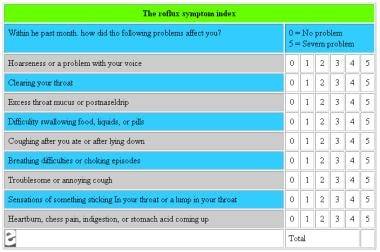
The RSI documents the presence and degree of nine LPR symptoms both before and after treatment; maximum score: 45.
Click to see larger pictureClick to see detailView Full Size Image
Media type:  Image

Media file 2:  The RFS documents the presence and degree of eight LPR findings during fiberoptic laryngoscopy; maximum score: 26.
Click to see larger pictureClick to see detailView Full Size Image
Media type:  Image


  • Ahmed TF, Khandwala F, Abelson TI. Chronic laryngitis associated with gastroesophageal reflux: prospective assessment of differences in practice patterns between gastroenterologists and ENT physicians. Am J Gastroenterol. Mar 2006;101(3):470-8. [Medline].
  • Altman KW, Stephens RM, Lyttle CS. Changing impact of gastroesophageal reflux in medical and otolaryngology practice. Laryngoscope. Jul 2005;115(7):1145-53. [Medline].
  • Belafsky PC, Postma GN, Koufman JA. The association between laryngeal pseudosulcus and laryngopharyngeal reflux. Otolaryngol Head Neck Surg. Jun 2002;126(6):649-52. [Medline].
  • Burton LK, Murray JA, Thompson DM. Ear, nose, and throat manifestations of gastroesophageal reflux disease. Complaints can be telltale signs. Postgrad Med. Feb 2005;117(2):39-45. [Medline].
  • Cadiere GB, Houben JJ, Bruyns J, et al. Laparoscopic Nissen fundoplication: technique and preliminary results. Br J Surg. Mar 1994;81(3):400-3. [Medline].
  • Chambers DW, Davis WE, Cook PR, et al. Long-term outcome analysis of functional endoscopic sinus surgery: correlation of symptoms with endoscopic examination findings and potential prognostic variables. Laryngoscope. Apr 1997;107(4):504-10. [Medline].
  • Cherry J, Margulies SI. Contact ulcer of the larynx. Laryngoscope. Nov 1968;78(11):1937-40. [Medline].
  • Curran AJ, Barry MK, Callanan V, Gormley PK. A prospective study of acid reflux and globus pharyngeus using a modified symptom index. Clin Otolaryngol. Dec 1995;20(6):552-4. [Medline].
  • Deveney CW, Benner K, Cohen J. Gastroesophageal reflux and laryngeal disease. Arch Surg. Sep 1993;128(9):1021-5; discussion 1026-7. [Medline].
  • Dobhan R, Castell DO. Normal and abnormal proximal esophageal acid exposure: results of ambulatory dual-probe pH monitoring. Am J Gastroenterol. Jan 1993;88(1):25-9. [Medline].
  • Farkkila MA, Ertama L, Katila H, et al. Globus pharyngis, commonly associated with esophageal motility disorders. Am J Gastroenterol. Apr 1994;89(4):503-8. [Medline].
  • Ford CN. Evaluation and management of laryngopharyngeal reflux. JAMA. Sep 28 2005;294(12):1534-40. [Medline].
  • Gallup Organizational National Survey. Heartburn Across America. Princeton, NJ:. The Gallup Organization Inc;1988.
  • Gill GA, Johnston N, Buda A. Laryngeal epithelial defenses against laryngopharyngeal reflux: investigations of E-cadherin, carbonic anhydrase isoenzyme III, and pepsin. Ann Otol Rhinol Laryngol. Dec 2005;114(12):913-21. [Medline].
  • Graham DY, Smith JL, Patterson DJ. Why do apparently healthy people use antacid tablets?. Am J Gastroenterol. May 1983;78(5):257-60. [Medline].
  • Grossman TW, Toohill RJ, Mushtag E, et al. Pharyngeal, laryngeal and tracheobronchial manifestations of gastroesophageal reflux. Proceedings of the XXIV World Congress of Otolaryngology Head and Neck Surgery. Berkley, Calif:. Kugler and Ghedini Publishing.
  • Hanson DG. Diagnosis and management of chronic irritative laryngitis. Am J Med. 1999.
  • Hanson DG, Jiang JJ, Chen J, Pauloski BR. Acoustic measurement of change in voice quality with treatment for chronic posterior laryngitis. Ann Otol Rhinol Laryngol. Apr 1997;106(4):279-85. [Medline].
  • Hanson DG, Kamel PL, Kahrilas PJ. Outcomes of antireflux therapy for the treatment of chronic laryngitis. Ann Otol Rhinol Laryngol. Jul 1995;104(7):550-5. [Medline].
  • Helm JF, Shaker R, Dodds WJ. Revelations about ambulatory esophageal pH monitoring. Gastroenterology. 1988;94(5):A421.
  • Hill J, Stuart RC, Fung HK, et al. Gastroesophageal reflux, motility disorders, and psychological profiles in the etiology of globus pharyngis. Laryngoscope. Oct 1997;107(10):1373-7. [Medline].
  • Hogan WJ, Hinder R, Dent JD. First multi-disciplinary international symposium on supraesophageal complications of gastroesophageal reflux disease. Workshop consensus reports. Am J Med. Nov 24 1997;103(5A):149S-150S. [Medline].
  • Howden GF. Erosion as the presenting symptom in hiatus hernia. A case report. Br Dent J. Nov 16 1971;131(10):455-6. [Medline].
  • Jacob P, Kahrilas PJ, Herzon G. Proximal esophageal pH-metry in patients with ''reflux laryngitis''. Gastroenterology. Feb 1991;100(2):305-10. [Medline].
  • Jamieson GG, Watson DI, Britten-Jones R, et al. Laparoscopic Nissen fundoplication. Ann Surg. Aug 1994;220(2):137-45. [Medline].
  • Jarvinen V, Meurman JH, Hyvarinen H, et al. Dental erosion and upper gastrointestinal disorders. Oral Surg Oral Med Oral Pathol. Mar 1988;65(3):298-303. [Medline].
  • Jindal JR, Milbrath MM, Shaker R, et al. Gastroesophageal reflux disease as a likely cause of "idiopathic" subglottic stenosis. Ann Otol Rhinol Laryngol. Mar 1994;103(3):186-91. [Medline].
  • Jones NS, Lannigan FJ, McCullagh M, et al. Acid reflux and hoarseness. J Voice. 1990;4(4):355-58.
  • Kambic V, Radsel Z. Acid posterior laryngitis. Aetiology, histology, diagnosis and treatment. J Laryngol Otol. Dec 1984;98(12):1237-40. [Medline].
  • Kamel PL, Hanson D, Kahrilas PJ. Omeprazole for the treatment of posterior laryngitis. Am J Med. Apr 1994;96(4):321-6. [Medline].
  • Katz PO. Ambulatory esophageal and hypopharyngeal pH monitoring in patients with hoarseness. Am J Gastroenterol. Jan 1990;85(1):38-40. [Medline].
  • Katzka DA, Paoletti V, Leite L, Castell DO. Prolonged ambulatory pH monitoring in patients with persistent gastroesophageal reflux disease symptoms: testing while on therapy identifies the need for more aggressive anti-reflux therapy. Am J Gastroenterol. Oct 1996;91(10):2110-3. [Medline].
  • Koufman JA. The otolaryngologic manifestations of gastroesophageal reflux disease (GERD): a clinical investigation of 225 patients using ambulatory 24-hour pH monitoring and an experimental investigation of the role of acid and pepsin in the development of laryn. Laryngoscope. Apr 1991;101(4 Pt 2 Suppl 53):1-78. [Medline].
  • Lazarchik DA, Filler SJ. Effects of gastroesophageal reflux on the oral cavity. Am J Med. Nov 24 1997;103(5A):107S-113S. [Medline].
  • Locke GR 3rd, Talley NJ, Fett SL, et al. Prevalence and clinical spectrum of gastroesophageal reflux: a population-based study in Olmsted County, Minnesota. Gastroenterology. May 1997;112(5):1448-56. [Medline].
  • Luostarinen M, Isolauri J, Laitinen J, et al. Fate of Nissen fundoplication after 20 years. A clinical, endoscopical, and functional analysis. Gut. Aug 1993;34(8):1015-20. [Medline].
  • Lussi A, Schaffner M, Hotz P, Suter P. Dental erosion in a population of Swiss adults. Community Dent Oral Epidemiol. Oct 1991;19(5):286-90. [Medline].
  • Mair IW, Natvig K, Maurer HJ, Odegaard HI. The globus symptom. ORL J Otorhinolaryngol Relat Spec. 1973;35(2):104-10. [Medline].
  • Maronian NC, Azadeh H, Waugh P. Association of laryngopharyngeal reflux disease and subglottic stenosis. Ann Otol Rhinol Laryngol. Jul 2001;110(7 Pt 1):606-12. [Medline].
  • McNally PR, Maydonovitch CL, Prosek RA, et al. Evaluation of gastroesophageal reflux as a cause of idiopathic hoarseness. Dig Dis Sci. Dec 1989;34(12):1900-4. [Medline].
  • Metz DC, Childs ML, Ruiz C, Weinstein GS. Pilot study of the oral omeprazole test for reflux laryngitis. Otolaryngol Head Neck Surg. Jan 1997;116(1):41-6. [Medline].
  • Meurman JH, Toskala J, Nuutinen P, Klemetti E. Oral and dental manifestations in gastroesophageal reflux disease. Oral Surg Oral Med Oral Pathol. Nov 1994;78(5):583-9. [Medline].
  • Miko TL. Peptic (contact ulcer) granuloma of the larynx. J Clin Pathol. Aug 1989;42(8):800-4. [Medline].
  • Milstein CF, Charbel S, Hicks DM. Prevalence of laryngeal irritation signs associated with reflux in asymptomatic volunteers: impact of endoscopic technique (rigid vs. flexible laryngoscope). Laryngoscope. Dec 2005;115(12):2256-61. [Medline].
  • Moloy PJ, Charter R. The globus symptom. Incidence, therapeutic response, and age and sex relationships. Arch Otolaryngol. Nov 1982;108(11):740-4. [Medline].
  • Moser G, Vacariu-Granser GV, Schneider C, et al. High incidence of esophageal motor disorders in consecutive patients with globus sensation. Gastroenterology. Dec 1991;101(6):1512-21. [Medline].
  • Ott DJ, Ledbetter MS, Koufman JA, Chen MY. Globus pharyngeus: radiographic evaluation and 24-hour pH monitoring of the pharynx and esophagus in 22 patients. Radiology. Apr 1994;191(1):95-7. [Medline].
  • Peters JH, Heimbucher J, Kauer WK, et al. Clinical and physiologic comparison of laparoscopic and open Nissen fundoplication. J Am Coll Surg. Apr 1995;180(4):385-93. [Medline].
  • Pindborg JJ. Chemical and physical injuries. In: Pindborg JJ, ed. Pathology of the Dental Hard Tissues. Philadelphia, Pa:. WB Saunders Co;1970:312-25.
  • Reavis KM, Morris CD, Gopal DV. Laryngopharyngeal reflux symptoms better predict the presence of esophageal adenocarcinoma than typical gastroesophageal reflux symptoms. Ann Surg. Jun 2004;239(6):849-56; discussion 856-8. [Medline].
  • Richter JE. Ambulatory esophageal pH monitoring. Am J Med. Nov 24 1997;103(5A):130S-134S. [Medline].
  • Ruth M, Carlsson S, Mansson I, et al. Scintigraphic detection of gastro-pulmonary aspiration in patients with respiratory disorders. Clin Physiol&#