Practice Essentials
Many classifications, both clinical and radiological, have been proposed in the literature to define acute sinusitis. [1] Although no consensus on the precise definition currently exists, acute sinusitis may be defined as a bacterial or viral infection of the sinuses of fewer than 4 weeks' duration that resolves completely with appropriate treatment. Subacute sinusitis represents a temporal progression of symptoms for 4-12 weeks. Recurrent acute sinusitis [2] is diagnosed when 2-4 episodes of infection occur per year with at least 8 weeks between episodes, and, as in acute sinusitis, the sinus mucosa completely normalizes between attacks. Chronic sinusitis is the persistence of insidious symptomatology beyond 12 weeks, with or without acute exacerbations, and is discussed in Chronic Sinusitis. [3]
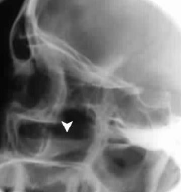 Air-fluid level (arrow) in the maxillary sinus suggests sinusitis.
Signs and symptoms of acute sinusitis
A history of purulent secretions and facial or dental pain are specific symptoms that may point to a bacterial etiology in acute sinusitis. In a patient in intensive care, acute sinusitis should be suspected in the presence of sepsis of unknown origin.
Workup in acute sinusitis
Some authors have reported on the use of laboratory tests including sedimentation rate, white blood cell counts, and C-reactive protein levels to help diagnose acute sinusitis. [4] These tests, however, appear to add little to the predictive value of clinical findings in the diagnosis.
Imaging studies are not necessary when the probability of sinusitis is either high or low but may be useful when the diagnosis is in doubt, based upon a thorough history and physical examination. Plain sinus radiographs may demonstrate mucosal thickening, air-fluid levels, and sinus opacification.
Computed tomography (CT) scanning has poor specificity for the diagnosis of acute sinusitis, demonstrating sinus air-fluid levels in 87% of individuals with simple upper respiratory tract infections (URTIs) and 40% of asymptomatic individuals. CT scanning is the modality of choice, however, in specific circumstances such as in the evaluation of a patient in intensive care, when complications are suspected, or in the preoperative evaluation of surgical candidates. CT scanning can give valuable information regarding the anatomical and mechanical contributions in the development of acute sinusitis.
Management of acute sinusitis
The primary goals of management of acute sinusitis are to eradicate the infection, decrease the severity and duration of symptoms, and prevent complications. The clinician seeks to provide adequate drainage and appropriate systemic treatment of the likely bacterial pathogens.
Drainage of the involved sinus can be achieved both medically and surgically. Aggressively treat patients in intensive care who develop acute sinusitis in order to avoid septic complications. Consider removal of nasotracheal and nasogastric tubes, and promote drainage either medically or surgically.
Sinus puncture and irrigation techniques allow for a surgical means of removal of thick purulent sinus secretions. The purpose of surgical drainage is to enhance mucociliary flow and provide material for culture and sensitivity. A surgical means of sinus drainage should be used when appropriate medical therapy has failed to control the infection and prolonged or slowly resolving symptoms result or when complications of sinusitis occur. Another indication for sinus puncture is to obtain culture material to guide antibiotic selection if empiric therapy has failed or antibiotic choice is limited.
In today's era of minimally invasive surgical techniques, sinus endoscopy is commonly used to achieve sinus drainage. It offers the advantages of (1) being able to open multiple sinuses or to decompress the orbit in cases of complications and (2) allowing the surgeon to open the natural ostia of the involved sinuses.
Embryology
To properly diagnose and treat infectious disorders of the paranasal sinuses, the clinician should have knowledge of the developmental milestones. The development of the paranasal sinuses begins in the third week of gestation and continues until early adulthood.
During the third week of embryonic development, proliferation and medial migration of ectodermal cells form the notochord. After the heart tube and pericardium have rotated from the cranial position to lie anteriorly, the notochord, which is initially in the caudal region of the embryonic disc, rotates to lie posterior to the primitive foregut. The paraxial layer of mesenchyme, which lies adjacent to the notochord, differentiates into the somite ridges, intermediate cell mass, and lateral plate mesoderm. From these mesodermal structures the branchial arches develop, the first of which gives rise to internal nasal structures.
The paranasal sinuses develop in conjunction with the palate from changes in the lateral wall of the nasal cavity. At 40 weeks' gestation, 2 horizontal grooves develop in the mesenchyme of the lateral wall of the nasal cavity. Proliferation of maxilloturbinate mesenchyme between these grooves results in an outpouching of tissue medially into the nasal lumen. This outpouching is the precursor of the middle and inferior meatus as well as the inferior turbinate. Ethmoidoturbinate folds develop superiorly to give rise to the middle and superior turbinates. Once the turbinate structures are established, sinus development begins and continues until early adult life.
Anatomy
The paranasal sinuses are air-filled bony cavities that extend from the skull base to the alveolar process and laterally from the nasal cavity to the inferomedial aspect of the orbit and the zygoma. They are lined with pseudostratified columnar epithelium that is contiguous, via ostia, with the lining of the nasal cavity. This epithelium contains a number of mucous-producing goblet cells. The arterial supply of the paranasal sinuses is from branches of the internal and external carotid arteries, while the venous and lymphatic drainage path is through the sinus ostia into the nasal cavity plexus. In addition, venous drainage occurs through valveless vessels corresponding to the arterial supply. The focal point of sinus drainage is the ostiomeatal complex, which is located in the middle meatus and is composed of the maxillary, frontal, and anterior ethmoid ostia. The posterior ethmoids empty into the superior meatus, and the sphenoids empty into the sphenoethmoidal recess.
The exact function of the paranasal sinuses is not well understood. The possible roles of the sinuses may include reducing the weight of the skull; dampening pressure; humidifying and warming inspired air; absorbing heat and insulating the brain; aiding in sound resonance; providing mechanical rigidity; and increasing the olfactory surface area.
The sinus mucosa has less secretory and vasomotor function than the nasal cavity does. Cilia are concentrated near and beat toward the natural sinus ostia. Blockage of the ostium results in stasis of mucous flow, which can lead to development of disease.
Pathophysiology
The sinuses are normally sterile under physiologic conditions. Purulent sinusitis can occur when ciliary clearance of sinus secretions decreases or when the sinus ostium becomes obstructed, which leads to retention of secretions, negative sinus pressure, and reduction of oxygen partial pressure. This environment is then suitable for growth of pathogenic organisms. Factors that predispose the sinuses to obstruction and decreased ciliary function are allergic, nonallergic, or viral insults, which produce inflammation of the nasal and sinus mucosa and result in ciliary dysmotility and sinus obstruction. Approximately 90% of patients who have viral upper respiratory tract infections (URTIs) have sinus involvement, but only 5-10% of these patients have bacterial superinfection requiring antimicrobial treatment.
Anatomical variations that narrow the ostiomeatal complex, including septal deviation, paradoxical middle turbinates, and Haller cells, make this area more sensitive to obstruction from mucosal inflammation. Mechanical obstruction of the ostiomeatal complex from foreign bodies, polyps, or tumors can also result in acute sinus disease. Systemic diseases that result in decreased mucociliary clearance, including cystic fibrosis and Kartagener syndrome, can be predisposing factors for acute sinusitis in rare cases. Patients with immunodeficiencies (eg, agammaglobulinemia, combined variable immunodeficiency, and immunodeficiency with reduced immunoglobulin G [IgG]– and immunoglobulin A [IgA]–bearing cells) are also at increased risk of developing acute sinusitis.
Acute sinusitis in the intensive care population is a distinct entity, occurring in 18-32% of patients with prolonged periods of intubation, and is usually diagnosed during the evaluation of unexplained fever. Cases in which the cause is obstruction are usually evident and can include the presence of prolonged nasogastric or nasotracheal intubation. Moreover, patients in an intensive care setting are generally debilitated, predisposing them to septic complications, including sinusitis.
Ciliary function is also reduced in the presence of low pH, anoxia, bacterial toxins, smoking, dehydration, foreign bodies, and drugs (eg, atropine, antihistamines, phenylephedrine). Approximately 10% of cases of acute sinusitis result from direct inoculation of the sinus with a large amount of bacteria. Dental abscesses or procedures that result in communication between the oral cavity and sinus can produce sinusitis by this mechanism. Facial trauma or large inoculations from swimming can produce sinusitis as well.
A study by Santee et al suggested that acute sinusitis in children causes changes in the nasopharyngeal microbiota, with these changes being linked to increased frequency of upper respiratory tract infections. Changes found included a reduction in the relative abundance of certain taxa, such as Faecalibacterium prausnitzii and Akkermansia spp, as well as enrichment of Moraxella nonliquefaciens. [5]
Epidemiology
Frequency
United States
About 1 out of every 8 adults in the United States is affected by sinusitis, either acute or chronic. [6]
-
Air-fluid level (arrow) in the maxillary sinus suggests sinusitis.
Tables
Antibiotic |
Dosage |
Streptococcus pneumoniae |
Haemophilus influenzae |
Moraxella catarrhalis |
Anaerobic bacteria |
||
Sensitive |
Intermediate |
Resistant |
|||||
Amoxicillin |
500 mg PO tid |
+++ |
++ |
+ |
++ |
+ |
+ |
Clarithromycin |
250-500 mg PO bid |
++ |
++ |
+ |
++ |
+++ |
+ |
Azithromycin |
500 mg PO first day, then 250 mg/d PO for 4 days |
++ |
++ |
+ |
++ |
+++ |
+ |
Antibiotic |
Dosage |
Streptococcus pneumoniae |
Haemophilus influenzae |
Moraxella catarrhalis |
Anaerobic bacteria |
||
Sensitive |
Intermediate |
Resistant |
|||||
Amoxicillin/clavulanate |
500 mg PO tid |
+++ |
++ |
+ |
+++ |
+++ |
+++ |
Cefuroxime |
250-500 mg PO bid |
+++ |
++ |
+ |
+++ |
++ |
++ |
Cefpodoxime + cefixime |
200 mg PO bid 400 mg/d PO |
- ++ |
+++ - |
++ - |
+ +++ |
+++ +++ |
+++ - |
Ciprofloxacin |
500-750 mg PO bid |
++ |
+ |
+ |
++ |
+++ |
+ |
Levofloxacin |
500 mg/d PO |
+++ |
+++ |
+++ |
+++ |
+++ |
+++ |
Trovafloxacin |
200 mg/d PO |
+++ |
+++ |
+++ |
+++ |
+++ |
+++ |
Clindamycin |
300 mg PO tid |
+++ |
+++ |
+++ |
- |
- |
+++ |
Metronidazole |
500 mg PO tid |
- |
- |
- |
- |
- |
+++ |
Antibiotic |
Dosage |
Streptococcus pneumoniae |
Haemophilus influenzae |
Moraxella catarrhalis |
Gram-negative |
Anaerobic bacteria |
Piperacillin |
3-4 g IV q4-6h |
+++ |
+ |
- |
+++ |
+++ |
Piperacillin/tazobactam |
3.375 g IV q6h |
+++ |
+++ |
+++ |
+++ |
++ |
Ticarcillin |
3 g IV q4h |
+++ |
- |
- |
+++ |
++ |
Ticarcillin/clavulanate |
3.1 g IV q4h |
+++ |
+++ |
- |
+++ |
++ |
Imipenem |
500 mg IV q6h |
+++ |
+++ |
+++ |
+++ |
+++ |
Meropenem |
1 g IV q8h |
+++ |
+++ |
+++ |
+++ |
++ |
Cefuroxime |
1 g IV q8h |
+++ |
+++ |
+++ |
++ |
++ |
Ceftriaxone |
2 g IV bid |
+++ |
+++ |
+++ |
+++ |
++ |
Cefotaxime |
2 g IV q4-6h |
+++ |
+++ |
+++ |
+++ |
++ |
Ceftazidime |
2 g IV q8h |
+++ |
+++ |
+++ |
+++ |
++ |
Gentamicin |
1.7 mg/kg IV q8h |
- |
+++ |
+++ |
++ |
- |
Tobramycin |
1.7 mg/kg IV q8h |
- |
+++ |
+++ |
++ |
- |
Vancomycin |
1 g IV q6-12h |
+++ |
- |
- |
- |
++ |


