Overview
Human eyes are highly sensitive in discerning minimal differences in the structure of human faces and in distinguishing persons. Discerning eyes can also detect many facial anomalies associated with systemic congenital anomalies. A classification of these facial anomalies based on embryological bases is helpful for understanding the morphogenesis of each anomaly.
The moment the human embryo is fertilized to the week of the baby’s birth is an important period for human appearance in the normally developing embryo. During preimplantation stages, differentiation occurs between precursors of embryonic and extraembryonic structures. A fore-hind axis begins within the inner cell mass at the time of implantation. By the end of the eighth week of gestation, the appearance of the head, face, hands, and feet suggest the embryo’s species but is not yet definitive.
When nonhuman mammalian development is compared with human development, the study subjects must be compared at the same developmental stage (fetal, perinatal, postnatal) When collected appropriately, data from experimental studies of nonhuman mammalian embryos elucidate important aspects of human facial development.
For example, using comparative morphologic and developmental analyses, Higashiyama et al demonstrated that “the therian mammal’s premaxilla (rostral-most upper jawbone) is derived from the maxillary prominence of the mandibular arch.” The investigators also state that the developmental primordia from which the premaxilla of nonmammalian tetrapods arises only rarely is involved in forming the upper jaw in therian mammals, instead developing into a motile nose. [1]
This article briefly highlights the key aspects of facial development. [2, 3, 4, 5, 6] Congenital anomalies of the face are discussed in further detail elsewhere (eg, see the Medscape Drugs & Diseases article Craniofacial Syndromes).
Images depicting face embryology can be seen below.
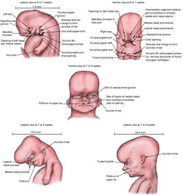 Face embryology.
Facial Morphogenesis
Craniofacial development is an extraordinarily complex process that requires the orchestrated integration of multiple specialized tissues, such as the surface ectoderm, neural crest, mesoderm, and pharyngeal endoderm, in order to generate the central and peripheral nervous systems, axial skeleton, musculature, and connective tissues of the head and face. Understanding the development of the structures of the face also requires knowledge of the pharyngeal or branchial arches. These arches form on either side of the foregut and correspond to the primitive branchial arches. The pharyngeal arch consists of a core of mesenchyme covered externally by ectoderm and covered internally by endoderm. [7]
The ectoderm is well around the stomodeum by the fourth week of embryonic development and contributes to the formation of the face and the nasal and oral cavities.
The mesenchyme that fills the pharyngeal arches is derived from the following 3 origins: the paraxial mesoderm, the lateral plate mesoderm, and the neural crest cells. Although paraxial mesoderm and lateral plate mesoderm contribute to the musculature that develops in each particular arch, neural crest cells contribute to the skeletal portion of each arch.
At the early stages of embryonic development, the vertebrate face has a common plan. A series of small buds of tissue called the facial primordia forms around the stomodeum, which forms the primitive mouth. The facial primordia are made up mainly of neural crest cells that have migrated from the cranial crest and settled.
The upper jaw develops from the following 5 main buds of tissue: a single median frontonasal mass (sometimes present as the median nasal processes or frontonasal prominences), the 2 lateral nasal prominences on both sides, and, flanking these, the 2 maxillae (maxillary prominences). The lower jaw develops from the paired mandibular primordia (mandibular prominences). Paired maxillary and mandibular prominences are derivatives of the first pair of branchial, or pharyngeal, arches. All of these prominences are produced by the proliferation of the neural crest cells that migrate into the arches from the neural crest during the fourth week of gestation.
The neural crest cells give rise to the connective tissue components, including cartilage, bone, and ligaments in the facial and oral regions. The myogenic cells of the muscles constitute a separate cell lineage. These cells originate from the paraxial mesoderm and migrate into the facial primordia. Prior to emigration, the neural crest cells in the head are formed according to which facial primordium they belong.
The individual facial primordia are populated by neural crest cell populations that arise in different regions of the head neural folds. The neural crest cells that settle to form the frontonasal mass first migrate from the prosencephalic region (forebrain) and are later joined by other migrating cells, mainly from the anterior mesencephalic region (midbrain). The cells of the maxillae come from the posterior mesencephalic region, whereas the cells of the mandibular primordia come mainly from the region of the anterior rhombencephalon (hindbrain). Cells that arise in the posterior mesencephalon also contribute. In the trunk, exchanges between different regions of the neural crest almost invariably lead to normal development.
The frontonasal prominence surrounds the ventrolateral part of the forebrain, which gives rise to the optic vesicles. These vesicles project from the sides of the forebrain into the mesenchyme and form the eyes. The frontal portion of the frontonasal prominence forms the forehead, whereas the nasal part of the frontonasal prominence forms the rostral boundary of the stomodeum and nose.
A summary of the derivatives of the prominences is as follows:
-
Frontonasal prominence - Forehead and the dorsum apex of the nose
-
Lateral nasal prominences - Sides (alae) of the nose
-
Medial nasal prominences - Nasal septum
-
Maxillary prominences - Upper cheek region and most of the upper lip
-
Mandibular prominences - Chin, lower lip, and lower cheek regions
-
Mesenchyme in the facial prominences - Fleshy derivatives and various bones
A summary of the derivatives of the first and second pharyngeal (ie, branchial) arches is as follows:
-
Pharyngeal arch I
Cranial nerve - Maxillary and mandibular division of the trigeminal nerve (cranial nerve V)
Artery - Maxillary (terminal branch)
Muscles - Muscles of mastication (ie, temporalis, masseter, pterygoids), mylohyoid, anterior belly of digastric, tensor tympani, and tensor veli palatini
Skeleton - Maxillary cartilage (incus, alisphenoid), mandibular or Meckel cartilage (malleus), and arch dermal mesenchyme (maxilla, zygomatic, squamous portion of temporal bone, mandible)
-
Pharyngeal arch II (hyoid)
Facial nerve - Cranial nerve VII
Artery - Stapedial
Muscles - Muscles of facial expression (ie, orbicularis oculi, orbicularis oris, risorius, buccinator, platysma, auricularis, frontalis), stapedius muscle, posterior belly of digastric, and stylohyoid muscle
Skeleton - Stapes, styloid process, stylohyoid ligament, lesser cornu of hyoid, and the upper part of the body of the hyoid bone
Early Development of the Face
Facial development occurs mainly between the fourth and eighth weeks of gestation.
-
Fourth week of development (stages 12 and 13)
Primordia of the face appear at the cephalic end of the embryo.
Two nasal placodes cap the bulbous frontal prominence.
The optic discs appear posterolateral to the frontal prominence.
Three paired branchial arches have formed.
The first arches split into maxillary and mandibular prominences. The hyoid arches are the second pair.
Between the first arches and frontal prominence, the buccopharyngeal membrane becomes fenestrated.
-
Fifth week of development (stages 14 and 15)
Nasal pits develop in the nasal placodes, and the rims of the placodes differentiate into medial and lateral nasal prominences.
The lens vesicles invaginate and close within the optic discs.
The mesenchyme of the mandibular arch fills in across the midline.
The caudal end of the medial nasal prominences begins to fuse with the maxillary prominences.
-
At the beginning of the sixth week of development (stage 16)
The nasals have shifted to a more ventral, central position.
Growing and shifting subectodermal mesenchyme smooths out the furrows between prominences and arches, and the second arch becomes more massive.
Six auricular hillocks, which will become the pinna of the ears, form on the mandibular and hyoid arches.
-
By the end of the sixth week of development (stage 17)
Medial and lateral nasal prominences fuse.
Maxillary prominences begin the formation of the upper jaw.
The midline approximation of the medial nasal prominences forms the nasal septum. [8]
-
At the beginning of the seventh week of development (stage 18)
The tip of the nose is elevated between the medial nasal prominences and is visible in profile.
Eyelids become prominent.
The pinna of the ear takes shape.
-
End of the seventh week of development (stage 18)
The pattern of facial features has taken on a human appearance. However, facial proportions develop during the fetal period.
The fusion of the medial nasal prominences, which forms the central axis of the nose and the philtrum of the lip, is complete.
Final Development of the Face
From the beginning of the eighth week of development to birth, the final facial development occurs slowly and consists mainly of changes in the proportion and relative positions of the facial components. During the early fetal period, the nose is flat and the mandible is underdeveloped. They obtain their characteristic form while facial development is being completed. As the brain enlarges, it creates a prominent forehead, the eyes move medially, and the external ears rise.
The prenatal face is small because of (1) the rudimentary upper and lower jaws, (2) the unerupted primary teeth, and (3) the small size of the nasal cavities and maxillary sinuses.
Conclusion
The development of the vertebrate face is a dynamic multistep process that starts with the formation of neural crest cells in the developing brain and their subsequent migration to form, together with mesodermal cells, the facial primordia. Patterning and morphogenesis of neural crest–derived tissues within a developing vertebrate embryo rely on a complex balance between signals acquired by neural crest cells in the neuroepithelium during their formation and signals from the tissues that the neural crest cells contact during their migration. Neural crest cells carry information that directs the axial pattern and species-specific morphology of the head and face. Signaling interactions coordinate the outgrowth of the facial primordia from buds of undifferentiated mesenchyme into the intricate series of bones and cartilage structures that, together with muscle and other tissues, form the adult face.
Some of the molecules thought to be involved have been identified through the use of mouse mutants, data from human craniofacial syndromes, and expression studies of signaling molecules during facial development.
However, the way in which these molecules control the epithelial-mesenchymal interactions, which mediate facial outgrowth and morphogenesis, is unclear. The role of neural crest cells in these processes has yet to be well defined. Similarly, the complex interaction of all these processes during face development and the candidate signaling molecules and their possible target genes have not been clearly defined.
-
Face embryology.


