Practice Essentials
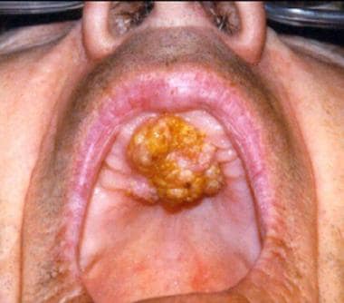
The palate is divided anatomically into the hard palate (part of the oral cavity) and the soft palate (part of the oropharynx). Cancer of the soft palate accounts for approximately 2% of head and neck mucosal malignancies. Half of all hard palate cancers are squamous cell carcinomas (SCCs) as seen in the image below. Nonsquamous cell cancers, including minor salivary gland cancers, sarcomas, and melanomas, account for the other half (see the histologic distribution of hard palate malignant neoplasms and the histologic types and frequencies of minor salivary gland neoplasms of the palate, below). [1]
However, in the soft palate, 80% of cancers are SCCs. Nonsquamous malignancies account for the other 20%. The prevalence of oral cavity and oropharyngeal cancer has geographic variations, with the highest rate reported in India, accounting for 50% of all cancer cases in that country.
The histologic distribution of hard palate malignant neoplasms is as follows:
-
Squamous cell carcinoma - 53%
-
Adenoid cystic carcinoma - 15%
-
Mucoepidermoid carcinoma - 10%
-
Adenocarcinoma - 4%
-
Anaplastic carcinoma - 4%
-
Other - 14%
The histologic types and frequencies of minor salivary gland neoplasms of the palate is as follows:
-
Benign - 26%
-
Malignant - 74% overall
Adenoid cystic carcinoma - 30%
Mucoepidermoid carcinoma - 16%
Adenocarcinoma - 18%
Malignant mixed tumor - 8%
Other - 2%
Workup in malignant tumors of the palate
Radiology
Radiologic evaluation helps to increase the accuracy of staging. Computed tomography (CT) scanning and magnetic resonance imaging (MRI) are the imaging modalities of choice.
Biopsy
Biopsy of an ulcerative lesion may be easily obtained in the office transorally using biopsy forceps, with the patient under local anesthesia. Alternatively, fine-needle aspiration cytology studies may be performed if an experienced cytopathologist is available.
For ulcerative lesions, obtaining a biopsy specimen from closer to the edge of the tumor is important to avoid the necrotic central component.
Staging
Perform staging of the tumor according to the American Joint Committee on Cancer staging protocol because this is of critical importance to the patient's prognosis.
Management of malignant tumors of the palate
Specific treatment of palate cancer depends on the location of the tumor (hard vs soft palate), stage of the tumor, and pathologic type of the cancer.
Individuals with small T1 and T2 SCC lesions of the hard palate can be managed with either surgery or radiation therapy. If the pathologic stage of the neck in these patients is N2 or higher, initiate postoperative radiotherapy.
Treatment of an N1 neck is controversial. A pathologic N1 node is considered adequately treated with neck dissection alone when no extracapsular extension is present. However, in many centers, any pathologic N1 node is treated with postoperative radiotherapy; this is recommended. Definitely initiate postoperative radiation therapy for patients with extracapsular extension.
T3 and T4 SCC lesions of the hard palate frequently require combined oncologic treatment, including surgery and radiation therapy to both the primary site and the neck.
Regarding patients with SCC of the soft palate, because of difficulties in adequate reconstruction, radiation therapy has been the recommended treatment for soft palate cancers in the past. Although advances in reconstructive techniques and prosthetic reconstruction have allowed for more effective surgical resection and rehabilitation for patients with soft palate cancers, radiation therapy remains the primary treatment modality in many centers for T1, T2, and T3 lesions, with results comparable with those of surgery.
Etiology
Although a strong correlation is established between tobacco and alcohol consumption and SCC of the oral cavity and soft palate, the relationship to hard palate cancer is not as clear. Reverse smoking is a specific etiologic factor for SCC of the hard palate. In reverse smoking, the lit end of the cigarette is placed in the mouth so that an intense heat is generated during smoking. Other factors, including ill-fitting dentures, poor oral hygiene, mechanical irritation, and mouthwash, are implicated in oral cavity SCC; however, the evidence is less convincing.
Pathophysiology
A thorough history and physical examination help to assess the extent of tumor.
SCC extension beyond the hard palate occurs in up to 70% of lesions. Posterior extension involves the soft palate, with possible velopharyngeal insufficiency and hypernasal speech. Palatal hypesthesia indicates trigeminal nerve involvement in the sphenopalatine foramen or pterygopalatine fossa extension.
An absent corneal reflex is indicative of skull-base extension through the foramen rotundum, foramen ovale, or inferior orbital fissure.
Dental numbness may indicate perineural invasion. Middle ear effusion is suggestive of nasopharyngeal extension or invasion of the tensor veli palatini muscle.
Involvement of the mandibular division of the trigeminal nerve may manifest as hypesthesia along the mandible or wasting of the temporalis or masseter muscles. This is indicative of infratemporal fossa involvement. Trismus, malocclusion, and pain are symptoms of invasion of the pterygoid muscles. Extension to the gingiva requires assessment. Dental sockets provide a pathway of invasion to the alveolar process of the maxillary bone and into the maxillary sinus. Nasal floor involvement may occur by direct extension through the palate.
Lymph node involvement is of special concern in SCC and high-grade mucoepidermoid cancer. It is rare in other salivary gland carcinomas. Approximately 30% of patients have cervical node metastasis at the time of presentation. [2] The submandibular nodes (level I) and upper deep jugular lymph nodes (level II) are the first echelon of nodal drainage. However, in tumors with posterior soft palate extension, retropharyngeal nodes may be involved. Soft palate carcinomas are staged as oropharyngeal cancers according to the American Joint Committee on Cancer (see Staging).
Almost half of patients present with extension of the tumor beyond the soft palate. Common sites of extension include the tonsils, retromolar trigone, inferior or superior alveolar process, hard palate, and base of tongue. Extension into the sphenopalatine foramen may result in palatal hypostasis. In extensive lesions extending into the nasopharynx, middle ear effusion is common. The tumor may extend anterosuperiorly into the pterygomaxillary and infratemporal fossa.
Presentation
SCCs of the palate manifest as ulcerative surface lesions. Often, patients are asymptomatic in the early stages, but they may experience pain in advanced stages. A palate mass, bleeding, a foul odor, ill-fitting dentures in edentulous patients, or loose teeth may be the presenting symptoms for patients with hard palate cancer. In persons with advanced-stage soft palate cancers, velopharyngeal insufficiency, altered speech, difficulty swallowing, referred otalgia, trismus, or a neck mass may be present. Because the area is easily visualized, tumors are often found at early stages incidentally by the patient or the physician.
On the other hand, minor salivary gland tumors manifest as submucosal lesions, as depicted in the image below, with a smooth, normal mucosal covering. Melanomas are smooth, black lesions but may be brown or brownish gray. Kaposi sarcomas are bluish lesions that are commonly seen in patients with HIV infection.
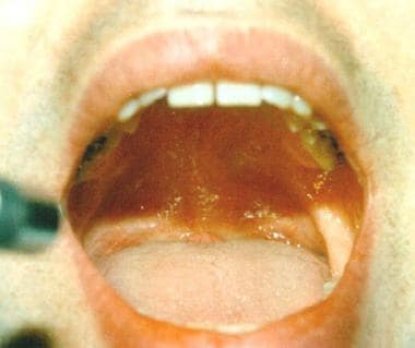 Thirty-two-year-old man with a submucosal lesion at the junction of the hard and soft palate.
Pseudoepitheliomatous hyperplasia and necrotizing sialometaplasia are benign self-limited lesions that can mimic SCC and need to be distinguished histologically. Torus palatina (ie, bony hyperplasia of the palate) are hard midline masses that produce no symptoms and should not be confused with tumors.
A study by Zhurakivska et al found that two of eight patients with necrotizing sialometaplasia of the minor salivary glands of the palate had associated tumors (pleomorphic adenoma and adenoid cystic carcinoma), indicating that previous reports may have underestimated the frequency with which such neoplasms occur in relation to the condition. The investigators also suggested that the obscurement of minor salivary gland tumors by necrotizing sialometaplasia may cause treatment delays. [3]
Relevant Anatomy
The palate separates the oral cavity from the nasal cavity and the maxillary sinuses. The mucosa of the palate is a keratinizing pseudostratified squamous epithelium. However, the submucosa has numerous minor salivary glands, especially in the hard palate. The periosteal covering of the hard palate becomes a relative barrier to the spread of cancer into the palatine bone.
The neurovascular supply to the palate comes from the palatine foramina, located medial to the third molar teeth. These foramina provide a pathway for the tumor to spread. Descending palatine arteries from the internal maxillary artery provide the blood supply. Vessels pass anteriorly through the nasopalatine foramen to the nose. Sensory and secretomotor fibers from the maxillary (V-II) branch of the trigeminal nerve and pterygopalatine ganglion traverse to the hard palate via the greater and lesser palatine nerves.
Anatomically, the soft palate is part of the oropharynx. It consists of mucosa on both surfaces. Intervening between the 2 mucosal surfaces are the connective tissue, muscle fibers, aponeurosis, numerous blood vessels, lymphatics, and minor salivary glands. Functionally, the soft palate serves to separate the oropharynx from the nasopharynx during swallowing and speech. The soft palate approximates with the posterior pharyngeal wall during swallowing to prevent nasopharyngeal regurgitation and approximates during speech to prevent air escape into the nose.
Contraindications
Contraindications to surgical correction of malignant palate tumors are based on the patient's comorbidities and his or her ability to tolerate surgery. Coexisting medical conditions may put the patient at risk during anesthesia. Additionally, tumors may be deemed inoperable because of their size or extent of involvement. Tumors that have intracranial extension are considered inoperable if they involve the brain parenchyma. Radiation therapy is a treatment option in these rare cases. See the Treatment section for recommended treatments for various types and stages of palate cancer.
-
Squamous cell carcinoma of the hard palate.
-
Thirty-two-year-old man with a submucosal lesion at the junction of the hard and soft palate.
-
Coronal CT scan revealing intranasal extension of the tumor.
-
Sagittal MRI revealing a mass confined to the palate, without sinonasal extension.
-
Coronal MRI.
-
Transoral resection of a mucoepidermoid carcinoma of the palate.
-
Schematic per-oral approach to the palate using a Dingman mouth retractor.


