Continually Updated Clinical Reference
 
 
  All Sources     eMedicine     Medscape     Drug Reference     MEDLINE
 
eMedicine - Ranulas and Plunging Ranulas : Article by

Quick Find
Authors & Editors
Introduction
Indications
RELEVANT ANATOMY
Contraindications
Workup
Treatment
Complications
Outcome And Prognosis
Multimedia
References


Patient Education
Click here for patient education.


Author: Ryan L Van De Graaff, MD, Consulting Staff, Ear, Nose and Throat Specialties

Ryan L Van De Graaff is a member of the following medical societies: Alpha Omega Alpha, American Academy of Otolaryngology-Head and Neck Surgery, and American Medical Association

Coauthor(s): Gregory C Allen, MD, Assistant Professor, Department of Otolaryngology-Head and Neck Surgery, University of Colorado School of Medicine

Editors: Daniel J Kelley, MD, Consulting Staff, Eastern Shore ENT & Allergy Associates; Francisco Talavera, PharmD, PhD, Senior Pharmacy Editor, eMedicine; Peter S Roland, MD, Chair, Professor, Department of Otolaryngology, University of Texas Southwestern Medical Center; Christopher L Slack, MD, Otolaryngology-Facial Plastic Surgery, Private Practice, Associated Coastal ENT; Medical Director, Treasure Coast Sleep Disorders; Arlen D Meyers, MD, MBA, Professor, Department of Otolaryngology-Head and Neck Surgery, University of Colorado School of Medicine

Author and Editor Disclosure

Synonyms and related keywords: ranulas and plunging ranulas, deep diving cervical ranula, deep plunging ranula, oral ranula with cervical extension, mucus escape reaction, MER, salivary gland cyst, swelling in the floor of the mouth, sublingual gland excision

History of the Procedure

The term ranula is derived from the Latin word rana, meaning frog, and describes a blue translucent swelling in the floor of the mouth reminiscent of the underbelly of a frog. Hippocrates described ranulas and thought that they were secondary to inflammation. Paré thought that ranulas may represent descent of brain or pituitary matter.

Frequency

Ranulas rarely occur. In one study of 1303 salivary gland cysts, only 42 were ranulas. The reported male-to-female ratio is 1:1.3, without significant side preference. Presentation is most frequently in the second and third decades of life, with an age range of 3-61 years.

Plunging ranulas occur less commonly than ranulas. Only slightly more than 100 well-documented cases of plunging ranulas have been reported in the English literature.

Etiology

Ranulas

Congenital ranulas can arise secondary to an imperforate salivary duct or ostial adhesion. These are very rare and have been known to spontaneously resolve.

Posttraumatic ranulas arise from trauma to the sublingual gland, leading to mucus extravasation and formation of a pseudocyst. The more appropriate term for this may be mucus escape reaction (MER).

Plunging ranulas

Other terms include deep, diving, cervical, or deep plunging ranula and oral ranula with cervical extension.

Plunging ranulas generally appear in conjunction with an oral ranula. Rarely, they can arise independently of the oral component. Patients present first with an oral swelling in up to 45% of cases, with associated oral swelling in 34%, and without any oral involvement in 21% of cases.

Pathophysiology

Ranulas

Ranulas are formed from 1 of 2 processes:

  • Partial obstruction of a sublingual duct can lead to formation of an epithelial-lined retention cyst. This is unusual, occurring in less than 10% of all ranulas.
  • Trauma can lead to formation of ranulas. Experimentally, partial severance or ligation of the sublingual duct leads to ranula formation, whereas ligation of the submandibular duct does not. The ligation of the parotid duct ultimately leads to atrophy. The difference lies in the fact that the sublingual gland secretes continuously in the interdigestive period, whereas the other two major salivary glands only secrete in response to stimuli, such as eating. Therefore, with trauma, if a duct is obstructed, secretory backpressure builds and acini rupture, leading to mucus extravasation. Alternately, trauma causes direct damage to the duct or acini, leading to mucus extravasation. A pseudocyst then forms.

Plunging ranulas

Plunging ranulas arise in the neck by 3 mechanisms:

  • The sublingual gland may project through the mylohyoid, or an ectopic sublingual gland may exist on the cervical side of the mylohyoid. This explains most plunging ranulas that exist without an oral component.
  • The cyst may penetrate through the mylohyoid. Up to 27-45% of mylohyoid muscles in cadavers are found to be dehiscent, usually in the anterior two thirds of the muscle. These sites of dehiscence provide a route of egress for the cyst. In some instances, surgical trauma from initial ranula operations may scar or fibrose the superior surface of a ranula. When the ranula recurs, the path of least resistance is through a dehiscent mylohyoid, and a plunging ranula forms when only a simple ranula was present initially. Up to 44% of all plunging ranulas are iatrogenically induced in this manner.
  • A duct from the sublingual gland may join the submandibular gland or its duct, allowing ranulas to form in continuity with the submandibular gland. Therefore, the ranula accesses the neck from behind the mylohyoid muscle.

Clinical

Ranulas

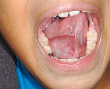
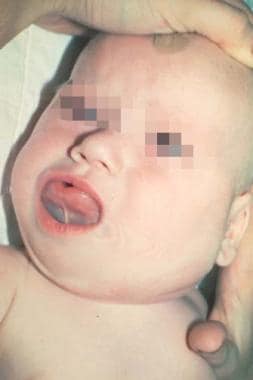
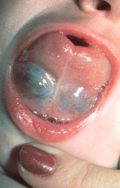
A ranula is most commonly observed as a bluish cyst located below the tongue (see Images 1-2). It may fill the mouth and raise the tongue. Typically, these are painless masses that do not change in size in response to chewing, eating, or swallowing. Occasionally, pain may be involved.

Plunging ranulas

Plunging ranulas can manifest as neck swelling in conjunction with, or independent of, a floor-of-mouth cyst. Occasionally, squeezing the mass causes swelling in the floor-of-mouth cyst. Most reported plunging ranulas are 4-10 cm in size and are usually found in the submandibular space. They have been reported to extend into the submental region, the contralateral neck, the nasopharynx up to the skull base, the retropharynx, and even into the upper mediastinum.


See Surgical therapy.


The sublingual gland lies against the sublingual depression of the mandible and directly on the mylohyoid. The submandibular duct (Wharton duct) and the lingual nerve lie medial to the gland. The genioglossus muscle is medial to these structures. No posterior fascial limits to the sublingual space exist, which allows lesions to exit the sublingual space and enter into the submandibular or parapharyngeal space.


Although some have advocated surgical management of congenital ranulas, recent literature supports observation in asymptomatic patients. Many congenital ranulas resolve on their own and do not require surgical intervention.


Imaging Studies

  • CT scanning
    • Ranulas on CT scanning (see Image 3) are described as cystic masses in the submandibular or parapharyngeal space that extend into or abut the sublingual space. On CT scanning, they are noted to be sharply demarcated lesions of low attenuation that conform to their local fascial boundaries. They are generally unilocular in nature. With the exception of a sublingual epidermoid, the appearance of a simple ranula on CT scanning is distinctive.
    • Plunging ranulas are occasionally noted on CT scanning to have a small tail extending into the sublingual space. This finding is almost pathognomonic for plunging ranulas. If this is absent, the presence of a homogeneous cyst in the submandibular or parapharyngeal space that abuts the sublingual space is highly indicative of a plunging ranula.
  • MRI
    • MRI is the most sensitive imaging study to evaluate the sublingual gland and its pathologic states.
    • On T1-weighted MRI, the gland appears as an area of intermediate signal intensity, lower than adjacent fat but higher than muscle.
    • T2-weighted images help discriminate cysts from surrounding normal structures.
  • Ultrasonography: Sublingual glands and their pathologic states are difficult to visualize on ultrasonography because of their location.

Diagnostic Procedures

  • Needle aspiration
    • Analysis of fluid from ranulas demonstrates mucus with prominent histiocytes.
    • The biochemistry of this fluid shows high amylase and protein content.
  • Differential diagnosis
    • Lymphadenopathy
    • Cystic hygroma
    • Pleomorphic adenoma
    • Abscess
    • Thyroglossal duct cyst
    • Dermoid or epidermoid cyst
    • Laryngocele
    • Lipoma
    • Hemangioma
    • Cervical thymic cyst
    • Cysts of the parathyroid or thyroid gland
    • Tumor

Histologic Findings

Most ranulas demonstrate a cyst devoid of epithelial lining, with the wall composed of vascular fibroconnective tissue resembling granulation tissue. Histiocytes predominate the pseudocyst wall. Mucin and foamy macrophages are often observed. Occasionally, partial epithelial linings are observed.


Medical therapy

A recent study evaluated the effectiveness of orally administered Nickel Gluconate-Mercurius Heel-Potentised Swine Organ Preparations D10/D30/D200, a homotoxicological agent. This medication acts to stimulate pseudocyst reabsorption and glandular repairing, and aids in improving the physiologic functioning of the gland. In this study, Nickel Gluconate-Mercurius Heel-Potentised Swine Organ Preparations D10/D30/D200 was administered regularly from 6 weeks to 6 months. Eight out of 9 ranulas responded to medical therapy.1

Surgical therapy

Congenital ranulas

Some have advocated that all patients with submandibular duct obstruction leading to ranula formation need early marsupialization and ductoplasty to prevent complications such as sialoadenitis.

Evidence exists that imperforate ducts may spontaneously resolve if rupture takes place during feeding. Therefore, observation for spontaneous resolution of congenital ranulas is reasonable. If airway obstruction or feeding problems arise, surgery is indicated.

Ranulas

  • Marsupialization: Simple marsupialization is the oldest and most widely reported treatment for ranulas. It involves unroofing the cyst and tacking the edges of the cyst to adjacent tissue. Failure rates range from 61-89%, with cysts recurring anywhere from 6 weeks to 12 months later. Inferior compression on the cyst from the tongue leads to premature closure of the opened cyst. This increases the risk of the cyst recurring. Packing the cyst cavity with gauze for 7-10 days improves the success rate. In one report, 11 of 12 patients had resolution of their ranula with marsupialization and subsequent packing of the cavity.
  • Placement of suture or Seton: A silk suture or Seton can be placed through the surface of the cyst under local anesthesia. This is left in place while an epithelial tract forms to allow for mucus drainage between the surface and the underlying salivary glandular tissue. Morbidity is minimal to nonexistent, and success has been good in limited studies. This can also be performed in the office.
  • Sclerosing agents: Bleomycin and OK-432 have been used with success in treatment of ranulas. In one study, 31/32 patients (97%) achieved a disappearance or marked reduction in ranula size with injection of OK-432. Nearly half of all patients experienced local pain or fever, which resolved over several days.
  • Carbon dioxide laser: The carbon dioxide laser has been used with limited patients with good success to remove the cyst and scar the gland enough to decrease risk for recurrence. A tissue biopsy is recommended first to confirm the diagnosis of ranula.
  • Radiation therapy: In the rare patient who cannot tolerate surgery, radiation therapy is a viable alternative. Low doses, from 20-25 grays (Gy), are effective. Xerostomia can be avoided with low-dose therapy and shielding of the contralateral parotid gland. The risk of radiation-induced malignancy is real but small.
  • Sublingual gland excision: The criterion standard for treatment of ranulas is excision of the sublingual gland. This removes the source of the mucus and thus significantly decreases the risk for recurrence. A review of 580 patients with ranulas and plunging ranulas found that recurrence rates varied greatly depending on the surgical method chosen. Marsupialization, excision of the ranula alone, and excision of the sublingual gland combined with the ranula resulted in recurrence rates of 66.67%, 57.69%, and 1.20% respectively.

Plunging ranulas

  • Transoral approach: This provides better access for complete removal of the sublingual gland. If ectopic sublingual gland is present on the cervical surface of the mylohyoid, this may be missed without exploring the undersurface of the muscle. Some surgeons advocate simply draining the cervical portion of the ranula and excising the gland transorally. Complete excision of the cyst is not necessary if the gland itself is excised. A biopsy of the cyst wall is recommended for tissue confirmation.
  • Transcervical approach: Complete removal of the sublingual gland is difficult with this approach, requiring division of the mylohyoid muscle and dissection up to the floor of the mouth. Some surgeons recommend a transoral excision of the gland with drainage of the cyst first. If that is unsuccessful, complete excision of the cyst via a transcervical approach is indicated. A transcervical approach is also indicated for ranulas located exclusively in the neck.

Intraoperative Details

Some authors advocate the injection of methylene blue into the ranula at the start of the procedure to improve the preservation of vital surrounding structures. Care must be taken in injecting the dye to avoid extravasation into surrounding tissue.2


Ranulas

Risks include paraesthesia of the lingual nerve (up to 25% in some studies), injury to the Wharton duct with the possibility of obstructive sialadenitis, and ductal laceration leading to salivary leakage. In a study of 571 patients who underwent 606 procedures for ranulas, the most common complications included recurrence of the ranula (5.78%), lingual nerve injury resulting in sensory deficit of the tongue (4.89%), and damage to Wharton's duct (1.82%).3 Other complications included hematoma, infection, and dehiscence of the wound, all of which were uncommon. The tongue numbness generally resolves over the course of six months.

Plunging ranulas

Risk for paresis and paralysis of the marginal mandibular nerve is increased because the nerve often lies just on the surface of the cyst. Drainage of the cyst following identification of the nerve can often reduce risk for postoperative complications.


The overall risk for recurrence when the sublingual gland is not excised has been reported to be in excess of 50%. This rate drops to as low as 2% if the gland is excised. Because the risk to adjacent structures is higher for gland-excising procedures, a trial of less-invasive procedures is advocated by some. Smaller cysts (<1.5 cm) are usually more superficial in nature and may respond more readily to marsupialization. Larger cysts are more closely associated with the gland and usually require gland excision in association with cyst removal.

Obtaining a specimen for pathology is essential, not only for histologic confirmation but also because the presence of squamous cell carcinoma arising in the cyst wall of a ranula and papillary cystadenocarcinoma of the sublingual gland presenting as a ranula have been reported.


Media file 1:  Ranula. Image courtesy of Sylvan Stool, MD.
Click to see larger pictureClick to see detailView Full Size Image
Media type:  Photo

Media file 2:  Ranula. Image courtesy of Sylvan Stool, MD.
Click to see larger pictureClick to see detailView Full Size Image
Media type:  Photo

Media file 3:  CT scan of ranula.
Click to see larger pictureClick to see detailView Full Size Image
Media type:  CT


  1. Garofalo S, Briganti V, Cavallaro S, Pepe E, Prete M, Suteu L. Nickel Gluconate-Mercurius Heel-Potentised Swine Organ Preparations: a new therapeutical approach for the primary treatment of pediatric ranula and intraoral mucocele. Int J Pediatr Otorhinolaryngol. Feb 2007;71(2):247-55. [Medline].
  2. Cochran CS, Zhou CQ, DeFatta RJ, Adelson RT. An innovative method of facilitating ranula excision with methylene blue injection. Ear Nose Throat J. March 2006;85(3):159, 163. [Medline].
  3. Zhao Y, Jia J, Jia Y. Complications Associated with Surgical Management of Ranulas. J Oral Malillofac Surg. 2005;63:51-54. [Medline].
  4. Ali MK, Chiancone G, Knox GW. Squamous cell carcinoma arising in a plunging ranula. J Oral Maxillofac Surg. Mar 1990;48(3):305-8. [Medline].
  5. Baurmash HD. Marsupialization for treatment of oral ranula: a second look at the procedure. J Oral Maxillofac Surg. Dec 1992;50(12):1274-9. [Medline].
  6. Baurmash, HD. A case against sublingual gland removal as primary treatment of ranulas. J Oral Maxillofac Surg. January 2007;65(1):117-21. [Medline].
  7. Coit WE, Harnsberger HR, Osborn AG. Ranulas and their mimics: CT evaluation. Radiology. Apr 1987;163(1):211-6. [Medline].
  8. Danford M, Eveson JW, Flood TR. Papillary cystadenocarcinoma of the sublingual gland presenting as a ranula. Br J Oral Maxillofac Surg. Aug 1992;30(4):270-2. [Medline].
  9. Davison MJ, Morton RP, McIvor NP. Plunging ranula: clinical observations. Head Neck. Jan 1998;20(1):63-8. [Medline].
  10. Fukase S, Ohta N, Inamura K. Treatment of Ranula with Intracystic Injecton of the Streptococcal Preparation OK-432. Ann Otol Rhinol Laryngol. 2003;112(3):214-20. [Medline].
  11. Ichimura K, Ohta Y, Tayama N. Surgical management of the plunging ranula: a review of seven cases. J Laryngol Otol. Jun 1996;110(6):554-6. [Medline].
  12. Ikarashi T, Inamura K, Kimura Y. Cystic lymphangioma and plunging ranula treated by OK-432 therapy: a report of two cases. Acta Otolaryngol Suppl. 1994;511:196-9. [Medline].
  13. Langlois NE, Kolhe P. Plunging ranula: a case report and a literature review. Hum Pathol. Nov 1992;23(11):1306-8. [Medline].
  14. Matt BH, Crockett DM. Plunging ranula in an infant. Otolaryngol Head Neck Surg. Sep 1988;99(3):330-3. [Medline].
  15. McGurk M. Management of the ranula. J Oral Maxillofac Surg. January 2007;65(1):115-6. [Medline].
  16. Mintz S, Barak S, Horowitz I. Carbon dioxide laser excision and vaporization of nonplunging ranulas: a comparison of two treatment protocols. J Oral Maxillofac Surg. Apr 1994;52(4):370-2. [Medline].
  17. Morton RP, Bartley JR. Simple sublingual ranulas: pathogenesis and management. J Otolaryngol. Aug 1995;24(4):253-4. [Medline].
  18. Shimm DS, Berk FK, Tilsner TJ. Low-dose radiation therapy for benign salivary disorders. Am J Clin Oncol. Feb 1992;15(1):76-8. [Medline].
  19. Steelman R, Weisse M, Ramadan H. Congenital ranula. Clin Pediatr (Phila). Mar 1998;37(3):205-6. [Medline].
  20. Sumi M, Izumi M, Yonetsu K. Sublingual gland: MR features of normal and diseased states. AJR Am J Roentgenol. Mar 1999;172(3):717-22. [Medline].
  21. Yoshimura Y, Obara S, Kondoh T. A comparison of three methods used for treatment of ranula. J Oral Maxillofac Surg. Mar 1995;53(3):280-2; discussion 283. [Medline].
  22. Zhao Y, Jia Y, Chen X. Clinical Review of 580 Ranulas. Oral Surg Oral Med Oral Pathol Oral Radiol Endod. 2004;98(3):281-7. [Medline].

Ranulas and Plunging Ranulas excerpt

Article Last Updated: Dec 3, 2007