You are in: eMedicine Specialties >
Otolaryngology and Facial Plastic Surgery > MIDDLE EAR AND MASTOID
Middle Ear, Tympanic Membrane, Infections
Article Last Updated: Mar 14, 2008
AUTHOR AND EDITOR INFORMATION
Section 1 of 11
Author: John Schweinfurth, MD, Associate Professor, Department of Otolaryngology, University of Mississippi Medical Center
John Schweinfurth is a member of the following medical societies: American Academy of Otolaryngic Allergy, American Academy of Otolaryngology-Head and Neck Surgery, and American Medical Association
Coauthor(s):
Yuri P Uliyanov, MD, PhD, Director, Department of Ear, Nose, and Throat, Agami Medical Center
Editors: Michael E Hoffer, MD, Director, Spatial Orientation Center, Department of Otolaryngology, Naval Medical Center of San Diego; Francisco Talavera, PharmD, PhD, Senior Pharmacy Editor, eMedicine; Gerard J Gianoli, MD, Clinical Associate Professor, Department of Otolaryngology-Head and Neck Surgery, Tulane University School of Medicine; Vice President, The Ear and Balance Institute; Chief Executive Officer, Ponchartrain Surgery Center; Christopher L Slack, MD, Otolaryngology-Facial Plastic Surgery, Private Practice, Associated Coastal ENT; Medical Director, Treasure Coast Sleep Disorders; Arlen D Meyers, MD, MBA, Professor, Department of Otolaryngology-Head and Neck Surgery, University of Colorado School of Medicine
Author and Editor Disclosure
Synonyms and related keywords:
tympanic membrane infection, otitis, acute otitis media, AOM, chronic otitis media, COM, TM, myringitis, acute myringitis, chronic myringitis, earache, ear ache, ear pain, hearing impairment, inflammation of the tympanic membrane, inflammation of the TM, external auditory canal, EAC, TM perforation, myringoplasty, tympanometry
Background
The extremely thin and delicate tympanic membrane (TM) is the first component of the middle ear conductive system. The TM is easily traumatized, and diseases of the TM deprive patients of their ability to work and to enjoy life. Myringitis, or inflammation of the TM, may be accompanied by hearing impairment and a sensation of congestion and earache. After 3 weeks, acute myringitis becomes subacute and, within 3 months, chronic. The TM lies across the end of the external auditory canal (EAC) and looks like a flattened cone with its apex pointed inward. The diameter of the TM is about 8-10 millimeters. Its outer surface is slightly concave (see Image 1). The edge of the membrane is thickened and attached to a groove in an incomplete ring of bone, the tympanic annulus, which almost encircles it and holds the membrane in place (see Image 2). In newborns, the angle of incline of the TM is more than 30° relative to the horizontal plane. In addition, the TM in newborns is thicker than in adults; consequently, in the newborn, examining the TM is sometimes difficult. The uppermost small area of the membrane, where the ring is open, is under no tension; this part is known as the pars flaccida. The majority of the membrane is tightly stretched; this is called the pars tensa. The loose part of the TM, or the pars flaccida, borders on the pars tensa from above and is considerably smaller, about one-eighth the size of the pars tensa (see Image 3). The physiologic function of the TM involves conduction of sound to the middle ear through a system of small bones, the ossicles. The surface of the TM is approximately 25 times larger than that of the stapes footplate, with the resulting amplification of sound to 45 decibels, or 27 times ambient volume levels. At the same time, the TM forms a safe shield with the round window of the labyrinth against direct sound waves. This window is necessary for movement of the liquid in the cochlea, providing for transmission of the sound to the acoustic receptors in the organ of Corti. In addition, the TM protects the gentle mucosa of the middle ear from the external environment.
Pathophysiology
Regarding diseases of the TM, discussion of pathophysiology includes the concrete causes, mechanisms, and common regularities of source, development, and termination of myringitis. Myringitises can develop as self-maintained primary disease of the TM (primary myringitis) or as an effect of an inflammatory process of adjacent tissues of the external or middle ear (secondary myringitis). The etiology and pathogenesis of primary myringitis and secondary myringitis are significantly different, and they require different treatments. Therefore, they should be considered separately. Etiology of primary myringitis - Acute myringitis can occur because of direct trauma to the TM through penetration by a foreign body.
- Primary myringitis may also be caused by unsuccessful removal of a foreign body, such as a live insect, or it may occur during self-cleaning of the ear.
- An explosion, a change in the pressure in an airplane cabin, a blow to the ear with the palm, or even a kiss in the ear can cause trauma to the TM.
- Acute bullous myringitis can be the consequence of a bacterial infection such as Streptococcus pneumoniae or a viral infection such as influenza, herpes zoster, and others.
- Acute hemorrhagic myringitis can also be the consequence of a bacterial or a viral infection.
- Fungal myringitis can be the consequence of a fungal infection of the TM's epidermis.
- Eczematous myringitis can occur in cases of dermal eczema of the TM's epidermis.
- Myringitis granulosa occurs when the TM is covered with granulation tissue. The causes of this destruction of the TM's epidermis are rarely clear, except when a similar case is demonstrated during a myringoplasty, when the epidermis perishes, or when the mucosa, expanding from a tympanic membrane perforation, erases an epidermis.
Etiology of secondary myringitis - Acute myringitis with acute otitis media
- The TM is involved in the initial stage of acute otitis media (AOM), the stage when negative pressure is formed in the middle ear space.
- During this time, the handle of the malleus, the lateral process of the malleus, and the TM bulge outward. The pars flaccida is also noticeably affected.
- With the appearance of fluid in the middle ear, these phenomena disappear from the surface of the TM, so observing the fluid is possible.
- The inflammatory process of an upper respiratory tract infection affects the TM in the form of myringitis. The TM becomes red and thickened, and the light reflex disappears.
- Increased inflammation in the middle ear results in bulging of the TM with possible perforation. This is accompanied by intense earache and by typical clinical manifestations of AOM.
- Acute myringitis with acute otitis externa
- Acute myringitis can occur in cases of posttraumatic acute otitis externa.
- Myringitis can be the consequence of bacterial acute otitis externa.
- Myringitis can also be the consequence of viral acute otitis externa.
- Fungal myringitis can occur in cases of fungal otitis externa.
- Eczematous myringitis can occur in cases of dermal eczema of the external acoustic canal.
- Acute myringitis can occur in cases of an exacerbation of chronic inflammation of the EAC.
- See also Middle Ear, Acute Otitis Media, Surgical Treatment and Middle Ear, Acute Otitis Media, Medical Treatment.
- Perforation of the tympanic membrane
- In untreated patients, as middle ear pressure increases, the TM eventually perforates, pain decreases, and mucopurulent discharge with blood appears in the EAC.
- In cases of favorable cessation of AOM, the inflammatory process and all the inflammatory phenomena gradually regress with restoration of the TM and normal hearing.
- Similar phenomena occur in cases of viral myringitis (influenza). With viral myringitis, as with acute bullous myringitis, bubbles filled with blood form on the surface of the TM and burst with effusing blood; however, the TM is not perforated.
- Chronic myringitis with chronic otitis media
- In cases of adverse courses of the inflammatory process, perforation of the TM persists. Persisting perforation is one characteristic sign of chronic otitis media (COM).
- Chronic inflammation of the TM accompanies inflammation of the middle ear. On the surface of the TM, the epidermis is actively displaced into the ear canal, and, in 15% of cases, it may penetrate through small perforations of the TM. As a result, the middle ear is isolated, with resulting long-term hearing impairment.
- This takes place in cases in which the speed of mucosal displacement and that of epidermal displacement coincide. If perforation of the TM is considerable, the epidermis and the mucous membrane meet on the edge of the perforation.
- Some evidence exists that chronic otitis media may be related to extra-esophageal reflux.1
- Mucoepidermal conflict
- If the speed of the epidermis displacement and that of the mucosal displacement are different, conflict, which is typical for chronic myringitis, develops.
- When the redundant mucous membrane penetrates the perforation and extends over the edges of the dermis, growth of the epithelium ceases.
- The maceration of the dermis and its tendency to grow into the injured tissue become the main sources of inflammation. Lacking the necessary support, the mucous membrane forms granulation tissue and polyps.
- When redundant tissue has formed within the middle ear mucosa, the epidermis penetrates into the middle ear space through the edge of the perforated TM and expands there; it is exposed to desquamation, and cholesteatoma is formed.
- All types of dermatitis of the external ear affect the TM, involving it in similar inflammation.
- Chronic myringitis with chronic otitis externa
Frequency
United States
Approximately 8% of children age 6 months to 12 years with AOM have acute bullous myringitis.
Mortality/Morbidity
Morbidity from myringitis is correlated with morbidity in cases of otitis media, external otitis, and foreign bodies in the ear.
Race
Data on racial distributions of TM diseases have not been collected. See also Middle Ear, Otitis Media with Effusion.
Sex
Males and females are affected by diseases of the TM with equal frequency.
Age
People of all ages are affected.
History
Generally, the patient presents with a 2- to 3-day history of ear congestion and mild hearing loss. Patients often have a history of self-cleaning of the EAC, trauma, or penetration of water into the EAC. Sensations of heaviness and slight pain in the ear are common. Sometimes an itch is present in the EAC, or discharge from it is noted.
Physical
The TM has long been recognized as the true mirror of the middle ear, with all its changes reflected on the surface of the TM. In the case of AOM, examining the changes related to all stages of inflammation on the surface of the TM is possible. Otoscopy allows examination of the tensed grey-blue membrane with reflected light directed into the lower front section. The TM has identifiable items, such as the light reflect, the umbo, the handle of the malleus, the lateral process of the malleus, the lenticular process of the incus, and the anterior and posterior plicae of the TM. Typical otoscopic examination results are as follows:
- In cases of acute myringitis, the TM is evidently altered by the inflammatory process; it is red and deformed, and the light reflex is shortened or disappears completely.
- Acute hemorrhagic myringitis can be the consequence of a bacterial infection such as S pneumoniae or a viral infection. Differential diagnoses for a red tympanic membrane are widely varied and include malformations, traumas, infections, and even tumors and other degenerative pathologies.
- Acute bullous myringitis can also be the consequence of a bacterial or viral infection.
- Myringitis granulosa, when the TM is covered with granulation tissue, may be observed.
- When acute otitis has resolved, recognizing perforations of the TM is possible. These perforations are characterized by scarring (myringosclerosis) and areas of calcification.
- In cases of chronic myringitis, the TM is perforated, with inflamed edges and granulation tissue.
- Hearing loss may be noted.
- Discharge from the EAC is present in some cases.
- Some children experience pain upon traction of the pinna.
Causes
Determining the cause of the TM inflammation is important to treat both it and the accompanying and subsequent processes of otitis media and external otitis. Causes include the following:
- Bacterial infection
- Staphylococcus pyogenes and Staphylococcus aureus
- Escherichia coli and Klebsiella species
- S aureus and Streptococcus epidermidis
- Haemophilus influenzae, Streptococcus pneumoniae, and Moraxella catarrhalis (causes of about 70% of cases)
- Bacillus fragilis and Peptostreptococcus species
- Pseudomonas aeruginosa, Proteus mirabilis, and S aureus
- Mycobacterium pneumoniae (bullous myringitis)
- Trichophyton rubrum in the external auditory meatus
- Mycobacterium tuberculosis
- Fungal infection
- Viral infection (eg, herpes zoster, influenza)
- Eczematous otitis externa, which can cause eczematous myringitis
- Granulation tissue covering the TM
- Extra-esophageal reflux1
- Chronic myringitis, which is often accompanied by chronic inflammation of the middle ear or the EAC (Chronic myringitis is often mistaken for chronic otitis media. Such confusion prolongs the initiation of appropriate management and sometimes leads to needless tympanomastoid surgery. The ENT specialist should be aware of this clinical entity and its varied presentation.)
- Chronic inflammation of the TM with perforation, which may also occur as a result of a condition developing at the junction between the skin and the mucous membrane (Retraction of the TM is clinically important because failure to do so is a possible cause of atelectasis, ossicular erosion, and cholesteatoma.)
- For more information, see Otitis Media, External Ear, Infections, External Ear, Malignant External Otitis, and External Ear, Inflammatory Diseases.
Complications of Otitis Media
External Ear, Infections
External Ear, Inflammatory Diseases
External Ear, Malignant External Otitis
Middle Ear, Otitis Media with Effusion
Other Problems to be Considered
Sensorineural hearing loss
Chronic myringitis
Cholesteatoma
TM perforations
Lab Studies
- No laboratory tests are needed to make the diagnosis of myringitis. Cultures may be obtained from middle ear fluid. In addition, some recent evidence suggests that examination of middle ear fluid for the ratio of albumin to immunoglobulin G may determine whether the fluid is a transudate or exudate.2
- Also see the following articles:
Imaging Studies
- Otomicroscopy with microscope or otoendoscopy with imaging display
- Pneumatic otoscopy - Provides information on the appearance and mobility of the TM and is the preferred method for diagnosis
- High-resolution computed tomography (CT) scanning of the temporal bones
- Magnetic resonance imaging (MRI) - Useful for the evaluation of intracranial complications from otitis, but, otherwise, this modality tends to overestimate middle ear inflammatory processes.
- Acoustic otoscopy - A method to examine the TM, using concurrent otoscopy and tympanometry, and that is especially useful for children
Other Tests
- Pure tone and speech audiometry: This consists of an oscillator, or signal generator; an amplifier; and an attenuator, which controls and specifies the intensity of tones produced. The shape of the audiogram for an individual with hearing loss can provide the otologist or audiologist with important information for determining the nature and cause of the hearing defect. The audiogram configuration of air conduction hearing loss can be used as an additional test for diagnosis of myringitis.
- Tympanometry: Tympanometry can provide evidence of fluid behind the eardrum, while multifrequency tympanometry has become an accepted objective method to determine the status of the middle ear, especially in regard to diagnosis of effusion.
- Infrared emission detection tympanic thermometry
Procedures
- Gentle cleaning of the EAC
- Irrigation of the EAC for removal of the debris (may be contraindicated if the status of the TM is unknown)
- Tympanocentesis: A small puncture is made in the TM with a needle to permit entry into the middle ear. This procedure permits culture and identification of the offending agent in situations in which this information is vital.
- Myringotomy: In cases of AOM, myringotomy and removal of fluid prevents bursting of the TM when it bulges. It contributes to faster relief of systems, and the resulting incision usually heals quickly.
- Tympanostomy with insertion of a tube into the middle ear to allow drainage: This is the most frequently performed otolaryngologic procedure in the United States; however, permanent perforation is possible.
Medical Care
- Seek emergency department or primary care when a patient presents with acute myringitis, suspected otitis media, external otitis, or foreign bodies in the ear.
- Analgesics, anti-inflammatory medications, antipruritics, and antihistamines may be prescribed.
- In case of suppurative complications, perforated TM, or suspicion of mastoiditis, consultation with an otolaryngology (ENT) specialist is imperative.
- The advice of the skilled ENT specialist is required to choose appropriate medication and to ensure successful treatment of chronic myringitis accompanied by perforation of the TM.
- Specific treatment of TM perforation includes the following:
- Solutions of alcohol containing salicylic acid stimulate growth of the epithelium, which is very useful if the growth rate of the epithelium is diminished. However, when in contact with the mucosae of the middle ear, alcohol can cause earache and excessive irritation of the mucosae with subsequent increased secretion of mucus.
- Aqueous solutions may help to eliminate inflammation of the mucosae in the middle ear, but they cause maceration of the epidermis in the auditory canal. In addition, granulation tissue or polyps must be removed.
Surgical Care
Untreated chronic perforation may result in exacerbation of COM and myringitis. Closure of perforations is also indicated in patients who enjoy water activities. Surgical closure of the TM perforation is called myringoplasty. Today, myringoplasty has made such viable progress that, in 70-90% of cases, a new TM is actually formed. Methods of partial surgical closure of TM perforations have been proposed. They consist of removing the epithelium from the edges of the perforation, covering it with film or paper on which the epidermis and the mucosa continue to grow, and, occasionally, blocking the perforation. However, such film is very thin and can be destroyed merely by sneezing. This procedure is typically reserved for perforations of less than 10%. A useful method of myringoplasty, described by Heermann, uses a cartilaginous framework. The TM is supported by the cartilaginous palisade without affecting mobility. Other techniques have used temporalis fascia and loose areolar tissue as graft material. - Preoperative details: The basic condition for preparation of the TM for myringoplasty is absence of moisture and infection.
- Intraoperative details: Intraoperative details are related to the anatomic features of the ear canal, the range of abnormalities to the middle ear, and the method of myringoplasty chosen by the surgeon.
- Postoperative details: The ear should be kept dry. The patient should avoid positions and activities that place undue pressure on the graft. An antibiotic-soaked packing is left in the external canal through days 7-14. Remove at follow-up visitation and begin administration of eardrops for 7-10 days.
Activity
Many surgeons postpone swimming until the ear is completely healed, or up to 6 months. In addition, some surgeons recommend water precautions during bathing for several weeks.
Controlled studies of effective antibiotics in various countries demonstrate 80-90% efficacy. See also Otitis Media. Myringitis is quite painful, and patients frequently request analgesics. Ortophenum, or acetaminophen with codeine (Tylenol #3), is commonly prescribed. See also Otitis Media; External Ear Infections; External Ear, Malignant External Otitis; and External Ear, Inflammatory Diseases. Good results occur with use of acidifying agents such as acetic acid solution. See External Ear Infections; External Ear, Malignant External Otitis; and External Ear, Inflammatory Diseases.
Drug Category: Analgesics
Pain control is essential to quality patient care. Analgesics ensure patient comfort and have sedating properties, which are beneficial for patients who experience pain.
| Drug Name | Diclofenac (Cataflam, Voltaren) |
| Description | Has anti-inflammatory, antipyretic, and analgesic effects. In terms of anti-inflammatory and antipyretic activity, this is much stronger than salicylic acid derivatives, ibuprofen, and butadionum. |
| Adult Dose | 25-50 mg PO bid/tid |
| Pediatric Dose | 15 mg tab PO or 3 mL 2.5% topically bid/tid |
| Contraindications | Documented hypersensitivity; ulcer of the stomach or duodenum |
| Interactions | Coadministration with aspirin increases risk of inducing serious NSAID-related side effects; probenecid may increase concentrations and, possibly, toxicity of NSAIDs; may decrease effect of hydralazine, captopril, and beta-blockers; may decrease diuretic effects of furosemide and thiazides; may increase PT when taking anticoagulants (instruct patients to watch for signs of bleeding) may increase risk of methotrexate toxicity; phenytoin levels may be increased when administered concurrently |
| Pregnancy | B - Fetal risk not confirmed in studies in humans but has been shown in some studies in animals D - Fetal risk shown in humans; use only if benefits outweigh risk to fetus
|
| Precautions | Acute renal insufficiency, hyperkalemia, hyponatremia, interstitial nephritis, and renal papillary necrosis may occur; increases risk of acute renal failure in patients with preexisting renal disease or compromised renal perfusion; low white blood cell counts occur rarely, and usually return to normal in ongoing therapy; discontinuation of therapy may be necessary if there is persistent leukopenia, granulocytopenia, or thrombocytopenia |
| Drug Name | Acetaminophen with codeine (Tylenol #3, Ortophenum) |
| Description | Indicated for the treatment of mild-to-moderate pain. |
| Adult Dose | 30-60 mg/dose based on codeine content PO q4-6h or 1-2 tab q4h; not to exceed 4 g/d of acetaminophen |
| Pediatric Dose | 0.5-1 mg/kg/dose based on codeine content PO q4-6h; 10-15 mg/kg/dose based on acetaminophen content; not to exceed 2.6 g/d of acetaminophen |
| Contraindications | Documented hypersensitivity |
| Interactions | Toxicity of codeine increases with CNS depressants, tricyclic antidepressants, MAOIs, neuromuscular blockers, CNS depressants, phenothiazines, and narcotic analgesics; rifampin can reduce analgesic effects of acetaminophen; coadministration with barbiturates, carbamazepine, hydantoins, and isoniazid may increase hepatotoxicity of acetaminophen |
| Pregnancy | C - Fetal risk revealed in studies in animals but not established or not studied in humans; may use if benefits outweigh risk to fetus
|
| Precautions | Caution in patients dependent on opiates because this substitution may result in acute opiate withdrawal symptoms; caution in severe renal or hepatic dysfunction; hepatotoxicity with acetaminophen possible in people with chronic alcoholism following various dose levels; severe or recurrent pain or high or continued fever may indicate a serious illness; acetaminophen is contained in many OTC products, and combined use with these products may result in cumulative acetaminophen doses exceeding recommended maximum dose |
Drug Category: Keratolytic agents
These agents cause cornified epithelium to swell, soften, macerate, and then desquamate. CAUTION: Any use of ototopical medications should be used with the knowledge of whether a tympanic membrane perforation exists and whether the medication has any ototoxic potential.
| Drug Name | Salicylic acid 1% drops |
| Description | May be used as local antiseptic and keratolytic with some benefit. By dissolving the intercellular cement substance, salicylic acid produces desquamation of the horny layer of skin, while not affecting the structure of the viable epidermis. |
| Adult Dose | 2-3 gtt q4-6h in auditory canal |
| Pediatric Dose | Administer as in adults |
| Contraindications | Documented hypersensitivity; prolonged use in infants, diabetics, and patients with impaired circulation; use on moles, birthmarks or warts with hair growing from them, genital or facial warts or warts on mucous membranes, irritated skin or any area infected or reddened |
| Interactions | None reported |
| Pregnancy | C - Fetal risk revealed in studies in animals but not established or not studied in humans; may use if benefits outweigh risk to fetus
|
| Precautions | Avoid contact with mucous membranes, normal skin surrounding warts, and eyes; immediately flush with water for 15 min if contact with eyes or mucous membranes occurs; avoid inhaling vapors |
Drug Category: Acidifying agents
These agents lower pH levels, which makes the environment unfavorable to microbial growth. CAUTION: Any use of ototopical medications should be used with the knowledge of whether a tympanic membrane perforation exists and whether the medication has any ototoxic potential.
| Drug Name | Acetic acid (VoSol) |
| Description | Works well in superficial bacterial infections of otitis externa. |
| Adult Dose | 1-2 gtt q4-6h in affected canal or on ear wick |
| Pediatric Dose | Administer as in adults |
| Contraindications | Documented hypersensitivity |
| Interactions | None reported |
| Pregnancy | C - Fetal risk revealed in studies in animals but not established or not studied in humans; may use if benefits outweigh risk to fetus
|
| Precautions | For external use only; systemic acidosis may result from absorption |
Further Outpatient Care
- Carry out general antibacterial and anti-inflammatory therapy on outpatient basis.
- Remove the packing from the canal after 7-14 days. Clean out the canal and recommend eardrops 3 times per day until healing is complete.
In/Out Patient Meds
Deterrence/Prevention
- Advise patients to protect ears from water in pools or in the shower and to avoid trauma to the EAC and the TM from removal of earwax. Patients who have recurrent episodes of myringitis should be taught to use 70% propyl alcohol or acidifying drops after every exposure to water.
Complications
Prognosis
- In most cases, patients with myringitis have a favorable prognosis.
- As a rule, the prognosis is favorable. In case of destruction of the new TM, the surgeon may assess the reasons for the failure and repeat myringoplasty with necessary corrections.
Patient Education
- Instruct patients to protect the EAC from penetration of water while washing hair or taking a shower.
- For excellent patient education resources, visit eMedicine's Ear, Nose, and Throat Center. Also, see eMedicine's patient education articles Earache and Earwax.
Medical/Legal Pitfalls
Special Concerns
- See specifics on treatment for TM perforation.
- Parents should prevent children from inserting foreign bodies into their ear canals. Parents should report any suspicion of foreign bodies in a child's ear canal to an ENT specialist.
| Media file 1:
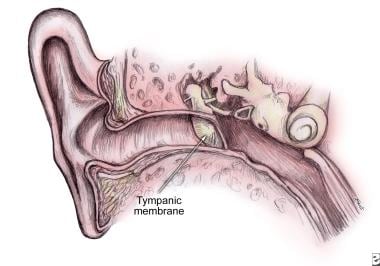
Tympanic membrane (TM) as continuation of the upper wall of external auditory canal (EAC) with angle of incline up to 45 degrees on the border between middle ear and the EAC. |
 | View Full Size Image | |
Media type: Image
|
| Media file 2:
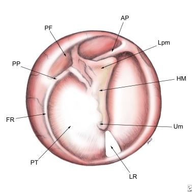
Normal tympanic membrane. Pars tensa (PT), pars flaccida (PF), light reflex (LR), fibrous ring (FR), umbo (Um), handle of malleus (HM), lateral process of malleus (Lpm), anterior plica (AP), posterior plica (PP). |
 | View Full Size Image | |
Media type: Image
|
| Media file 3:
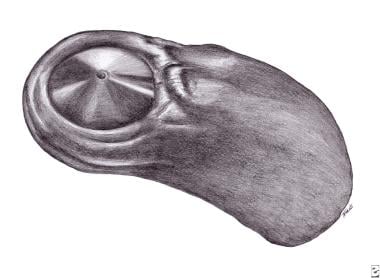
Mirror display of a tympanic membrane surface on the polymeric masc from external acoustical canal of healthy man. Masc of tympanic membrane surface (MtmS). |
 | View Full Size Image | |
Media type: Image
|
- Crapko M, Kerschner JE, Syring M, Johnston N. Role of extra-esophageal reflux in chronic otitis media with effusion. Laryngoscope. Aug 2007;117(8):1419-23. [Medline].
- Rezes S, Késmárki K, Sipka S, Sziklai I. Characterization of otitis media with effusion based on the ratio of albumin and immunoglobulin G concentrations in the effusion. Otol Neurotol. Aug 2007;28(5):663-7. [Medline].
- Arnold DJ, Bressler KL. Permeability of tympanotomy tubes to ototopical preparations. Otolaryngol Head Neck Surg. Jul 1999;121(1):35-7. [Medline].
- Blevins NH, Karmody CS. Chronic myringitis: prevalence, presentation, and natural history. Otol Neurotol. 2001;Jan;22(1):3-10. [Medline].
- Cox L, Macdonald CB. Further Study of the Acoustic Otoscope. Otolaryngology HNS. 1998;119(2):160.
- Dawes PJ, Bingham BJ, Rhys R. Aspirating middle ear effusions when inserting ventilation tubes: does it influence post-operative otorrhoea, tube obstruction or the development of tympanosclerosis?. Clin Otolaryngol. Oct 1991;16(5):457-61. [Medline].
- Franklin DJ, Starke JR, Brady MT, et al. Chronic otitis media after tympanostomy tube placement caused by Mycobacterium abscessus: a new clinical entity?. Am J Otol. May 1994;15(3):313-20. [Medline].
- Hoshino T, Yano J, Ichimura K, Hashimoto H, Nozue M. Chronic myringitis and chronic suppurative otitis media. Arch Otorhinolaryngol. 1982;234(3):219-23. [Medline].
- Jaisinghani VJ, Hunter LL, Li Y, Margolis RH. Quantitative analysis of tympanic membrane disease using video-otoscopy. Laryngoscope. 2000;Oct;110(10 Pt 1):1726-30. [Medline].
- Jones WS, Kaleida PH. How helpful is pneumatic otoscopy in improving diagnostic accuracy?. Pediatrics. 2003;Sep;112(3 Pt 1):510-3. [Medline].
- Jung HH, Cho SD, Yoo CK, Lim HH, Chae SW. Vinegar treatment in the management of granular myringitis. J Laryngol Otol. 2002;Mar;116(3):176-80. [Medline].
- Kotikoski MJ, Palmu AA, Nokso-Koivisto J, Kleemola M. Evaluation of the role of respiratory viruses in acute myringitis in children less than two years of age. Pediatr Infect Dis J. 2002;Jul;21(7):636-41. [Medline].
- Kotikoski MJ, Palmu AA, Puhakka HJ. The symptoms and clinical course of acute bullous myringitis in children less than two years of age. Int J Pediatr Otorhinolaryngol. 2003;Feb;67(2):165-72. [Medline].
- Li Y, Hunter LL, Margolis RH. Prospective study of tympanic membrane retraction, hearing loss, and multifrequency tympanometry. Otolaryngol Head Neck Surg. Nov 1999;121(5):514-22. [Medline].
- Ott PM. [The red tympanic membrane] Schweiz Rundsch Med Prax. Review. German. 2004;Oct 13;93(42):1733-7. [Medline].
- Palmu AA, Kotikoski MJ, Kaijalainen TH, Puhakka HJ. Bacterial etiology of acute myringitis in children less than two years of age. Pediatr Infect Dis J. 2001;Jun;20(6):607-11. [Medline].
- Pichichero ME, Poole MD. Assessing diagnostic accuracy and tympanocentesis skills in the management of otitis media. Arch Pediatr Adolesc Med. 2001;Oct;155(10):1137-42. [Medline].
- Ramsay HA, Heikkonen EJ, Laurila PK. Effect of epidermal growth factor on tympanic membranes with chronic perforations: a clinical trial. Otolaryngol Head Neck Surg. Oct 1995;113(4):375-9. [Medline].
- Romain O. Pneumatic otoscopy in pediatrics. Arch Pediatr. 2002;Jun;9(6):644-5. French. [Medline].
- Schilder AG, Zielhuis GA, Haggard MP. Long-term effects of otitis media with effusion: otomicroscopic findings. Am J Otol. May 1995;16(3):365-72. [Medline].
- Uliyanov YP. Method of reception impression of meatus acusticus externus and prints of a membrana tympani. Bulletin of Russian Inventions and Discoveries USSR. 1965;19:Patent: 175173.
- van Staaij BK, Rovers MM, Schilder AG, Hoes AW. Accuracy and feasibility of daily infrared tympanic membrane temperature measurements in the identification of fever in children. Int J Pediatr Otorhinolaryngol. 2003;Oct;67(10):1091-7. [Medline].
- Weymuller EA Jr. Contemporary antimicrobial therapy. In: Gluckman JL. Renewal of Certification Study Guide in Otolaryngology HNS. 1998:38-45.
- Wielgosz R, Fronz Th, Lamprecht J. The Heermann-technique of cartilage palisade tympanoplasty. Long-term results from more than 15,300 operated clinical cases. XYI World Congress of Otorhinolaryngology HNS. 1997;2:1085-8.
- Yinglin Z. Three cases of chronic granular myringitis cured by formalin. J Otolaryngol. Dec 1985;14(6):379-80. [Medline].
Middle Ear, Tympanic Membrane, Infections excerpt Article Last Updated: Mar 14, 2008
|