Continually Updated Clinical Reference
 
 
  All Sources     eMedicine     Medscape     Drug Reference     MEDLINE
 
eMedicine - External Ear, Malignant External Otitis : Article by

Quick Find
Authors & Editors
Introduction
Clinical
Differentials
Workup
Treatment
Medication
Follow-up
Miscellaneous
References

Related Articles
Malignant Tumors of the Temporal Bone


Patient Education
Click here for patient education.


Author: Brian Nussenbaum, MD, FACS, Assistant Professor, Co-director of Fellowship in Microvascular Head/Neck Oncology, Department of Otolaryngology-Head and Neck Surgery, Washington University School of Medicine

Brian Nussenbaum is a member of the following medical societies: Alpha Omega Alpha, American Academy of Otolaryngology-Head and Neck Surgery, American College of Surgeons, American Head and Neck Society, Phi Beta Kappa, Society of University Otolaryngologists-Head and Neck Surgeons, and Triological Society

Coauthor(s): Peter S Roland, MD, Professor, Department of Neurological Surgery, Professor and Chairman, Department of Otolaryngology-Head and Neck Surgery, Director of Clinical Center for Auditory, Vestibular and Facial Nerve Disorders, Chief of Pediatric Otology, University of Texas Southwestern Medical Center; Adjunct Professor of Communicative Disorders, School of Human Development, Chief of Medical Services at Callier Center for Communicative Disorders, University of Texas at Dallas

Editors: Jack A Shohet, MD, Chairman of Otolaryngology, Hoag Hospital; Francisco Talavera, PharmD, PhD, Senior Pharmacy Editor, eMedicine; Gerard J Gianoli, MD, Clinical Associate Professor, Department of Otolaryngology-Head and Neck Surgery, Tulane University School of Medicine; Vice President, The Ear and Balance Institute; Chief Executive Officer, Ponchartrain Surgery Center; Christopher L Slack, MD, Otolaryngology-Facial Plastic Surgery, Private Practice, Associated Coastal ENT; Medical Director, Treasure Coast Sleep Disorders; Arlen D Meyers, MD, MBA, Professor, Department of Otolaryngology-Head and Neck Surgery, University of Colorado School of Medicine

Author and Editor Disclosure

Synonyms and related keywords: malignant external otitis of the external ear, MEO, invasive external otitis, necrotizing external otitis, progressive external otitis, skull base osteomyelitis, ear infection, pseudomonal osteomyelitis of the temporal bone, Pseudomonas aeruginosa, P aeruginosa, diabetes, malignant external otitis

Background

Toulmouche was probably the first physician to report a case of malignant external otitis (MEO), in 1838.

In 1959, Meltzer reported a case of pseudomonal osteomyelitis of the temporal bone.

In 1968, Chandler discussed the clinical characteristics of MEO and defined it as a distinct clinical disease. He described this external otitis as malignant because he observed an aggressive clinical behavior, poor treatment outcome, and a high mortality rate for the patients affected by this disease.

The subsequent development of effective antibiotics for treating pseudomonal infections has improved the treatment outcomes for patients with MEO. Thus, some physicians have suggested that the term malignant should be abandoned in order to provide a more accurate description of the disease process.
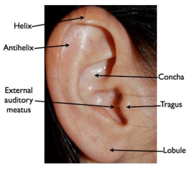
Pathophysiology

MEO is an infection that affects the external auditory canal and temporal bone. The causative organism is usually Pseudomonas aeruginosa, and the disease commonly manifests in elderly patients with diabetes. The infection begins as an external otitis that progresses into an osteomyelitis of the temporal bone. Spread of the disease outside the external auditory canal occurs through the fissures of Santorini and the osseocartilaginous junction.

Frequency

United States

MEO is more common in humid and warm climates than in other climates.

Mortality/Morbidity

  • Cranial neuropathy
    • Cranial nerves can be affected by inflammation along the skull base or by a neurotoxin produced by Pseudomonas species. The facial nerve (VII) is affected most commonly, usually at the stylomastoid foramen. As the disease progresses, cranial nerves IX, X, and XI can be affected at the jugular foramen, followed by XII at the hypoglossal canal. Cranial nerves V and VI can be affected if the disease extends to the petrous apex.
    • In 1977, Chandler reported a 32% incidence of facial nerve paralysis. The incidence of facial nerve paralysis appears to have decreased with the development of more effective medical therapy. The development of cranial neuropathy generally was thought to reflect advanced-stage disease associated with a worse prognosis. More recently, Corey et al suggested that the presence of facial nerve paralysis alone does not worsen the prognosis. The other cranial nerves are affected much less frequently than the seventh cranial nerve, but these neuropathies still are associated with an 80-100% mortality rate. The recovery of nerve function is unpredictable and should not be used as an indicator of successful treatment.
  • Intracranial complications: These complications rarely occur in the absence of cranial nerve palsies. Meningitis, brain abscess, and dural sinus thrombosis may ensue. Cranial neuropathies related to the jugular foramen should raise concern for sigmoid sinus thrombosis. Cavernous sinus thrombosis should be considered if cranial nerves V or VI are affected. Intracranial complications reflect severe disease and are commonly fatal.
  • Comorbid conditions: Patients with MEO almost always have diabetes, often with other multiple medical problems. During the course of therapy, Chandler found some deaths related to pneumonia, uremia, myocardial infarction, strokes, and liver failure.

Sex

MEO is more common in males than in females.

Age

MEO has been reported in all age groups but is most common in patients who are elderly (age, >60 y).


History

  • Diabetes (90%) or immunosuppression (illness or treatment related)
  • Severe, unrelenting, deep-seated otalgia
  • Temporal headaches
  • Purulent otorrhea
  • Possibly dysphagia, hoarseness, and/or facial nerve dysfunction

Physical

  • Inflammatory changes are observed in the external auditory canal and the periauricular soft tissue.
  • The pain is out of proportion to the physical examination findings.
    • Marked tenderness is present in the soft tissue between the mandible ramus and mastoid tip.
    • Granulation tissue is present at the floor of the osseocartilaginous junction. This finding is virtually pathognomonic of MEO. Otoscopic examination may also reveal exposed bone.
  • The cranial nerves (V-XII) should be examined.
  • Mental status examination should be performed. Deterioration of the mental status may indicate intracranial complication.
  • The tympanic membrane is usually intact.
  • Fever is uncommon.

Causes

  • Diabetes (90% of patients)
    • Diabetes is the most significant risk factor for developing MEO.
    • Small-vessel vasculopathy and immune dysfunction associated with diabetes are primarily responsible for this predisposition.
    • The cerumen of patients with diabetes has a higher pH and reduced concentration of lysozyme, which may impair local antibacterial activity.
    • No difference in predisposition is found between diabetes types I and II.
    • The predisposition is not necessarily related to the severity of glucose intolerance or periods of hyperglycemia.
  • Immunodeficiencies, such as lymphoproliferative disorders or medication-related immunosuppression
  • AIDS
    • MEO associated with AIDS may have a different pathophysiology than classic MEO.
    • Patients present with similar symptoms but are generally younger and do not have diabetes.
    • Granulation tissue may be absent in the external auditory canal.
    • Pseudomonas is not necessarily the dominant causative organism.
    • Patients with AIDS generally have a poorer outcome than patients with diabetes.
  • Aural irrigation: As many as 50% of cases of MEO have been reported to be preceded by traumatic aural irrigation in patients with diabetes.


Malignant Tumors of the Temporal Bone


Lab Studies

  • Leukocyte count
    • The leukocyte count is usually normal or mildly elevated.
    • A left shift is not commonly found.
  • Erythrocyte sedimentation rate
    • Erythrocyte sedimentation rate (ESR) is invariably elevated, with an average of 87 mm/h.
    • It begins to decrease within 2 weeks of initiating therapy but takes many months to return to normal.
    • ESR can be used to support the clinical diagnosis since acute external otitis or ear canal malignancy usually does not cause a rate elevation in this lab test.
  • Serum chemistry
    • Patients with known diabetes need an evaluation of the serum chemistry to determine if the infection is affecting their baseline glucose intolerance.
    • Patients without a history of diabetes should be tested for glucose intolerance.
  • Culture and sensitivities from the external auditory canal
    • Culture from the ear drainage should be performed ideally before antimicrobial therapy is initiated.
    • The most common causative organism is P aeruginosa (95%). This organism is an aerobic, gram-negative rod. Pseudomonas species has a mucoid coating that deters phagocytosis. Exotoxins (ie, exotoxin A, collagenase, elastase) can cause tissue necrosis, and some strains produce a neurotoxin that may be partially responsible for cranial neuropathies.
    • Less common organisms identified include Aspergillus and Proteus species, Staphylococcus aureus, and Staphylococcus epidermidis.

Imaging Studies

  • These are important adjuncts for determining the presence of osteomyelitis, the extent of disease, and response to therapy.
  • Technetium Tc 99 methylene diphosphonate bone scanning is based on binding to osteoblasts.
    • This scan depicts as little as a 10% increase in osteoblastic activity. However, this test is not specific since tumors or bony dysplasias, in addition to osteomyelitis, can cause osteoblastosis.
    • It is useful in the initial evaluation because a positive finding in the correct clinical context can lead to confirmation of the diagnosis.
    • The test is not useful for assessing the response to therapy since results remain persistently positive long after clinical improvement because of continuous bone remodeling and reformation.
    • This test may also have limited usefulness for patients with a prior history of mastoiditis or otologic surgery.
    • The application of single-photon emission computed tomography (SPECT) technology has improved the poor spatial resolution traditionally associated with this test.
  • Gallium citrate Ga 67 scan is very sensitive but is not specific because gallium binds to actively dividing cells, including inflammatory cells, tumor cells, and osteoblasts.
    • Uncertainty is possible regarding whether a positive test result represents an inflammatory condition, soft tissue, or bone disease.
    • This test is most helpful when used as a monitor of successful treatment. Improvement of a positive test result correlates with therapeutic response.
    • A baseline test is usually obtained at the initial diagnosis for comparison with follow-up studies during treatment.
    • A quantitative comparison of the lesion to the nonlesion side may improve the interpretation of these studies for distinguishing acute external otitis from MEO and for determining the efficacy of therapy.
    • The application of SPECT technology has improved the poor spatial resolution traditionally associated with this test.
  • Indium In 111–labeled leukocyte scan attempts to provide the same sensitivity as a gallium citrate Ga 67 scan but is more specific to an inflammatory process.
    • It does not appear to provide an improvement in scintographic technique for helping to establish the diagnosis.
    • It may be better than gallium citrate Ga 67 scans for assisting in establishing the correct timing of disease resolution.
    • This test can be unreliable for imaging chronic osteomyelitis in other areas of the body. Thus, the accuracy of this application needs further study.
  • CT scanning and MRI are both useful for evaluating the anatomic extent of soft tissue inflammation, abscess formation, and intracranial complications.
    • CT scanning fails to diagnose early osteomyelitis because 30-50% of bone destruction is required to detect osteomyelitis by CT scanning.
    • MRI provides poor bone resolution.
    • The soft tissue manifestations regress on CT scanning and MRI with response to therapy.
    • Bone changes remain persistently abnormal on CT scans for at least one year and are not well demonstrated by MRI studies. Thus, neither of the tests can be used to determine osteomyelitis resolution.
    • Most authors advocate obtaining a CT scan with the initial evaluation for all patients, whereas Benecke advocates obtaining this test selectively for patients with cranial neuropathy, extensive bone changes on technetium scan, or poor clinical response to treatment. Grandis et al and Okpala et al support obtaining a CT scan early in the diagnostic/treatment algorithm.
    • MRI and CT scanning are equally sensitive in detecting the soft tissue extent of the disease, but MRI is more sensitive for detecting intracranial complications.

Procedures

  • Obtain a biopsy of the external auditory canal to exclude carcinoma or other etiologies.

Histologic Findings

Nadol described the histopathology of 2 temporal bones affected by MEO. The infection did not spread through the pneumatized air tracts of the temporal bone. Rather, it spread along the vascular and fascial planes on exiting the temporal bone through the external auditory canal osseocartilaginous junction or fissures of Santorini. The otic capsule appeared to be resistant to the disease process. Linthicum described histopathologic findings in 5 temporal bones. Extensive destruction in the wall of the bony external auditory canal and osteomyelitic destruction of the wall of the fallopian canal in the descending portion of the facial nerve was seen. The infection spread beneath the otic capsule to erode the wall of the carotid canal and then extended into the central skull base.

Staging

  • Levenson et al, Corey et al, Benecke, and Davis et al have proposed staging systems for MEO.
    • These staging systems are generally based on extent of soft tissue/bony involvement or development of neurologic complications.
    • None of these staging systems has been widely adopted.


Medical Care

Treatment includes meticulous glucose control, aural toilet, systemic and ototopic antimicrobial therapy, and hyperbaric oxygen therapy.

  • Systemic antibiotic choice: Until the development of third-generation antipseudomonal cephalosporins, long-term intravenous antibiotics using an antipseudomonal penicillin and aminoglycoside were the mainstay of medical treatment.
    • Several authors have demonstrated the effectiveness of intravenous ceftazidime monotherapy in the treatment of malignant external otitis (MEO).
    • Fluoroquinolones that attain high soft tissue and bone levels with oral doses were then developed. Subsequently, several authors have demonstrated the efficacy of oral ciprofloxacin monotherapy.
    • Although no established treatment guidelines are available, case series and anecdotal experience suggest that initial outpatient therapy with oral ciprofloxacin is efficacious for patients without a fluoroquinolone allergy, cranial neuropathy, or intracranial complication and who do not require hospital admission for diabetes or pain management.
    • The widespread use of fluoroquinolones for upper respiratory infections and simpler ear infections is beginning to confound the typical clinical spectrum of MEO. Ciprofloxacin-resistant P aeruginosa has been increasingly isolated in patients with MEO, accounting for as many as 33% of isolates in patients who failed outpatient management in a study by Berenholz et al. Most notably in this patient population, 63% of isolates from 1998-2001 were resistant to ciprofloxacin, whereas only 15% of isolates were found to be resistant in the 10 years before this 3-year period. No increased morbidity or mortality was found in patients with ciprofloxacin-resistant Pseudomonas. Patients with resistant P aeruginosa require parenteral antibiotics with antipseudomonal beta-lactam antibiotics with or without an aminoglycoside.
  • Duration of therapy
    • Symptoms and examination findings improve with appropriate therapy, but these changes do not correlate with the length of needed therapy. Despite symptom relief, prolonged antimicrobial treatment as indicated for osteomyelitis is still indicated.
    • Imaging studies are helpful in determining the adequate length of treatment for each patient.
    • Treatment response should be evaluated with a gallium citrate Ga 67 scan every 4-6 weeks during treatment. Benecke recommended ending treatment 1 week after the gallium citrate Ga 67 scan findings return to normal and confirming this with a repeat scan 1 month after the treatment is stopped. Using this protocol for 13 patients, the average duration of treatment was 8.8 weeks with a range of 4-17 weeks.
  • Hyperbaric oxygen therapy
    • This should be used only as an adjunct to antimicrobial therapy; it should not be used alone.
    • Hyperbaric oxygen therapy may be helpful for patients with complications, experiencing a poor response to therapy, or with recurrent cases.

Surgical Care

  • Chandler advocated surgery in his original report when appropriate antimicrobials were not available; he had very poor results, with a 50% mortality rate.
  • Surgical removal of the lesion requires resection of large portions of the temporal bone.
  • Because of the histopathology of MEO, removal of contiguous areas of bone may not be sufficient because of the spread of infection through vascular and fascial planes.
  • Surgery is now reserved for local debridement, removal of bony sequestrum, or abscess drainage.
  • Facial nerve decompression is not indicated for patients with facial paralysis.

Consultations

  • Consultation with internal medicine specialists is required for the management of diabetes and other comorbidities.
  • Infectious-disease consultants may help with the choice of antibiotics in complicated cases.


The goals of pharmacotherapy are to reduce morbidity and prevent complications.

Drug Category: Antibiotics

The choice of antimicrobial therapy and whether treatment can be delivered PO versus IV, as monotherapy, or on an outpatient basis need to be carefully individualized to each patient. Refer to Medical Care for further discussion of these issues.

Drug NameCiprofloxacin (Cipro)
DescriptionA DNA gyrase inhibitor that prevents DNA replication.
Adult Dose1500-2250 mg/d PO/IV divided bid/tid
Pediatric Dose<18 years: Not recommended
>18 years: Administer as in adults
ContraindicationsDocumented hypersensitivity
InteractionsAntacids, iron salts, and zinc salts may reduce serum levels; administer antacids 2-4 h before or after taking fluoroquinolones; cimetidine may interfere with metabolism of fluoroquinolones; ciprofloxacin reduces therapeutic effects of phenytoin; probenecid may increase ciprofloxacin serum concentrations
May increase toxicity of theophylline, caffeine, cyclosporine, and digoxin (monitor digoxin levels); may increase effects of anticoagulants (monitor PT)
PregnancyC - Safety for use during pregnancy has not been established.
PrecautionsCaution in epilepsy or other conditions that have a low seizure threshold; in prolonged therapy, perform periodic evaluations of organ system functions (eg, renal, hepatic, hematopoietic); adjust dose in renal function impairment; superinfections may occur with prolonged or repeated antibiotic therapy; dehydration or urine alkalinization should be avoided

Drug NameCeftazidime (Fortaz, Tazicef, Tazidime)
DescriptionThird-generation cephalosporin that inhibits bacterial cell wall synthesis by adhering to penicillin-binding proteins. Can be either bactericidal or bacteriostatic depending on the organism, tissue penetration, and drug dosage.
Adult Dose1-2 g IV q8h
Pediatric DoseNot established
ContraindicationsDocumented hypersensitivity
InteractionsNephrotoxicity may increase with aminoglycosides, furosemide, and ethacrynic acid; probenecid may increase ceftazidime levels; concomitant use with aminoglycosides can provide synergistic activity against P aeruginosa
PregnancyB - Usually safe but benefits must outweigh the risks.
PrecautionsAdjust dose in renal impairment

Drug NameTicarcillin/clavulanate (Timentin)
DescriptionExtended-spectrum penicillin/beta-lactamase inhibitor. Ticarcillin binds to penicillin-binding proteins, thus inhibiting bacterial cell wall synthesis. Clavulanate binds irreversibly to beta-lactamases, thus preventing inactivation of ticarcillin.
Adult Dose3.1 g IV q6h
Pediatric DoseNot established
ContraindicationsDocumented hypersensitivity
InteractionsTetracyclines may decrease effects of ticarcillin; high concentrations of ticarcillin may physically inactivate aminoglycosides if administered in same IV line; effects when administered concurrently with aminoglycosides are synergistic; probenecid may increase penicillin levels
PregnancyB - Usually safe but benefits must outweigh the risks.
PrecautionsAdjust dose in renal impairment; use cautiously in patients with sodium restrictions

Drug NameGentamicin (Garamycin, Jenamicin)
DescriptionBinds to 30S ribosomal subunit, thus inhibiting bacterial protein synthesis. Aminoglycosides have bactericidal activity against P aeruginosa. Bacterial strains resistant to one aminoglycoside still may be sensitive to other antibiotics within this category.
Adult Dose3-5 mg/kg/d IV in equally divided doses q8h or 5-7 mg/kg/d IV administered qd; dose is adjusted based on serum drug levels
Pediatric DoseNot established
ContraindicationsDocumented hypersensitivity; non–dialysis-dependent renal insufficiency
InteractionsCoadministration with other aminoglycosides, cephalosporins, penicillins, and amphotericin B may increase nephrotoxicity; aminoglycosides enhance effects of neuromuscular blocking agents, and prolonged respiratory depression may occur; coadministration with loop diuretics may increase auditory toxicity of aminoglycosides; possible irreversible hearing loss of varying degrees may occur (monitor regularly); concomitant use with penicillin can provide synergistic activity against P aeruginosa
PregnancyD - Unsafe in pregnancy
PrecautionsNarrow therapeutic index (not intended for long-term therapy); caution in renal failure (not on dialysis), myasthenia gravis, hypocalcemia, and conditions that depress neuromuscular transmission; adjust dose in renal impairment; dehydration can increase risk of ototoxicity and nephrotoxicity; can aggravate muscle weakness in patients with myasthenia gravis or parkinsonism


Prognosis

  • Disease recurrence
    • Disease recurrence is reported in 9-27% of patients.
    • It usually is related to inadequate length of therapy and manifests as recurrent headaches and otalgia, not as otorrhea.
    • The ESR begins increasing again.
    • MEO can recur as long as one year after treatment is completed; thus, a patient should not be considered cured until that time.
  • Mortality
    • Chandler reported a mortality rate of 50% in the original series.
    • The mortality rate has decreased to 20% with the introduction of appropriate antibiotics, improved imaging modalities, and increased awareness of the disease.
    • Most current studies report a mortality rate of less than 10%, but mortality remains high for patients with cranial neuropathies (other than VII) or intracranial complications.


Medical/Legal Pitfalls

  • Failure to exclude a malignant neoplastic process

Special Concerns

  • Infants and children
    • In 1988, Rubin et al reported MEO in 15 children. Most did not have diabetes, and the more common predisposing risk factor was immune dysfunction.
    • The facial nerve was affected more frequently than in adults, and other cranial neuropathies were not found. This is likely related to the underdeveloped mastoid process and the more medial location of the fissures of Santorini in children. Tympanic membrane involvement was common, and bacteremia was more common than in adults.
    • Long-term morbidity was uncommon.
    • Fluoroquinolones are not approved by the Food and Drug Administration (FDA) for use in the management of MEO in the pediatric population, but they have been used widely and safely in the management of cystic fibrosis in children.
  • Possible impact of managed care and delayed diagnosis because of late referrals: Insufficient duration of therapy by primary care physicians treating MEO as an uncomplicated otitis externa may lead to emergence of drug resistance.


  • Amorosa L, Modugno GC, Pirodda A. Malignant external otitis: review and personal experience. Acta Otolaryngol Suppl. 1996;521:3-16. [Medline].
  • Benecke JE Jr. Management of osteomyelitis of the skull base. Laryngoscope. Dec 1989;99(12):1220-3. [Medline].
  • Berenholz L, Katzenell U, Harell M. Evolving resistant pseudomonas to ciprofloxacin in malignant otitis externa. Laryngoscope. Sep 2002;112(9):1619-22. [Medline].
  • Chandler JR. Malignant external otitis and osteomyelitis of the base of the skull. Am J Otol. Mar 1989;10(2):108-10. [Medline].
  • Chandler JR. Malignant external otitis. Laryngoscope. Aug 1968;78(8):1257-94. [Medline].
  • Chandler JR. Malignant external otitis: further considerations. Ann Otol Rhinol Laryngol. Jul-Aug 1977;86(4 Pt 1):417-28. [Medline].
  • Chandler JR. Pathogenesis and treatment of facial paralysis due to malignant external otitis. Ann Otol Rhinol Laryngol. Oct 1972;81(5):648-58. [Medline].
  • Corey JP, Levandowski RA, Panwalker AP. Prognostic implications of therapy for necrotizing external otitis. Am J Otol. Jul 1985;6(4):353-8. [Medline].
  • Davis JC, Gates GA, Lerner C, et al. Adjuvant hyperbaric oxygen in malignant external otitis. Arch Otolaryngol Head Neck Surg. Jan 1992;118(1):89-93. [Medline].
  • Driscoll PV, Ramachandrula A, Drezner DA, et al. Characteristics of cerumen in diabetic patients: a key to understanding malignant external otitis?. - Schaffer SR. Oct 1993;109(4):676-9. [Medline].
  • Epstein JS, Ganz WI, Lizak M, et al. Indium 111-labeled leukocyte scintigraphy in evaluating head and neck infections. Ann Otol Rhinol Laryngol. Dec 1992;101(12):961-8. [Medline].
  • Gehanno P. Ciprofloxacin in the treatment of malignant external otitis. Chemotherapy. 1994;40 Suppl 1:35-40. [Medline].
  • Gherini SG, Brackmann DE, Bradley WG. Magnetic resonance imaging and computerized tomography in malignant external otitis. Laryngoscope. May 1986;96(5):542-8. [Medline].
  • Gold S, Som PM, Lucente FE, et al. Radiographic findings in progressive necrotizing "malignant" external otitis. Laryngoscope. Mar 1984;94(3):363-6. [Medline].
  • Grandis JR, Curtin HD, Yu VL. Necrotizing (malignant) external otitis: prospective comparison of CT and MR imaging in diagnosis and follow-up. Radiology. Aug 1995;196(2):499-504. [Medline].
  • Johnson MP, Ramphal R. Malignant external otitis: report on therapy with ceftazidime and review of therapy and prognosis. Rev Infect Dis. Mar-Apr 1990;12(2):173-80. [Medline].
  • Kimmelman CP, Lucente FE. Use of ceftazidime for malignant external otitis. Ann Otol Rhinol Laryngol. Sep 1989;98(9):721-5. [Medline].
  • Lang R, Goshen S, Kitzes-Cohen R, Sade J. Successful treatment of malignant external otitis with oral ciprofloxacin: report of experience with 23 patients. J Infect Dis. Mar 1990;161(3):537-40. [Medline].
  • Levenson MJ, Parisier SC, Dolitsky J, Bindra G. Ciprofloxacin: drug of choice in the treatment of malignant external otitis (MEO). Laryngoscope. Aug 1991;101(8):821-4. [Medline].
  • Mendelson DS, Som PM, Mendelson MH, Parisier SC. Malignant external otitis: the role of computed tomography and radionuclides in evaluation. Radiology. Dec 1983;149(3):745-9. [Medline].
  • Meyers BR, Mendelson MH, Parisier SC, Hirschman SZ. Malignant external otitis. Comparison of monotherapy vs combination therapy. Arch Otolaryngol Head Neck Surg. Sep 1987;113(9):974-8. [Medline].
  • Nadol JB Jr. Histopathology of Pseudomonas osteomyelitis of the temporal bone starting as malignant external otitis. Am J Otolaryngol. Nov 1980;1(5):359-71. [Medline].
  • Okpala NC, Siraj QH, Nilssen E, Pringle M. Radiological and radionuclide investigation of malignant otitis externa. J Laryngol Otol. Jan 2005;119(1):71-5. [Medline].
  • Redleaf MI, Angeli SI, McCabe BF. Indium 111-labeled white blood cell scintigraphy as an unreliable indicator of malignant external otitis resolution. Ann Otol Rhinol Laryngol. Jun 1994;103(6):444-8. [Medline].
  • Ress BD, Luntz M, Telischi FF, et al. Necrotizing external otitis in patients with AIDS. Laryngoscope. Apr 1997;107(4):456-60. [Medline].
  • Rubin Grandis J, Branstetter BF, Yu VL. The changing face of malignant (necrotising) external otitis: clinical, radiological, and anatomic correlations. Lancet Infect Dis. Jan 2004;4(1):34-9. [Medline].
  • Rubin J, Yu VL, Stool SE. Malignant external otitis in children. J Pediatr. Dec 1988;113(6):965-70. [Medline].
  • Rubin J, Yu VL. Malignant external otitis: insights into pathogenesis, clinical manifestations, diagnosis, and therapy. Am J Med. Sep 1988;85(3):391-8. [Medline].
  • Seabold JE, Simonson TM, Weber PC, et al. Cranial osteomyelitis: diagnosis and follow-up with In-111 white blood cell and Tc-99m methylene diphosphonate bone SPECT, CT, and MR imaging. Radiology. Sep 1995;196(3):779-88. [Medline].
  • Slattery WH 3rd, Brackmann DE. Skull base osteomyelitis. Malignant external otitis. Otolaryngol Clin North Am. Oct 1996;29(5):795-806. [Medline].
  • Stokkel MP, Takes RP, van Eck-Smit BL, Baatenburg de Jong RJ. The value of quantitative gallium-67 single-photon emission tomography in the clinical management of malignant external otitis. Eur J Nucl Med. Nov 1997;24(11):1429-32. [Medline].
  • Stokkel MP, Boot CN, van Eck-Smit BL. SPECT gallium scintigraphy in malignant external otitis: initial staging and follow-up. Case reports. Laryngoscope. Mar 1996;106(3 Pt 1):338-40. [Medline].
  • Strauss M, Aber RC, Conner GH, Baum S. Malignant external otitis: long-term (months) antimicrobial therapy. Laryngoscope. Apr 1982;92(4):397-406. [Medline].
  • Sudhoff H, Linthicum FH. Malignant external otitis: temporal bone histopathology case of the month. Otol Neurotol. Mar 2003;24(2):346-7. [Medline].
  • Uri N, Gips S, Front A, et al. Quantitative bone and 67Ga scintigraphy in the differentiation of necrotizing external otitis from severe external otitis. Arch Otolaryngol Head Neck Surg. Jun 1991;117(6):623-6. [Medline].

External Ear, Malignant External Otitis excerpt

Article Last Updated: Apr 14, 2006