Practice Essentials
Otitis externa (OE) is an inflammation or infection of the external auditory canal (EAC), the auricle, or both. [1] This condition can be found in all age groups. [2] See the image below.
Classification
OE may be classified as follows:
-
Acute diffuse OE - Most common form of OE, typically seen in swimmers
-
Acute localized OE (furunculosis) - Associated with infection of a hair follicle
-
Chronic OE - Same as acute diffuse OE but is of longer duration (>6 weeks)
-
Eczematous (eczematoid) OE - Encompasses various dermatologic conditions (eg, atopic dermatitis, psoriasis, systemic lupus erythematosus, and eczema) that may infect the EAC and cause OE
-
Necrotizing (malignant) OE - Infection that extends into the deeper tissues adjacent to the EAC; occurs primarily in immunocompromised adults (eg, diabetics, patients with AIDS)
-
Otomycosis - Infection of the ear canal from a fungal species (eg, Candida, Aspergillus)
Signs and symptoms
The key physical finding of OE is pain upon palpation of the tragus (anterior to ear canal) or application of traction to the pinna (the hallmark of OE). Patients may also have the following signs and symptoms:
-
Otalgia - Ranges from mild to severe, typically progressing over 1-2 days
-
Hearing loss
-
Ear fullness or pressure
-
Erythema, edema, and narrowing of the EAC
-
Tinnitus
-
Fever (occasionally)
-
Itching (especially in fungal OE or chronic OE)
-
Severe deep pain - Immunocompromised patients may have necrotizing (malignant) OE
-
Discharge - Initially, clear; quickly becomes purulent and foul-smelling
-
Cellulitis of the face or neck or lymphadenopathy of the ipsilateral neck (occasionally)
-
Bilateral symptoms (rare)
-
History of exposure to or activities in water (frequently) (eg, swimming, surfing, kayaking)
-
History of preceding ear trauma (usually) (eg, forceful ear cleaning, use of cotton swabs, or water in the ear canal)
See Clinical Presentation for more detail.
Diagnosis
The patient’s history and physical examination, including otoscopy, usually provide sufficient information for the clinician to make the diagnosis of OE. Note that a patient who is diabetic or immunocompromised with severe pain in the ear should have necrotizing OE excluded by an otolaryngologist.
Laboratory testing
Typically, laboratory studies are not needed, but they may be helpful if the patient is immunocompromised, if the usual treatment measures are ineffective, or if a fungal cause is suspected. Tests may include the following:
-
Gram stain and culture of any discharge from the auditory canal
-
Blood glucose level
-
Urine dipstick
Imaging studies
Imaging studies are not required for most cases of OE. However, radiologic investigation may be helpful if an invasive infection such as necrotizing (malignant) OE is suspected or if the diagnosis of mastoiditis is being considered.
Imaging modalities may include the following:
-
High-resolution computed tomography (CT) - Preferred; better depicts bony erosion [3]
-
Radionucleotide bone scanning
-
Gallium scanning
-
Magnetic resonance imaging (MRI) - Not used as often as the other modalities; may be considered secondarily or if soft-tissue extension is the predominant concern [4]
See Workup for more detail.
Management
Most persons with OE are treated empirically. Primary treatment involves the following:
-
Pain management
-
Removal of debris from the EAC
-
Administration of topical medications to control edema and infection
-
Avoidance of contributing factors
Pharmacotherapy
-
Topical medications (eg, acetic acid in aluminum acetate, hydrocortisone and acetic acid otic solution, alcohol vinegar otic mix)
-
Analgesic agents (eg, acetaminophen, acetaminophen and codeine)
-
Antibiotics (eg, hydrocortisone/neomycin/polymyxin B, otic ofloxacin, otic ciprofloxacin, otic finafloxacin, gentamicin 0.3%/prednisolone 1% ophthalmic, dexamethasone/tobramycin, otic ciprofloxacin and dexamethasone, otic ciprofloxacin and hydrocortisone suspension)
-
Oral antibiotics (eg, ciprofloxacin)
-
Antifungal agents (eg, otic clotrimazole 1% solution, nystatin powder)
Surgery
-
Surgical debridement of the ear canal - Usually reserved for necrotizing OE or for complications of OE (eg, external canal stenosis); often necessary in more severe cases of OE or in cases where a significant amount of discharge is present in the ear; mainstay of treatment for fungal infections
-
Incision and drainage of an abscess
See Treatment and Medication for more detail.
Background
Otitis externa (OE) is an inflammation or infection of the external auditory canal (EAC), the auricle, or both. [1] It is a common disease that can be found in all age groups. [2] OE usually represents an acute bacterial infection of the skin of the ear canal (most commonly attributable to Pseudomonas aeruginosa or Staphylococcus aureus [5] ) but can also be caused by other bacteria, viruses, or a fungal infection (see Pathophysiology and Etiology).
Several factors can contribute to EAC infection and the development of OE, including the following [6] :
-
Absence of cerumen
-
High humidity
-
Retained water in ear canal
-
Increased temperature
-
Local trauma (eg, use of cotton swabs or hearing aids)
Aquatic athletes are particularly prone to the development of OE because repeated exposure to water results in removal of cerumen and drying of the EAC. Retained water in the ear canal can cause maceration of the skin and a milieu conducive to bacterial or fungal proliferation. OE occurs more often in the summer months, when swimming is more common, [2, 6] and it is also common in tropical areas. [7] Individuals with allergic conditions (eg, eczema, allergic rhinitis, and asthma) are also at significantly higher risk for OE. [8, 9]
Although OE rarely causes prolonged problems or serious complications, the infection is responsible for significant pain and acute morbidity (see Presentation). Prompt diagnosis (see DDx and Workup) and appropriate therapy (see Treatment) cure the majority of cases without complications; however, patients who are diabetic, immunocompromised, or untreated may develop necrotizing (malignant) OE, [10] a potentially life-threatening infection.
In 2014, the American Academy of Otolaryngology–Head and Neck Surgery Foundation (AAO-HNSF) released updated clinical practice guidelines for the diagnosis and treatment of acute OE (see Guidelines). [1]
Anatomy
The external ear (see the image below) consists of the auricle and the EAC.
The auricle is composed of elastic cartilage with the overlying skin attached directly to the perichondrium. It begins to form during week 6 of gestation through consolidation of portions of the mesoderm of the first and second branchial arches, giving rise to the His hillocks. The first three hillocks are derived from the first arch, the second three from the second arch. The auricle reaches adult shape by the week 20 of gestation, but the adult size is not reached until the age of 9 years.
The EAC begins to form during week 8 of gestation, when the surface ectoderm of the first pharyngeal groove thickens and grows toward the middle ear. This core of tissue begins to resorb by week 21 of gestation to form a channel that is complete by week 28. The canal reaches adult size by the age of 9 years and ossifies completely by the age of 3 years. The EAC is related to the mandibular fossa anteriorly, the mastoid air cells posteriorly, the middle cranial fossa superiorly, and the parotid gland inferiorly.
The EAC is lined with squamous epithelium and is approximately 2.5 cm long in adults. Its function is to transmit sound to the middle ear while protecting more proximal structures from foreign bodies and any changes in environmental conditions. The outer one third of the canal is primarily cartilaginous and is oriented superiorly and posteriorly; the inner two thirds of the canal is osseous, is covered with thinner skin that adheres tightly, and is oriented inferiorly and anteriorly; this portion of the canal is devoid of any apocrine glands or hair follicles.
The thicker skin over the outer (cartilaginous) portion of the EAC contains apopilosebaceous units comprising apocrine and eccrine glands that secrete their products around the base of a hair follicle. These secretions combine with sloughed squamous epithelium (cerumen) to coat the EAC and maintain an acidic pH (4-5). This cerumen coat migrates from the isthmus of the EAC to the lateral part, and its waxy nature protects the underlying epithelium from maceration or skin breakdown. The quantity of cerumen produced varies widely among individuals.
The acidity of the cerumen inhibits bacterial or fungal growth. Whereas a paucity of cerumen allows bacterial growth, an excess can create an environment ideal for bacterial invasion by allowing retention of water and debris (as when the EAC is regularly exposed to water). Localized trauma from foreign objects placed in the ear can also lead to direct bacterial invasion in the ear canal. Once an infection becomes established, localized maceration and inflammation occur, which lead to symptoms.
Pathophysiology
OE is a superficial infection of the skin in the EAC. It may be classified as follows:
-
Acute diffuse OE – This is the most common form of OE, typically seen in swimmers; it is characterized by rapid onset (generally within 48 hours) and symptoms of EAC inflammation (eg, otalgia, itching, or fullness, with or without hearing loss or jaw pain) as well as tenderness of the tragus or pinna or diffuse ear edema or erythema or both, with or without otorrhea, regional lymphadenitis, tympanic membrane erythema, or cellulitis of the pinna [7]
-
Acute localized OE – This condition, also known as furunculosis, is associated with infection of a hair follicle
-
Chronic OE – This is the same as acute diffuse OE but is of longer duration (>6 weeks)
-
Eczematous (eczematoid) OE – This encompasses various dermatologic conditions (eg, atopic dermatitis, psoriasis, systemic lupus erythematosus, and eczema) that may infect the EAC and cause OE
-
Necrotizing (malignant) OE – This is an infection that extends into the deeper tissues adjacent to the EAC; it primarily occurs in adult patients who are immunocompromised (eg, as a result of diabetes mellitus or AIDS) and is rarely described in children; it may result in cases of cellulitis and osteomyelitis (see Cellulitis, Osteomyelitis, and Chronic Osteomyelitis Imaging)
-
Otomycosis - Infection of the ear canal secondary to fungus species such as Candida or Aspergillus
The processes involved in the development of OE can be divided into the following four categories:
-
Obstruction (eg, cerumen buildup, surfer’s exostosis, or a narrow or tortuous canal), resulting in water retention
-
Absence of cerumen, which may occur as a result of repeated water exposure or overcleaning the ear canal
-
Trauma
-
Alteration of the pH of the ear canal
If moisture is trapped in the EAC, it may cause maceration of the skin and provide a good breeding ground for bacteria. This may occur after swimming (especially in contaminated water) or bathing—hence the common lay term “swimmer’s ear.” It may also occur in hot humid weather. Obstruction of the EAC by excessive cerumen, debris, surfer’s exostosis, or a narrow and tortuous canal may also lead to infection by means of moisture retention.
Trauma to the EAC allows invasion of bacteria into the damaged skin. This often occurs after attempts at cleaning the ear with a cotton swab, paper clip, or any other utensil that can fit into the ear.
Once infection is established, an inflammatory response occurs with skin edema. Exudate and pus often appear in the EAC as well. If severe, the infection may spread and cause a cellulitis of the face or neck.
Necrotizing (malignant) OE is a rare complication that occurs in patients who are immunocompromised or in those who have received radiotherapy to the skull base. In this condition, bacteria invade the deeper underlying structures of the soft tissues and cause osteomyelitis of the temporal bone. This is a life-threatening disorder with an overall mortality that historically has approached 50%.
Etiology
OE is most often caused by a bacterial pathogen; other varieties include fungal OE (otomycosis) and eczematoid (psoriatic) OE. [11] In one study, 91% of cases of OE were caused by bacteria. [5] Others have found that as many as 40% of cases of OE have no primary identifiable microorganism as a causative agent. The most common causative bacteria are Pseudomonas species (38% of all cases), [11] Staphylococcus species, and anaerobes and gram-negative organisms.
Fungal OE may result from overtreatment with topical antibiotics or may arise de novo from moisture trapped in the EAC. It is caused by Aspergillus 80-90% of the time; Candida and other organisms have also been isolated. This condition is characterized by long, white, filamentous hyphae growing from the skin surface. Besides otorrhea, erythema and edema of the EAC are common. In severe cases, soft tissue stenosis may be present. Extension of the infection may manifest as cellulitic skin changes involving the concha of the auricle and the tragus.
Eczematoid (psoriatic) OE is associated with the following conditions:
-
Eczema
-
Seborrhea
-
Neurodermatitis
-
Contact dermatitis from earrings or hearing aid use
-
Purulent otitis media with perforation of the tympanic membrane and drainage; this may mimic OE to an extent, but it is usually painless and does not cause any swelling of the ear canal
-
Sensitivity to topical medications
Chronic OE is a fairly common condition that is sometimes the result of incomplete treatment of acute OE. [12] More often, however, chronic OE is caused by overmanipulation of the ear canal as a consequence of cleaning and scratching. Such overmanipulation results in a low-grade inflammatory response that causes further itching of the skin. Eventually, the skin thickens, and canal stenosis may occur.
Necrotizing OE occurs in patients who are immunocompromised and represents a true osteomyelitis of the temporal bone.
Risk factors for OE include the following:
-
Previous episodes of OE
-
Swimming, diving, or participating in aquatic activities
-
Use of earplugs or probing of the EAC (possibly secondary to trauma to the EAC)
-
Hot, humid weather
-
Use of a hearing aid
-
Coexistence of eczema, allergic rhinitis, or asthma
-
Comorbidities such as diabetes mellitus, AIDS, leukopenia, or malnutrition
Epidemiology
United States and international statistics
OE is found in all regions of the United States, occurring in 4 of every 1000 people annually. [6, 8] The infection is believed to be more prevalent in hot and humid conditions such as prevail during the summer months, presumably because participation in aquatic activities is higher. [2, 6, 13] Acute, chronic, and eczematous OE are also common. Necrotizing OE is rare.
The international frequencies of OE have not been fully determined; however, the incidence is increased in tropical countries. [7]
Age-, sex-, and race-related demographics
Although the infection can affect all age groups, OE appears to be most prevalent in the older pediatric and young adult population, with a peak incidence in children aged 7-12 years. [11] A single epidemiologic study from the United Kingdom found a similar 12-month prevalence for individuals aged 5-64 years and a slight increase in prevalence for those older than 65 years. [2] This was postulated to occur secondary to an increase in comorbidities, as well as an increase in the use of hearing aids, which may cause trauma to the EAC.
OE affects both sexes equally. No racial predilection has been established, though people in some racial groups have small ear canals, which may predispose them to obstruction and infection.
Prognosis
Most incidents of OE resolve without difficulty. The majority of patients improve within 48-72 hours of antibiotic administration. Failure to improve within 2-3 days should call the diagnosis into question and prompt the physician to reevaluate the patient. OE usually resolves fully in 7-10 days. Resolution of eczematoid OE occurs with control of the primary skin condition. In some patients with OE, the ear must be debrided for full resolution. Surgical incision and drainage are sometimes necessary.
In some patients, OE can cause severe otalgia necessitating administration of narcotic pain relievers. Pain usually improves 2-5 days after initiating therapy. Temporary hearing loss is common secondary to canal occlusion. Severe infections may cause lymphadenitis or cellulitis of the face or neck.
If left untreated, the infection may invade the deeper adjacent structures and progress to necrotizing (malignant) OE, a serious condition that requires prolonged treatment and often results in severe morbidity or mortality. This complication is almost exclusively seen in immunocompromised patients, such as those with diabetes, AIDS patients, those undergoing chemotherapy, and patients taking immunosuppressant medications (eg, glucocorticoids). Pseudomonas is the inciting organism in the vast majority of cases.
When necrotizing OE develops, mortality is in the 20% range among adults, mostly because of the associated comorbidities and the rapid extension of the infection to include sepsis or intracranial extension. If left untreated, necrotizing OE has a mortality approaching 50%. This complication should be suspected if the patient’s pain and tenderness seem out of proportion to clinical appearance or if granulation tissue is seen in the ear canal.
Patient Education
OE is a common problem, with risk factors that are easily avoided. Education regarding ways of keeping the ear dry is helpful. Preventive use of acidifying drops is encouraged in patients with recurrent OE. Avoidance of the use of cotton-tipped swabs to remove ear cerumen should be discussed with patients. Improper use of cotton-tipped applicator sticks simply packs cerumen into the canal and can cause trauma to the tympanic membrane.
Patients should be made aware that when OE does strike, it can usually be resolved in a short time, with few if any complications.
For patient education resources, see the Ear, Nose, and Throat Center, as well as Swimmer’s Ear.
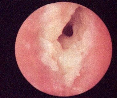
-
Acute otitis externa. Ear canal is red and edematous, and discharge is present.
-
Otitis externa with ear wick in place. Note discharge from canal and swelling of canal.
-
Anatomy of external and middle ear.


