Practice Essentials
Microstomia is the term used to describe a congenital or acquired reduction in the size of the oral aperture that is severe enough to compromise cosmesis, nutrition, and quality of life.
The development of improved surgical reconstructive techniques, particularly the transfer of regional flaps and vascularized free tissue, has made this disorder increasingly rare among adults who undergo lip resections. As a consequence, microstomia due to connective-tissue disorders has become more important in adults.
During the early 1900s, electrical service became available to most Americans. On the other hand, lye and other caustic substances were introduced into many homes as household cleaners. Coincidentally, microstomia due to accidental burns and subsequent scarring around the mouth was noted to affect an increasing number of children. Despite federal legislation that mandated protections incorporated into both electrical wiring and packaging for caustic materials, such accidents remain a frequent cause of microstomia among children.
Advances in prosthetic dentistry since the late 20th century have improved early management of pediatric patients with oral burns, but surgical correction is still sometimes indicated.
Less commonly, genetic disorders are associated with microstomia. Earlier identification of children with this problem has resulted in avoidance of complications and early intervention when necessary.
The image below shows a child with microstomia.
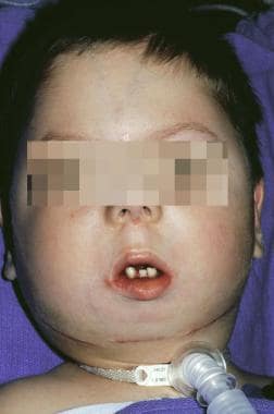 Child with craniofacial anomalies and microstomia. A tracheotomy is in place for airway control because of micrognathia. Advancement osteotomies have been performed in an effort to lengthen the mandible.
Workup in microstomia
Any systemic abnormalities that result from the underlying cause of microstomia may affect the patient's ability to heal from the inciting event or to undergo anesthesia and surgery; thus, such systemic abnormalities should be sought. For example:
-
Electrical burns - Patients with electrical burns should undergo an electrocardiogram (ECG) because myocardial injuries, conduction abnormalities, or both are possible and must be promptly diagnosed
-
Scleroderma - Computed tomography (CT) scanning or radiography of the chest may be useful in patients with scleroderma for determining the presence of pulmonary fibrosis
-
Epidermolysis bullosa - Endoscopic evaluation of the airway should be considered in patients with microstomia due to epidermolysis bullosa
Management of microstomia
Prevention
Various oral appliances that prevent microstomia have been developed. [1] The microstomia prevention apparatus (MPA) is widely available, simple to use for physicians and patients, and does not require intact dentition. However, as with all devices, this apparatus has disadvantages. It does not prevent lower lip eversion when the scar is adjacent to the vermilion borders, a potential for skin breakdown exists, and the patient is unable to retain oral secretions.
In cases of trauma, a splint should generally be instituted within 2 weeks of the injury. In general, the appliance is worn for 6 months; the patient may then transition to nighttime use only until the scar is mature.
In addition to preventive apparatus, the injection of chemical agents that inhibit fibroblast growth have shown potential in preventing scar formation. Exercise and massage improve patient outcome.
Surgical therapy
The surgical correction of microstomia is approached in the following two ways:
-
Commissuroplasty (also known as commissurotomy) - In this, the corners of the lips (commissures) are re-created; the essentials include reestablishing the intended location of the commissure, excising the scar tissue, and covering the area with mucosal flaps; establishing an intact orbicularis muscle clinically prior to proceeding with scar excision is crucial
-
Augmentation of the lips or commissures - The oral aperture may be widened with stair-step lengthening of the muscle in patients with a congenitally small orbicularis oris, such as those with Freeman-Sheldon syndrome; reconstruction using regional pedicled flaps and free flaps brings in distant tissue to expand the oral opening if inadequate tissue is present
Tissue expanders are sometimes feasible in patients in whom extensive skin scarring has occurred but muscle is intact. Dynamic slings with temporalis muscle can be used to improve lip and commissure movement when muscle damage has occurred.
Background
Definition and history
Microstomia is the term used to describe a congenital or acquired reduction in the size of the oral aperture that is severe enough to compromise cosmesis, nutrition, and quality of life.
The development of improved surgical reconstructive techniques, particularly the transfer of regional flaps and vascularized free tissue, has made this disorder increasingly rare among adults who undergo lip resections. As a consequence, microstomia due to connective-tissue disorders has become more important in adults.
During the early 1900s, electrical service became available to most Americans. On the other hand, lye and other caustic substances were introduced into many homes as household cleaners. Coincidentally, microstomia due to accidental burns and subsequent scarring around the mouth was noted to affect an increasing number of children. Despite federal legislation that mandated protections incorporated into both electrical wiring and packaging for caustic materials, such accidents remain a frequent cause of microstomia among children.
Advances in prosthetic dentistry over the past 30 years have improved early management of pediatric patients with oral burns, but surgical correction is still sometimes indicated.
Less commonly, genetic disorders are associated with microstomia. Earlier identification of children with this problem has resulted in avoidance of complications and early intervention when necessary.
The image below shows a child with microstomia.
Child with craniofacial anomalies and microstomia. A tracheotomy is in place for airway control because of micrognathia. Advancement osteotomies have been performed in an effort to lengthen the mandible.
Problem
Individuals with microstomia may experience problems with speech, oral intake, dental hygiene, facial expression, and difficulty inserting dental appliances. Additionally, this problem sometimes carries a cosmetic disfigurement that contributes to social isolation of the patient.
The severe tooth decay that may follow is then compounded by limited access for the dentist, possibly delaying treatment and leading to more extensive odontogenic infections. As a result, head-and-neck surgeons should be familiar with the management of microstomia and, more importantly, with its prevention.
Epidemiology
Frequency
The incidence and prevalence of microstomia are difficult to determine since no established criteria or classification for diagnosis is available.
With the development of innovative flap techniques and microvascular free-tissue transfer, microstomia following oral cavity tumor extirpation has become an uncommon condition. However, reports have shown that that 3.7-10.8% of thermal burn admissions and more than 30% of diffuse facial scleroderma cases are complicated by microstomia. [2, 3] Microstomia related to congenital syndromes is less common.
Etiology
In toddlers, orofacial burns occur when the child sucks on the female end of a live extension cord or the junction of two cords. The relatively high electrical resistance of the skin reduces injury to the skin and more distal structures; however, the low resistance of saliva-coated mucosa results in significant injury to the oral tissues. The lower lip is usually damaged more extensively than the upper lip, with most of the injury occurring at the vermilion border of the oral commissures.
Caustic injury from suicide attempts, assaults, and accidental ingestions can also cause chemical burns and resultant scarring. Lye and industrial cleaning solutions are the predominant items associated with suicide attempts and accidental ingestions that cause such burns. Since the advent of product safety laws, bleach and other household products rarely have a pH level greater than 12, and the resulting injuries are far less serious than in the past. Conversely, sulfuric acid from car batteries has reportedly been used as a weapon in domestic abuse cases. Burns of the lips may also result from explosions of volatile liquids, which spray a patient's face with burning fuel before he or she has time to react. Although uncommon, splash burns from flaming foods and alcoholic drinks have also been reported.
Microstomia is common following resection of masses of the lips; however, the problem is usually not functionally significant unless at least half of the lip is involved. Large resections of the lip that require local advancement and transposition flaps, such as those described by Gillies, Karapandzic, Estlander, and Abbe, use only the remaining lip tissue and often result in microstomia severe enough to cause functional compromise. [4] Flaps that mobilize cheek or mental-area tissue (ie, Grimm or Bernard reconstructions), incorporate pedicled distal tissue (ie, pectoralis myocutaneous flap and deltopectoral flap), or bring in vascularized free tissue (radial forearm or fibula free flaps) are less likely to result in narrowing of the oral aperture.
Autoimmune disease, mainly the calcinosis cutis, Raynaud phenomenon, esophageal dysmotility disorder, sclerodactyly, and telangiectasia (CREST) syndrome variant of scleroderma, can cause microstomia through contracture from severe sclerosis of the facial skin. These patients also have xerostomia, which places teeth at further risk due to loss of the protective effects of saliva. Decreased salivary flow and limited tongue mobility from fibrosis often leads to dysphagia. Ischemic changes in the gingiva lead to recurrent gingivitis and mobile teeth. Furthermore, gastroesophageal reflux, part of the esophageal dysmotility of CREST syndrome, promotes erosion of the enamel layer of the teeth.
A study by Khidir et al indicated that in patients with scleroderma, risk factors for the development of a smaller maximal mouth opening (MMO) at median 2-year follow-up include, at baseline, peripheral vasculopathy and pulmonary, renal, and gastrointestinal involvement. A more extended skin subtype of scleroderma also appeared to be associated with a smaller MMO at follow-up. [5]
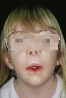
A few congenital and inherited disorders have been associated with microstomia. Perhaps the most dramatically small mouths appear in children with Freeman-Sheldon syndrome (ie, craniocarpotarsal dysplasia, whistling baby syndrome). [6] Other disorders that may cause microstomia include Hallermann-Streiff syndrome, oro-palatal dysplasia, Fine-Lubinsky syndrome, restrictive dermopathy, types of epidermolysis bullosa, and, occasionally, Down syndrome and hemifacial microsomia (see the images below).
 Child with craniofacial anomalies and microstomia. A tracheotomy is in place for airway control because of micrognathia. Advancement osteotomies have been performed in an effort to lengthen the mandible.
A genetic screening study by Patat et al of four patients indicated that mutations in the orthodenticle homeobox 2 (OTX2) gene are associated with otocephaly-dysgnathia complex, the characteristics of which also include microstomia. The paired related homeobox 1 (PRRX1) gene has been implicated in the complex as well. [7]
Pathophysiology
Oral burns are generally third degree, with a central area of necrosis surrounded by a pale elevation of the skin. The adjacent skin is usually hyperemic. In electrical burns, soft tissue injury is typically more extensive than initially appreciated, as the current follows the low-resistance paths of muscle, nerves, and blood vessels. Coagulation necrosis occurs, followed by a period of coagulative necrosis with inflammation of adjacent vital tissues. Over several weeks, the necrotic cells are removed by fragmentation and phagocytosis of the cellular debris by scavenger white cells and by the action of proteolytic lysosomal enzymes brought in by the immigrant white cells. Eventually, fibroblast formation and collagen deposition occur, along with scar tissue formation and contraction.
In scleroderma, endothelial alterations lead to stimulatory changes that involve many cells, including fibroblasts, T lymphocytes, macrophages, and mast cells. The activated cells secrete various substances that lead to deposition of extracellular matrix compounds, including fibronectin; proteoglycans; and collagen types I, III, V, and VII, in the skin and other tissues. The degree of sclerosis increases when profibrotic cytokine–induced fibrosis is also present. [8]
Presentation
The cause of microstomia in affected patients can usually be determined by the clinical history. Microstomia caused by disorders that are likely to chronically and progressively involve the perioral tissues must be differentiated from microstomia due to trauma or surgical scar that is characterized by a more correctable narrowing of the oral aperture. Microstomia due to the latter is more likely to be corrected with surgery.
Patients affected by isolated microstomia may be socially isolated for long periods before presentation to a physician because of their appearance. Asymmetry, lack of proportion, excessive dental show, and altered geometric shape can produce a mouth that draws the curious stares of others. As with other facial deformities, microstomia can render a patient socially disabled.
Often, patients with microstomia present with functional problems. Articulation abnormalities can lead to impaired communication. Patients may report difficulty brushing their teeth or inserting dentures. Some affected individuals may be referred by their dentists, since a limited oral opening can impair cleaning teeth and complicate extractions or restorative procedures. Caloric intake is limited only when the oral aperture size is drastically reduced. Patients with scleroderma may present with dysphagia or involvement of other systems before the oral aperture is affected.
Indications
Early intervention is indicated following burns and other perioral trauma in a prophylactic measure to reduce complications due to scarring. [9] It generally involves some sort of appliance therapy. In patients with microstomia of longer duration, impairment of functions such as those described in the Clinical section, including speech, swallowing, and oral hygiene, are indications for intervention. Patients should also be considered for management of their microstomia if the deformity is socially disabling.
Relevant Anatomy
The lips are composed of skin and mucosa that are not supported directly by any rigid framework. Externally, the facial skin extends to the vermilion border. Internally, the lips form the anterior boundary of the oral vestibule; here, they are lined with oral cavity mucosa that harbors minor salivary glands. The upper lip is bounded superiorly by the nose and is divided into 3 subunits, the philtrum and 2 lateral subunits that extend from the philtral columns to the melolabial folds laterally. The lower lip is one functional subunit that extends to the labiomental fold inferiorly and to the melolabial folds laterally.
The orbicularis oris is a circular muscle innervated by branches of the facial nerve. Its function is important in maintaining oral competence, normal speech articulation, and facial expression. The deep fibers of the orbicularis oris are oriented horizontally and act to compress the lips and to provide sphincter function, whereas the superficial fibers are responsible for finer movements. The oblique fibers act to evert the lips. The depressors of the lip include the depressor anguli oris, mentalis, depressor labii inferioris, and the platysma. The elevators of the lip include the levator anguli oris, zygomaticus, and risorius. Many of these muscles converge at the oral commissures, or corners of the mouth. Because the orientation of the muscle fibers at these locations is so variable, motion of the lip is normally most restricted at the commissures, and these areas are most severely affected by scarring and fibrosis in microstomia.
Sensory innervation to the upper lip is primarily via branches of the infraorbital nerve (cranial nerve [CN] V2), and innervation to the lower lip is via the buccal and mental branches of the mandibular nerve (CN V3). Innervation to the commissures is primarily from the buccal branch of the mandibular nerve.
The superior and inferior labial branches of the facial artery provide blood supply to the lips, and venous drainage is through corresponding veins that drain into the anterior facial vein. These vessels form a vascular ring that encircles the oral aperture. The lymphatic drainage of the lips is by cutaneous and mucosal lymphatics. The lateral portion of the lower lip drains into the submandibular lymph nodes; the central portion is drained by the submental lymph nodes. Lymphatic anastomoses between the 2 halves of the lower lip lead to bilateral drainage of the central portion. In contrast, the upper lip has little bilateral drainage, with lymphatics that lead to the preauricular, infraparotid, submandibular, and, sometimes, the submental lymph nodes.
Contraindications
Surgical repair is not advised for patients with the calcinosis cutis, Raynaud phenomenon, esophageal dysmotility disorder, sclerodactyly, and telangiectasia (CREST) syndrome because of the likelihood of poor tissue healing.
Oral appliances are to be used carefully in patients with epidermolysis bullosa, since they are likely to induce further oral trauma and ulceration.
-
Child with Freeman-Sheldon syndrome.
-
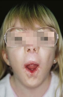
Child with Freeman-Sheldon syndrome demonstrating limited vertical expansion.
-
Child with craniofacial anomalies and microstomia. A tracheotomy is in place for airway control because of micrognathia. Advancement osteotomies have been performed in an effort to lengthen the mandible.
-
Microstomia prevention apparatus (MPA).
-
Commissuroplasty, as performed by Converse (1959). (A) Skin excision designed to accommodate elongation of the commissure. (B) Skin excision performed and mucosal incisions diagrammed. (C) Mucosal flaps incised and mobilized. (D) Mucosal flaps sutured in place. (Reprinted from Converse JM and Wood-Smith D. Techniques for repair of defects of the lips and cheeks. In Converse JM, ed. Reconstructive Plastic Surgery, 2nd edition, volume 3. WB Saunders; Philadelphia:1977. pp.1574, with permission from Elsevier).
-
Commissuroplasty, as performed by Gillies and Millard (1957). (A) Triangular skin excision designed to accommodate elongation of the commissure. A flap of vermilion is raised from the lower lip. (B) Vestibular mucosa is advanced externally to reconstruct the vermilion of the lower lip. (C) The vermilion flap is rotated and sutured laterally to reconstruct commissure. (Reprinted from Converse JM and Wood-Smith D. Techniques for repair of defects of the lips and cheeks. In Converse JM, ed. Reconstructive Plastic Surgery, 2nd edition, volume 3. WB Saunders; Philadelphia:1977. pp.1575, with permission from Elsevier).
-
Schematic representation of orbicularis oris muscle-lengthening procedure.
-
Schematic representation of orbicularis oris muscle-lengthening procedure.


