Practice Essentials
Carcinoma of the buccal mucosa is relatively uncommon in North America compared with other oral cavity cancers such as carcinomas of the tongue or floor of the mouth. Squamous cell carcinoma is the most common pathology and is more prevalent in those who use tobacco and alcohol. Surgery is the preferred treatment for early and advanced buccal carcinoma in North America.
The image below depicts buccal carcinoma.
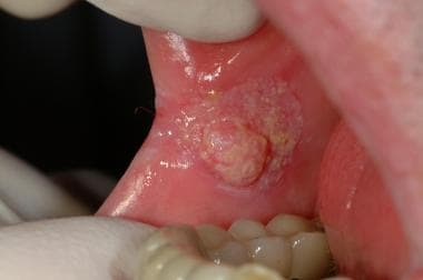 Early oral squamous cell carcinoma in the buccal mucosa arising from leukoplakia.
As the orifice of the upper aerodigestive tract, the oral cavity plays a critical role in breathing, speech, and swallowing. The buccal region is particularly important in bolus formation, preventing food from spilling into the lateral oral gutters or extraorally during the oral preparatory phase of swallowing. Cancer of the buccal mucosa and subsequent treatment of the disease may interfere with these functions.
Buccal carcinoma has the propensity to become aggressive, with high rates of locoregional recurrence. Diagnosis and treatment at an early stage leads to significantly improved prognosis and function over advanced disease.
Workup in buccal carcinoma
Useful laboratory tests in the preoperative workup of buccal carcinoma include the following:
-
Complete blood count (CBC), electrolytes, blood urea nitrogen (BUN), and creatinine
-
Prothrombin time (PT), activated partial thromboplastin time (aPTT), and international normalized ratio
-
Liver function and gamma-glutamyl transpeptidase (GGT) tests
-
Blood typing and cross-matching
Imaging studies include the following:
-
Chest radiography - To assess for pulmonary metastases, a synchronous lung tumor, or chronic lung disease
-
Contrast-enhanced computed tomography (CT) scanning or magnetic resonance imaging (MRI) of the primary site
-
Chest CT scanning and positron emission tomography (PET) scanning - May be used for metastatic workup
-
Panorex imaging (mandibular orthopantomography) - May be used to evaluate mandibular invasion in conjunction with CT scanning and clinical examination
With regard to diagnostic procedures, any suspicious or nonhealing lesion of the buccal mucosa should be biopsied for histopathologic examination. In addition, examination under anesthesia, employing panendoscopy, can be carried out for inspection and palpation of the oral cavity and oropharynx, direct laryngoscopic examination of the hypopharynx and larynx, and endoscopic evaluation of the esophagus, trachea, and nasopharynx.
Management of buccal carcinoma
Radiation therapy
Radiation has a limited role as primary therapy for buccal carcinoma in North America but is used as adjuvant therapy in advanced stage disease. For early T-stage disease, local-regional control and survival rates for primary radiation therapy are comparable to those for surgery.
Indications for radiation or chemoradiation therapy in the postoperative setting include large or deeply invasive tumors, close or positive margins, multiple lymph nodes with metastatic cancer, lymph node extracapsular spread, and perineural invasion. The results of using radiation therapy alone in patients with advanced buccal carcinoma have been dismal.
Chemotherapy
The role of induction chemotherapy in the treatment of advanced stage head and neck squamous cell carcinoma is controversial and is often recommended for clinical trials.
Chemotherapy given concomitantly with radiation therapy for the treatment of nonoperative head and neck squamous cell carcinoma has an overall and disease-free survival rate greater than that of radiation therapy alone.
Surgery
Surgery is the preferred treatment for early and advanced buccal carcinoma in North America. Patients with advanced disease should receive postoperative radiation or chemoradiation. The surgical approach depends on the size of the tumor. Small lesions can usually be treated via transoral wide local excision, whereas advanced lesions usually require excision via a cheek flap. Composite resection is indicated for mandibular invasion, while partial maxillectomy is used for superior alveolar ridge invasion. Complete resection of the tumor with negative margins confirmed by frozen section histopathology is the goal. Positive margins are associated with increased recurrence and decreased survival rates.
Metastatic neck disease (N+ disease) requires either a modified radical neck dissection or a radical neck dissection, depending on the extent of disease. Management of the clinically negative neck is controversial.
Epidemiology
Frequency
Squamous cell carcinoma of the buccal mucosa accounts for 5-10% of all cancers of the oral cavity in North America and Western Europe. It occurs more often in men, with a male:female ratio of 3-4:1, and most commonly in the 7th or 8th decade of life.
The incidence of buccal carcinoma is much higher in Asia. In Southeast Asia, the disease is the most common form of oral cavity cancer. In India, buccal carcinoma is the most common cancer in men and the third most common cancer in women.
The higher rate of buccal carcinoma in Asia is likely related to the widespread practice of betel nut chewing. Betel nut, composed mainly of the fruit of the Areca Palm and often mixed with tobacco, is placed along the buccal mucosa to induce a feeling of euphoria. Buccal carcinoma related to betel nut chewing tends to develop at an earlier age, with most cases occurring between the ages of 40-70.
Etiology
Tobacco and alcohol use are the main etiologic agents associated with the development of buccal carcinoma. In North America, a history of using tobacco is documented in 70% of patients. Although alcohol by itself is not thought to be a significant risk, tobacco and alcohol have a well-recognized synergistic effect in the development of carcinoma.
In Asia, betel nut is a significant etiologic agent, in addition to tobacco and alcohol. In India, over 90% of patients with buccal carcinoma have a history of using betel nut. [1]
Other suspected but not confirmed etiologic agents include human papilloma virus, poor oral hygiene, and chronic irritation.
Premalignant conditions include submucosal fibrosis and lichen planus. The latter has a reported transformation rate of 0.5-3%, whereas the former has a malignant transformation rate of 0.5%.
Presentation
Buccal carcinoma commonly presents as a slow-growing mass on the buccal mucosa. Small lesions tend to be asymptomatic and are often noted incidentally on dental examination. Pain commonly occurs as the lesion enlarges and ulceration develops. Oral intake may worsen the pain and lead to malnutrition and dehydration. Associated symptoms include bleeding, poor denture fit, facial weakness or sensory changes, dysphagia, odynophagia, and trismus.
A detailed medical history is important to determine the patient's candidacy for surgery or radiation therapy. The person often has a history of tobacco and alcohol use. A history of previous malignancies of the upper aerodigestive tract should be ascertained.
Comprehensive examination of the head and neck should be conducted with a focus on the oral cavity. The mucosa of all the subsites of the oral cavity and oropharynx should be examined systematically. Palpation is important to determine the depth of invasion. Mandibular or maxillary alveolar invasion should be noted on inspection and palpation. Dentition must also be assessed, especially if irradiation is part of the planned management. The larynx and hypopharynx should be assessed by means of examination with a mirror or flexible endoscopy to rule out a second primary tumor of the upper aerodigestive tract.
The ears should be examined in those patients with a history of otalgia because a lack of evidence of ear disease suggests referred pain due to malignancy.
The neck and parotid gland should be carefully examined for adenopathy. Diaz et al found that 27% of patients presented with clinically positive nodes. [2] The risk of nodal disease at presentation increases with advanced-stage disease. A meta-analysis of 4 studies with 223 cases of buccal carcinoma by Chhetri et al found that most presented with T2 or T3 disease (12% T1, 47% T2, 19% T3, 22% T4). [3] The rate of nodal metastases at presentation was 40% for T2 disease and 52% for T3 disease.
Signs of advanced disease on physical examination include bleeding, skin ulceration, facial swelling, neck mass, trismus, facial numbness, and paralysis of the facial musculature.
The lesion often has 1 of 3 morphologic types: exophytic, ulceroinfiltrative, or verrucous. The exophytic type is the most common, appearing as a papillary mass that becomes ulcerated when large. [4] The ulceroinfiltrative variety appears as an ulcer that penetrates deep into the underlying structures, with surrounding induration. Verrucous carcinomas are uncommon variants of oral-cavity carcinomas; among these, the buccal mucosa is the most common site. These lesions appear as papillary masses, and keratinization gives them a whitish appearance.
Indications
Any lesion of the buccal mucosa suggestive of malignancy should be biopsied for pathologic diagnosis. Once a malignant diagnosis is established, treatment options include surgery, irradiation, or combined-modality therapy. Treatment recommendations should be based upon multiple factors, including the stage of the tumor, the patient's general health, and patient desires.
In North America, surgery is the primary treatment for buccal carcinoma. Adjuvant radiation or chemoradiation is offered based upon disease stage, as well as histopathologic factors. Primary radiation or chemoradiation is reserved for those patients who are poor surgical candidates.
In some parts of the world, nonsurgical therapy is considered the standard of care. In India, irradiation is the mainstay of treatment, with surgery and postoperative radiation therapy reserved for cases of advanced disease.
Relevant Anatomy
The American Joint Commission on Cancer defines the buccal mucosa as the membrane lining of the inner surface of the cheeks from the line of contact of the opposing lips anteriorly to the line of the pterygomandibular raphe (lateral to retromolar trigone) posteriorly. The medial boundary is the line of attachment of the buccal mucosa to the upper and lower alveolar ridges.
The layers of the cheek from medial to lateral include the mucosa, pharyngobasilar fascia, buccinator muscle, buccinator fat pad, subcutaneous tissue and skin. Sensory innervation of the buccal mucosa and cheek skin is from the maxillary and mandibular branches of the trigeminal nerve. The buccinator muscle is innervated by the facial nerve. The parotid duct (Stenson duct) pierces the buccinator muscle and buccal mucosa opposite the maxillary second molar.
The buccal region lacks anatomic barriers beyond the buccinator muscle and its fascia to prevent the spread of cancer. Buccal cancer can spread laterally to extend through the skin of the cheek; medially to involve the alveoli, palate, tongue, and floor of the mouth; posteriorly to involve the retromolar trigone mucosa, the ascending ramus of the mandible, and the masseter and pterygoid muscles; and anteriorly to involve the oral commissure and lips.
The primary-echelon lymphatics of the buccal mucosa drain to the facial and submandibular lymph node basins prior to the upper jugular nodes. The lymphatics may occasionally drain to the upper jugular nodes via the parotid nodes.
Contraindications
Contraindications to surgery include poor medical status, a patient's refusal of surgery, unresectable disease (eg, skull-base fixation and carotid encasement), and the presence of distant metastases.
Contraindications to radiation therapy include previous irradiation (relative contraindication) and collagen vascular disorders. Reluctance of the patient to undergo the dental intervention frequently required to prevent osteoradionecrosis is another relative contraindication for radiation therapy.
-
Early oral squamous cell carcinoma in the buccal mucosa arising from leukoplakia.


