Background
Chemical peeling, also termed chemexfoliation, represents accelerated exfoliation or skin damage induced by caustic agents that cause controlled damage, followed by the release of cytokines and inflammatory mediators, resulting in thickening of the epidermis, deposition of collagen, reorganization of structural elements, and increases in dermal volume. This process decreases solar elastosis and replaces and reorients the new dermal connective tissue. The result is an improved clinical appearance of the skin, with fewer rhytides and decreased pigmentary dyschromia, and a more youthful appearance.
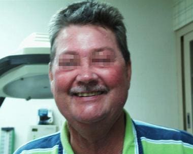
The images below depict a patient during and after a salicylic acid peel.
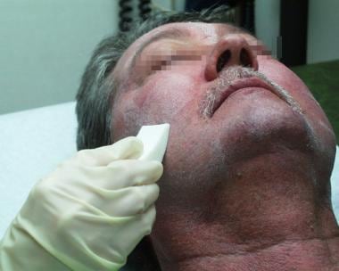 Men also request chemical peeling. This 56-year-old man is in the process of a salicylic acid peel.
In recent years, a large shift has occurred in the manner and depth to which peels are performed. Lasers largely have supplanted deep chemical peeling because of the control of depth they afford, their lesser effect on pigmentation, and their ease of use, with no chemical adverse effects. Superficial peels, in contrast, have increased in popularity. Various agents are used; for simple exfoliation, glycolic or lactic acids are now commonly found in almost all moisturizers and in many makeup bases.
Aging skin undergoes a number of changes. With time, it thins, falls, and creases along muscular and gravitational folds. Compared with the effects of simple aging on skin, sun damage leads to additional and different problems, including thickening, solar elastosis, and resultant pigmentary irregularities. Carcinogenic effects lead to actinic keratoses, basal and squamous cell cancers, and, less directly, to melanomas.
Scarring from trauma or acne contributes to an irregular skin surface. True skin laxity in the form of brow ptosis, eyelid bags, jowls and loss of neckline, ear lobule elongation, nasal tip drop, upper lip thinning, and other manifestations are currently treated surgically with facial rejuvenation procedures (eg, face lift, brow lift, rhinoplasty, lip augmentation). Conversely, these procedures do not help skin textural damage. Fine lines, pigmentary irregularities over a broad area, and aging skin are treated with peeling using either chemical or mechanical means.
A discussion of mechanical peeling is warranted for thoroughness. Lasers largely are used for skin ablation, with the carbon dioxide ultrapulsed laser currently used most often. The erbium-doped yttrium aluminium garnet (Er:YAG) laser is used more for superficial resurfacing when little tightening and superficial depth of peel are required. [1] Fractional laser resurfacing devices have shown encouraging results, especially for acne scarring. [2] Many surgeons still use dermabrasion. The lack of control and lack of ability to resurface evenly have resulted in the supplantation of this technique by those already discussed. Microdermabrasion is currently a popular technique because no downtime or discomfort is associated with the procedure. Medical devices tend to have stronger suction, and more abrasive crystals are used, while spa and lay devices tend to be gentler, with less overall effect but increased safety. In general, mechanical abrasion tends to improve scarring more than chemical peeling agents, given similar depth of penetration. [3]
Currently, a number of categories of chemical peeling agents available for rejuvenating the skin can be found. These range from superficial formulations available over the counter to deep chemical agents that should only be applied by a physician in a controlled setting. When used in the proper setting with appropriate technique, nearly all of these products have proven successful in improving quality and appearance of facial skin.
The goal of chemical peeling is to remove a predictable uniform thickness of damaged skin. Normal wound healing and skin rejuvenation follow, while complications such as scarring and pigmentary changes are minimized.
Indications
The indications for a chemical peel, since it is largely a cosmetic procedure, depend on the patient's tolerances and wishes for correcting skin textural problems. Many individuals do not wish to improve skin texture despite severe problems, and others desire marked improvement in relatively minor problem areas. Treatments vary with the severity of the condition and the wishes of the patient. These wishes should be tempered with information on what is possible and what is desirable for the patient in terms of treatment. Approach each patient truthfully, discussing possibilities, risks, benefits, and alternatives.
Indications related to pigmentary disorders are as follows:
-
Postinflammatory hyperpigmentation
-
Freckles
-
Lentigines
-
Facial melanoses
-
Periorbital hyperpigmentation
Indications related to acne are as follows [4, 5] :
-
Superficial acne scars
-
Postacne pigmentation
-
Comedonal acne [6]
-
Acne excoriée
-
Acne vulgaris - Mild to moderately severe acne
Indications for aesthetic concerns are as follows [4, 7] :
-
Photoaging
-
Fine superficial wrinkling
-
Dilated pores
-
Superficial scars
Indications related to epidermal growths are as follows:
-
Warts
Indications related to inflammatory conditions are as follows: [8]
-
Pseudofolliculitis barbae
Upper epidermal defects, such as melasma, can be treated with superficial peels, while deeper defects, such as deep wrinkles, may require a deep peeling agent. [9] Medium-depth (superficial dermis) defects, such as mild dermatoheliosis, require a medium-depth peel. Deep perioral rhytides may require a deep peel, such as the Baker-Gordon solution.
Aging faces are common to all and recognized as such. Distinguish between textural skin damage treatable by resurfacing or skin rejuvenation and actual ptosis or falling of major structures treatable by surgical interventions.
Contraindications
Relative contraindications are determined by the skin type of the patient and the defect being treated. To optimize the procedure, some classifications are very useful, such as the Fitzpatrick and the Glogau photoaging classifications. See Procedure Planning for detailed information on Fitzpatrick and the Glogau photoaging classifications.
Absolute contraindications include the following:
-
Active bacterial, viral, fungal, or herpetic infection
-
Open wounds
-
History of drugs with photosensitizing potential
-
Preexisting inflammatory dermatoses (eg, psoriasis, atopic dermatitis, pemphigus)
-
Facial cancers, especially facial melanoma
-
Uncooperative patient (patient is careless about sun exposure or application of medicine)
-
Patient with unrealistic expectations
-
For medium-depth and deep peels, history of abnormal scarring, keloids, atrophic skin, or isotretinoin use in the last 12 months
Other considerations
Degree of photoaging damage
Patients with either severely damaged skin or excellent skin may not be good candidates for chemical peels. Sun-damaged skin shows epidermal changes, elastosis, and collagen distortion in the midreticular dermis.
To eradicate photodamage, deep peels are required. More superficial peels, even when performed in repetitive fashion, do not reach the affected histological level and therefore have a minimal effect on photodamaged skin.
Smoking
Patients must understand the necessity for smoking cessation.
The dynamic action of puffing can worsen perioral rhytides, and the chemicals in the smoke can cause enzymatic reactions that weaken the skin and cause further wrinkling around the mouth and eyes.
Prior cosmetic surgery
Waiting several months following surgery that involves the face is recommended. Give the skin time to heal prior to subjecting it to chemexfoliation.
Compliance with prepeel and postpeel treatment must be assured. The patient must be motivated enough to adhere to a daily regimen for a few weeks before and after the procedure.
General health
With phenol peels, the patient should be in good general health because phenols can cause arrhythmias. Phenol is directly toxic to myocardium. Cardiac arrhythmias have been recorded in up to 23% of patients when a full-face peel was performed in less than 30 minutes. These arrhythmias have included tachycardia, premature ventricular beats, bigeminy, atrial tachycardia, and ventricular tachycardia. Adequate patient management reduces this complication rate to less than 7%.
Good kidney and liver function are necessary for adequate excretion and detoxification. A screening blood chemistry that includes blood urea nitrogen, creatinine, and liver function is wise. ECG monitoring is necessary during the peeling process. No hepatorenal or central nervous system toxicities have been reported in the literature with properly performed chemical peels.
Mental health
Patients who are mentally unstable may be overly self-conscious and may not be prepared for their aesthetic appearance immediately following the peel.
Medications
A thorough medical and drug history is very important.
Medical conditions such as cardiac, hepatic, or renal disease may influence treatment decisions and the choice of peeling agents.
Exogenous estrogens, oral contraceptives, and other medications may be photosensitizing and predispose patients to pigmentation complications after chemical peeling and worsening the skin discoloration that the chemical peel was intended to eradicate.
Patients taking blood thinners, such as warfarin, should avoid deep peels because of the possibility of blood oozing from the peel site. Patients taking aspirin usually do not have complications, but, if the medication is not necessary, advise them to stop taking it 1 week prior to a deep peel.
Samargandy and Raggio recommend that chemical peels not be performed on patients with isotretinoin use in the previous 6 months, particularly when a moderately-deep or deep peel is planned. [8]
Herpes
A history of herpes simplex requires antiviral prophylaxis from the immediate prepeel period until reepithelialization is complete. Acyclovir (400 mg) should be started 2 days prior to the peel and continued for 5 days after the peel to reduce the risk of recurrent herpes infection.
Some dermatologists advise prophylaxis in all patients to avoid the risks of a herpetic outbreak.
Any existing lesion must heal completely before undergoing a chemical peel.
History of scarring
Patients need to be asked if they have a history of hypertrophic scarring. Many people who have hypertrophic scarring can develop keloids. This usually is found in patients with Fitzpatrick skin types 5 and 6 but can develop in patients with skin types 1, 2, 3, and 4.
Medium and deep peels penetrate into the superficial and deep dermis, which may stimulate keloidal development in patients who are inclined to develop keloids. Weak superficial peels can be considered in patients with skin types 4 and 5 because the penetration is only into the epidermis. Patients with a history of scarring are not candidates for major skin resurfacing, such as laser or medium/deep peels.
Expectations
A discussion between the physician and patient is necessary prior to a chemical peel, especially a deep peel.
Examples of before-and-after results should be shown, and the possibility of complications must be explained to the patient.
Follicle unit density
Previous use of isotretinoin must be noted. Patients should wait until 6 months after the last dose of isotretinoin to reduce the risk of scarring.
Patients who have had recent radiation treatment need to have a skin biopsy performed to ascertain the existence of hair follicle units, because these follicle units are where the reepithelialization occurs.
Technical Considerations
Peeling agent concentration
When combination peels are used, better clinical results can be achieved with reduced risk of complications. [10] Peeling agent concentration can vary, even though the label indicates the same concentration. The different methods used to determine the concentration of an acid can produce some variation.
The most frequently used peeling agents are salicylic acid, glycolic acid, pyruvic acid, lactic acid, mandelic acid, Jessner solution, trichloroacetic acid, and phenol.
From strongest to weakest, these methods are dilutions of a saturated solution, the weight-to-weight method, the weight-to-volume method, and grams of acid crystal mixed to 100 mL of water.
Free acid availability
Molecules found in chemical peels are either alcohols that contain a carboxyl (-COOH) and hydroxyl (-OH) groups or regular acids. It has been suggested that according to their chemical properties, substances used in chemical peels are classified as metabolic, caustic, or toxic.
The pH of the agent, or free acid available (pKa), is another measurement. The pKa of the solution is the pH at which half is in acid form; therefore, a lower pKa means that more free acid is available. Many products advertise the acid percentage; however, pKa is a more accurate determinant of strength.
Outcomes
The outcome generally is excellent once patient expectations are adjusted to the procedure. The patient must know what to expect from the surgery and during the healing process. Giving the patient realistic expectations for each stage of healing is imperative to the success of the entire procedure.
After a deep peel, the skin is new and supple. Some of the rhytides return as the swelling resolves. Approximately 1 month after the peel, the skin appears wrinkled. After it begins to thicken, many of these rhytides resolve over the ensuing months. A repeat peel can be performed after 1 year if necessary. Superficial peels can be repeated every few weeks if needed. Many superficial peels do not produce the same effects as a deep peel. The skin continues to weather and age after the procedure, but the effects of the peel are permanent.
A noted decrease in the incidence of superficial skin cancers and actinic keratoses occurs after resurfacing procedures.
Etiology of Facial Aging
The etiology of facial aging is a broad subject. This article briefly discusses aging and contrasts it with sun and environmental damage.
When not compounded by extraneous factors, skin aging basically is the process of atrophy. Loss of subcutaneous tissue is the most obvious and recognizable sign of aging; however, skin, skin appendages, and cutaneous blood supply also atrophy with age. Both the epidermis and dermis thin, and cutaneous strength and elasticity are lost. Dermoepidermal adherence afforded by rete pegs is lost, and blistering or superficial epidermal loss commonly occurs with aged skin. Overall thinning and loss of integrity and wall strength of the cutaneous vasculature cause easy bruising.
Atrophy of the skin is a well-known process that occurs with aging. This process typically begins during the fourth decade of life. The outermost portion of the epidermis, the stratum corneum, becomes disorganized and less effective as a protective barrier to the external environment. A gradual decline in the number of melanocytes populating the basal layer of the epidermis also occurs. The dermoepidermal junction becomes flattened because of a decrease in the number of dermoepidermal papillae. More significant changes can be seen within the dermis, where an overall loss of organization occurs as this layer begins to thin with age. The amount of ground substance decreases, and elastic fibers degenerate, making the skin less resistant to deformational forces. Collagen is also lost, and the relative proportion of type I to type III collagen is reduced.
Environmental damage to skin often is explained incorrectly in the literature because of confusion between the short- and long-term changes that occur. Initially, as with most damage to the human body, the response is inflammatory. This tends to subside rather quickly in the skin, but continuous damage can result in prolonged inflammatory responses. Although postinflammatory hyperpigmentation is often considered a limited medical condition, most individuals express it to some extent, and prolonged exposure to damaging environmental factors results in tanning and prolonged hyperpigmentation. The increased volume of skin from inflammation tends to be transient and is caused by increased water volume from increased proteoglycans and glycosaminoglycans.
The true long-term damage to skin from environmental stresses is a decrease in the water volume and an increase in damaged cutaneous proteins. In particular, the elastic fibers tend to form tangled masses of nonelastic elastin remnants. This leads to increased volume of skin without functional elements. The solar elastosis or heliosis that is observed histologically is the end stage of this damage. In much the same manner that scarring or fibrosis is observed as the end stage of renal or hepatic disease, scarring and remnants of proteinaceous elements tend to be the end stage of cutaneous disease. Although contracture is present, the general trend in environmental damage of the skin is toward increased thickness, especially of the dermis. This thickening is with nonelastic and structurally weak skin. Sun damage, especially from ultraviolet (UV)–A wavelengths, causes ionization and oxidation of dermal elements and genetic information, resulting in premalignant and malignant skin lesions.
Many years of acne (both cystic and rosacea) increase the blood flow to skin and tend to hypertrophy the basic elements. Scar tissue also deposits and can contract, leading to uneven skin surfaces. True cysts and sinus tracts commonly result, and ice pick lesions usually are the visible manifestations of these processes. Actinic keratoses and lentigines are two examples of actinic damage, or photodamage. [11]
Skin Anatomy
Before embarking on chemical peeling, one must have a thorough knowledge of skin anatomy and normal wound healing. The skin serves as a protective barrier, preventing exposure of internal tissues to trauma, UV radiation, temperature extremes, toxins, and bacteria. Other important functions include sensory perception, immunologic surveillance, thermoregulation, and control of insensible fluid loss.
The skin is composed of two mutually dependent layers, the epidermis and dermis, which rest on a fatty subcutaneous soft tissue. The epidermis contains no blood vessels and is dependent entirely on the underlying dermis for nutrient delivery and waste disposal. This occurs by diffusion through the so-called dermoepidermal junction. The primary function of the dermis is to sustain and support the epidermis.
Epidermal appendages are intradermal epithelial structures lined with epithelial cells that have the potential for division and differentiation. These structures are named from the fact that they develop as down-growths or diverticula of the epidermis into the dermis. These appendages serve an important role as a source of epithelial cells, which are responsible for re-epithelialization when the overlying epidermis is removed or destroyed.
Epidermal appendages include sebaceous glands, sweat glands, apocrine glands, mammary glands, and hair follicles. Sebaceous glands are highest in concentration on the face and scalp, where as many as 900 glands/cm2 may be found. Epithelial appendages are located deep within the dermis. In the face, these appendages may also be located in the subcutaneous fat beneath the dermis. The deep location of these structures and their density in the face account for the remarkable ability of this area to re-epithelialize the deepest cutaneous wounds.
Mechanism of Action in Chemical Peeling
Chemical peeling is the process of applying chemicals to the skin to destroy the outer damaged layers. It causes controlled destruction of a part of or the entire epidermis, with or without the dermis, leading to exfoliation and removal of superficial lesions, followed by regeneration of new epidermal and dermal tissues. [10] The epidermis regenerates from the epidermal appendages located in the remaining dermis. This process begins within 24 hours of wounding and is usually complete in 5-10 days. The new epidermis shows greater organization and vertical polarity, with the disappearance of actinic keratoses and lentigines. Dermal regeneration is a slower process but is usually complete within several months. The regenerated dermis demonstrates less elastosis and improved organization, with compact horizontally arranged bundles of collagen interspersed with elastic fibers. Ground substance is decreased and telangiectasias are absent. The overall result is soft supple skin that appears more youthful with fewer rhytides and dyschromias.
Different agents and formulations are chosen based on their depth of penetration and risk profile. Destruction confined to the epidermis results in rapid healing without scarring, although some pigmentation change may be present if melanocytes are damaged. This superficial wounding has the disadvantage of producing less dramatic results but is very safe. Deeper wounding, extending into the papillary and, sometimes, reticular dermis, produces more dramatic results. However, deeper penetration eradicates a portion of the epidermal appendages, making healing slower and scarring more likely. Penetration into the reticular dermis entails a very high risk of scarring.
The weaker agents (eg, alpha-hydroxy acids) change the pH sufficiently to cause a superficial shock to the cells and, depending on many variables, cell injury or death. When used with a moisturizer, the acid acts simply to cause cellular and intercellular swelling and plumping, leading to a transient increase in cell and matrix size and lessening of fine lines and rhytides. Sequential treatments lead to exfoliation and a smoother complexion. Continued irritation can lead to many of the same effects of tretinoin or retinoid treatment (ie, increased thickness of dermis, increased blood flow to skin).
Stronger agents such as phenol (with various additives such as croton oil and glycerin) and trichloroacetic acid (TCA) produce a chemical necrosis of the skin to variable depths, depending on numerous controlled and uncontrolled variables.
Other factors that can affect the depth of peel include the following:
-
Presence of skin surface oils and dirt
-
Skin water content
-
Temperature of the room, skin, and solution
-
Humidity of the air
-
Length of time the solution is left in contact with the skin
-
Occlusion or nonocclusion
-
Batch of croton oil
-
Thickness of epidermis and dermis
-
Presence of hyperkeratotic lesions
-
The defatting process is important for the even penetration of the peeling agent. This patient is a Fitzpatrick type II, Glogau type I, using alcohol and acetone mixture to defat the skin.
-
This 45-year-old woman has just completed her salicylic acid peel and will neutralize the peel with cold water.
-
Men also request chemical peeling. This 56-year-old man is in the process of a salicylic acid peel.
-
Same patient as shown above following a successful chemical peeling.


