Practice Essentials
Merkel cell carcinoma (MCC) is the eponym for primary cutaneous neuroendocrine carcinoma, a dermal neoplasm with cytoplasmic, dense-core neuroendocrine granules and keratin filaments. A rare cancer, Merkel cell carcinoma (MCC) is an aggressive cutaneous neoplasm that lacks distinguishing clinical features. More than half of Merkel cell carcinomas (MCCs) occur in the head and neck of elderly people, in areas of actinically damaged skin. The most common site of occurrence is the periorbital region. Merkel cell carcinoma (MCC) has a propensity to recur and to cause local and distant metastases. Distant metastases indicate a condition that is nearly always fatal. [1]
Current treatment consists of wide local excision with adjuvant irradiation. Neck dissection is used for clinically positive nodes, and chemotherapy is given for advanced disease. [2]
If the prognosis of patients with (MCC) is to be improved, early diagnoses are needed, and further understanding of the roles of neck dissection, radiation therapy, and chemotherapy must be attained. [3]
Symptoms of Merkel cell carcinoma
Merkel cell carcinoma (MCC) commonly appears as a painless mass on or just under the skin surface. Appropriate clinical diagnosis is often delayed because of a lack of symptoms. The tumor may take on an erythematous or violaceous appearance. Bleeding and superficial ulceration are late findings suggestive of advanced disease. Regional lymph node metastasis is common, even with tumors smaller than 2 cm.
Workup in Merkel cell carcinoma
Baseline laboratory studies should include a complete blood count (CBC), a chemistry profile, and liver function tests.
No optimal imaging algorithm has been defined. However, because of the difficulties in distinguishing metastatic oat cell carcinoma from Merkel cell carcinoma (MCC), chest radiography should be performed. Obtain computed tomography (CT) scans of the chest, abdomen, and pelvis to rule out metastases.
Light microscopy, electron microscopy, and immunohistochemistry may be needed to confirm the diagnosis of Merkel cell carcinoma (MCC).
No universally accepted staging system for Merkel cell carcinoma (MCC) exists. Some have used the American Joint Committee on Cancer Staging System for skin cancer to stage Merkel cell carcinoma (MCC). Others employ a staging system developed by the Memorial Sloan Kettering Cancer Center or the system suggested by Yiengpruksawan et al (1991).
Management of Merkel cell carcinoma
For patients with operable disease, most agree that surgery is the treatment of choice. As with most tumors, treatment is based on the stage.
Guidelines released in 2015 by a collaborative group of multidisciplinary experts from the European Dermatology Forum (EDF), the European Association of Dermato-Oncology (EADO), and the European Organization of Research and Treatment of Cancer (EORTC) included the following with regard to the management of Merkel cell carcinoma (MCC) [4] :
-
The primary tumor should be excised with 1-2 cm margins
-
In patients with regional lymph node involvement, radical lymphadenectomy is recommended
-
Adjuvant radiation therapy may be considered in patients with multiple affected lymph nodes of extracapsular extension
-
In unresectable metastatic Merkel cell carcinoma, monochemotherapy or polychemotherapy achieve high remission rates; however, responses are usually short-lived
-
Treatment within clinical trials is regarded as a standard of care in disseminated Merkel cell carcinoma
Complications
In addition to metastasizing to the lymph nodes, Merkel cell carcinoma (MCC) can also spread to the brain, bones, liver, and lungs, thus affecting their function. [5] As stated above, distant metastases indicate a condition that is nearly always fatal. [1]
Avoidance
As with other skin cancers, the risk of developing Merkel cell carcinoma (MCC) can likely be decreased via protection from ultraviolet (UV) light. This can be accomplished through the following [5, 6] :
-
Reduction of sun exposure during peak daylight hours
-
Application and reapplication of sunscreen
-
Use of protective clothing during sun exposure
-
Avoidance of tanning beds and sunlamps
Individuals can also regularly check themselves for skin changes, contacting a doctor if any occur. [6] Nonetheless, it remains unproven whether Merkel cell carcinoma (MCC) is associated with sun exposure, [5] and the disease has been reported in areas not exposed to the sun, such as the nasal cavity, buccal mucosa, gingiva, hard palate, and postauricular skin.
History of the Procedure
Freidrich Sigmund Merkel, a German histopathologist, first described the Merkel cell in 1875. He fixed and stained the skin of geese and ducks and demonstrated touch cells in the snouts of pigs. These clear-staining cells at the dermoepidermal junction were near myelinated nerve fibers. Merkel postulated that these cells acted as mechanoreceptors in all animals.
Cyril Toker first described Merkel cell carcinoma (MCC) in 1972. [7] On the basis of the histologic characteristics of the tumor, he named it trabecular cell carcinoma of the skin.
Subsequent studies involving immunohistochemistry and electron microscopy revealed that these tumors originate from the Merkel cell.
Problem
Merkel cell carcinoma (MCC) is a deadly disease with a poor likelihood for survival. Local recurrence occurs in 44% of patients; multiple local recurrences occur in 15%. These tumors appear as rapidly growing, painless nodules in elderly Caucasian individuals or in young adults with ectodermal dysplasia syndromes. The mean age at presentation is 68 years, and no sex bias is observed. Merkel cell carcinomas (MCCs) usually appear as indurated plaques or violaceous (red or deep purple) solitary and dome-shaped nodules. The surface is typically shiny, with telangiectasias and possibly ulceration. Most tumors measure 0.7-1.2 cm in diameter.
Merkel cell carcinomas (MCCs) usually occur in sun-damaged skin. They are often found near other lesions of actinically damaged skin, including skin involved with Bowen disease, squamous cell carcinoma, basal cell carcinoma, solar keratoses, or lentigo maligna. Merkel cell carcinoma (MCC) has also been linked to previous radiation exposure and B-cell lymphoma.
Approximately 53% of Merkel cell carcinomas (MCCs) occur in the head and neck; 35% occur in the extremities. In the head and neck, 46% of tumors occur in the periorbital region; 29%, on the cheek; 18%, on the eyelid; and 17%, on the forehead. Other sites in the head and neck include the lips (9%), ears (7%), nose and neck (5.4%), and scalp (4%). Common distribution of Merkel cell carcinoma in the head and neck is shown in the image below.
Tumors have also been reported in areas not exposed to the sun, such as the nasal cavity, buccal mucosa, gingiva, hard palate, and postauricular skin.
About 3% of patients with Merkel cell carcinoma (MCC) have tumors at several sites. Approximately 11-15% of patients present with clinically positive nodes. About 75-83% of patients eventually develop regional nodal and distant metastases during their illness.
The nonspecific characteristics of Merkel cell carcinoma (MCC) lead to a lengthy differential diagnosis that includes basal cell carcinoma, squamous cell carcinoma, keratoacanthoma, amelanotic melanoma, epidermal cysts, lymphoma, and metastatic carcinoma of the skin. As a result, Merkel cell carcinoma (MCC) is rarely diagnosed until biopsy is performed.
Complications
In addition to metastasizing to the lymph nodes, Merkel cell carcinoma (MCC) can also spread to the brain, bones, liver, and lungs, thus affecting their function. [5] As stated above, distant metastases indicate a condition that is nearly always fatal. [1]
Epidemiology
Frequency
Since Merkel cell carcinoma (MCC) was first described in 1972, more than 600 cases have been reported in the literature; over 320 of these cases have involved the head and neck.
The reported annual incidence of Merkel cell carcinoma (MCC) is 0.2-0.45 case per 100,000 population. This rare cancer occurs 100 times less frequently than does melanoma.
Evidence suggests that the incidence of Merkel cell carcinoma (MCC) is increasing. In an analysis of the Surveillance, Epidemiology and End Results (SEER) database, Hodgson (2005) reported that the incidence of Merkel cell carcinoma (MCC) increased 3-fold between 1986 and 2001. [8] Moreover, a study by Uitentuis et al reported that in the Netherlands, between 1993 and 2016, the incidence of Merkel cell carcinoma (MCC) rose from 0.17 per 100,000 person-years to 0.59 per 100,000 person-years. [9]
Etiology
The Merkel cell is found in the skin of fish, amphibians, reptilians, avians, and mammals. It is an ovoid or round cell in the basal layer of the epidermis, lying parallel to the surface. The cell has scant cytoplasm and a round or oval nucleus with fine, evenly dispersed chromatin. The cells cluster in areas of sensory perception, such as fingertips, the tip of the nose, and tactile hair follicles.
Ultrastructural evaluation of the Merkel cell reveals desmosomal connections with surrounding keratinocytes; intracytoplasmic aggregates of intermediate filaments; and numerous, membrane-bound, dense core granules located in short, spinous, cytoplasmic processes that synapse with adjacent terminal nerve endings.
Immunohistochemical studies of the Merkel cell have demonstrated the presence of neuron-specific enolase (NSE), an amine precursor uptake and decarboxylation (APUD) cell marker. Studies have also shown staining for cytokeratins 8, 18, and 19.
The origin of the Merkel cell is still controversial. The cell has both epithelial and neuroendocrine elements. This finding has led some to hypothesize that the cell is derived from an epidermal stem cell in the basal layer of the epidermis that is capable of differentiation along either lineage. An alternative hypothesis, one stimulated by the presence of calcitonin and other hormones, suggests that the cell may be of neural crest origin.
The exact function of the Merkel cell has yet to be delineated, but most believe that it acts to modulate mechanoreception.
Recently, a polyomavirus was found to be integrated into the genome of MCC and has been postulated to play a role in the pathogenesis and progression of this disease. [10, 11, 12, 13, 14]
Presentation
Merkel cell carcinoma (MCC) commonly appears as a painless mass on or just under the skin surface. Appropriate clinical diagnosis is often delayed because of a lack of symptoms. The tumor may take on an erythematous or violaceous appearance. Bleeding and superficial ulceration are late findings suggestive of advanced disease. Regional lymph node metastasis is common, even with tumors smaller than 2 cm.
In a representative case that demonstrates common findings, a patient was an 89-year-old Caucasian woman with a 6-month history of an enlarging painless mass involving the right side of her nose. No pain or bleeding was associated with this mass. Her medical history included no previous cutaneous malignancies or sun exposure.
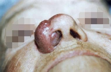
Physical examination revealed a smooth violaceous discolored mass measuring 2 X 3 cm involving the right nasal ala. The mass deeply invaded the full thickness of the nasal skin, with evidence of right nasal obstruction as seen in the image below. The rest of her facial skin contained no additional lesions. The bilateral intraparotid and jugulodigastric nodes were normally sized.
The patient underwent right-sided partial rhinectomy, with at least 5-mm margins from the visible borders of the tumor. Frozen sections revealed that all margins were free of disease. Reconstruction was accomplished immediately with a nasolabial flap. The patient's postoperative treatment included radiation therapy of 45 Gy for 5 weeks. The patient was free from recurrence at 2 years after surgery, when she died from causes unrelated to this mass.
-
Common distribution of Merkel cell carcinoma in the head and neck.
-
Merkel cell carcinoma affecting the right nasal ala.


