Practice Essentials
The diagnosis and treatment of foreign bodies in the airway are a challenge for otolaryngologists. Despite improvements in medical care and public awareness, approximately 3000 deaths occur each year from foreign body aspiration, with most deaths occurring before hospital evaluation and treatment. A high index of suspicion is needed for foreign body aspiration to allow for prompt treatment and avoidance of complications. [1, 2]
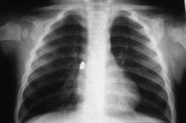 Aspirated foreign body (backing to an earring) lodged in the right main stem bronchus.
Workup in foreign bodies of the airway
Imaging
High-kilovolt anteroposterior and lateral radiographs of the airway are the tests of choice in patients in whom laryngeal foreign bodies are suspected.
Posteroanterior and lateral chest radiographs are an adjunct to the history and physical examination in patients in whom foreign body aspirations are suspected.
Lateral decubitus chest films may be helpful in children in whom the dependent lung remains inflated with bronchial obstruction. Typically, the dependent lung collapses.
Chest radiographs (inspiratory and expiratory films) demonstrate atelectasis on inspiration and hyperinflation on expiration with a foreign body obstructing the bronchus.
Biplane fluoroscopy uses intraoperative fluoroscopic evaluation while identifying and locating a foreign body within the lung periphery.
Other
Chest auscultation is critical in the evaluation of a patient in whom foreign body aspiration is suspected. Typically, these patients have wheezing, decreased breath sounds, or both on the side of the foreign body. Patients may have normal examination findings despite having a foreign body within the airway because it may partially obstruct the airway.
Management of foreign bodies of the airway
Use of the Heimlich maneuver has improved the mortality rate of patients with complete airway obstruction, but its employment in patients with partial obstruction may produce complete obstruction.
Surgical therapy
With laryngeal foreign bodies, use an insufflation catheter through the nose with the tip in the hypopharynx to maintain anesthesia and oxygenation. The laryngoscope tip is placed in the vallecula for exposure, and the foreign body is visualized in the larynx and removed with appropriate foreign body forceps.
In tracheobronchial foreign body removal, a bronchoscope is inserted into the airway after exposure to the larynx.
Larger objects unable to pass through the larynx can be broken into pieces and removed. If the object cannot pass through the larynx, a tracheotomy can be performed to remove the object through the tracheostoma.
Foreign bodies in the distal bronchial segments may be removed with the use of a Fogarty endovascular catheter through the suction port of a rigid bronchoscope.
History of the Procedure
Until the late 1800s, airway foreign body removal was performed by bronchotomy. The first endoscopic removal of a foreign body occurred in 1897. Chevalier Jackson revolutionized endoscopic foreign body removal in the early 1900s with principles and techniques still followed today. The development of the rod-lens telescope in the 1970s and improvements in anesthetic techniques have made foreign body removal a much safer procedure.
Epidemiology
Frequency
Most airway foreign body aspirations occur in children younger than 15 years; children aged 1-3 years are the most susceptible. Vegetable matter tends to be the most common airway foreign body; peanuts are the most common food item aspirated. The incidence of metallic foreign body aspirations, particularly of safety pins, has decreased in frequency secondary to the advent of disposable diapers.
Using the Healthcare Cost and Utilization Project’s Kids’ Inpatient Database (HCUP KID), a study by Wanstreet et al indicated that in pediatric patients (up to age 20 years), nonfood airway foreign bodies tend to be more often found in older children. [3]
A study by Cheng et al using the KID found that between 2000 and 2009, the mean annual hospital admission rate in the United States for children diagnosed with an airway foreign body was 6.6 per 10,000 pediatric patients. [4]
Using the National Electronic Injury Surveillance System database, a study by Saw-Aung et al reported that the incidence of foreign body aspiration among children in the United States underwent a significant decline between 2010 and 2020, the reduction being 23.6%. The incidence of foreign body ingestion, however, did not significantly fall over this period, the decline being 9.4%. Compared with White pediatric patients, Black children with aspirated foreign bodies were found to have less likelihood of same-hospital admission (odds ratio [OR] = 0.8), while the likelihood of transfer admission and mortality were higher (ORs = 1.6 and 9.2, respectively) in these patients. [5]
Mortality
Another report by Cheng and colleagues determined that between 2000 and 2009, US pediatric inpatients with airway foreign bodies had a weighted mortality rate of approximately 2.75%, with the rate staying relatively unchanged over that period. [6]
The aforementioned study by Wanstreet et al, however, indicated that the overall mortality rate among the pediatric patients associated with airway foreign bodies is 3.7%. The report also found evidence that, with regard to bronchial airway foreign bodies, the All Patients Refined Diagnosis Related Groups (APR-DRG) risk of mortality is significantly higher in pediatric patients over age 2 years. [3]
As stated above, the study by Saw-Aung and colleagues found that compared with White pediatric patients, the chance of mortality in Black children between 2010 and 2020 was greater in foreign body aspiration (OR = 9.2). [5]
Etiology
Young children make up the most common age group for foreign body aspiration because of the following:
-
They lack molars for proper grinding of food.
-
They tend to be running or playing at the time of aspiration.
-
They tend to put objects in their mouth more frequently.
-
They lack coordination of swallowing and glottic closure.
Pathophysiology
After foreign body aspiration occurs, the foreign body can settle into 3 anatomic sites, the larynx, trachea, or bronchus.
-
Of aspirated foreign bodies, 80-90% become lodged in the bronchi.
-
In adults, bronchial foreign bodies tend to be lodged in the right main bronchus because of its lesser angle of convergence compared with the left bronchus and because of the location of the carina left of the midline.
-
Several papers have demonstrated equal frequency of right and left bronchial foreign bodies in children.
-
Larger objects tend to become lodged in the larynx or trachea.
Presentation
In general, aspiration of foreign bodies produces the following 3 phases:
-
Initial phase - Choking and gasping, coughing, or airway obstruction at the time of aspiration
-
Asymptomatic phase - Subsequent lodging of the object with relaxation of reflexes that often results in a reduction or cessation of symptoms, lasting hours to weeks
-
Complications phase - Foreign body producing erosion or obstruction leading to pneumonia, atelectasis, or abscess
Clinical presentation depends on the location of the foreign body. A large foreign body lodged in the larynx or trachea can produce complete airway obstruction from either the dimensions of the object or the resulting edema.
-
Laryngeal foreign bodies present with airway obstruction and hoarseness or aphonia.
-
Tracheal foreign bodies present similarly to laryngeal foreign bodies but without hoarseness or aphonia. Tracheal foreign bodies can demonstrate wheezing similar to asthma.
-
Bronchial foreign bodies typically present with cough, unilateral wheezing, and decreased breath sounds, but only 65% of patients present with this classic triad.
Foreign body aspiration can mimic other respiratory problems, such as asthma. Foreign body aspiration differs in the presence of unilateral wheezing and decreased breath sounds.
Indications
Perform surgical intervention with rigid bronchoscopy on patients who have a witnessed foreign body aspiration, those with radiographic evidence of an airway foreign body, and those with the previously described classic signs and symptoms of foreign body aspiration. The history and physical examination are the most important aspects in the decision for surgical intervention. A strong history of suspected foreign body aspiration prompts an endoscopic evaluation, even if the clinical findings are not as conclusive or are not present.
Relevant Anatomy
Airway foreign bodies can become lodged in the larynx, trachea, and bronchus. The size and shape of the object determine the site of obstruction; large, round, or expandable objects produce complete obstruction, and irregularly shaped objects allow air passage around the object, resulting in partial obstruction.
Contraindications
No contraindications exist to the removal of an airway foreign body from a child. If necessary, health problems can be optimized before surgical intervention. However, even children who are at high risk due to health reasons still need surgical intervention to remove the foreign body.
-
Aspirated foreign body (backing to an earring) lodged in the right main stem bronchus.


