Background
A substernal goiter, also known as a retrosternal goiter, is an enlarged thyroid gland that grows inferiorly and passes through the thoracic inlet into the thoracic cavity. [1, 2] A substernal goiter is generally defined as a thyroid mass that has 50% or more of its volume located below the thoracic inlet. [1] The normal anatomic position of the thyroid is in the anterior neck, lying posterior to the sternothyroid and sternohyoid muscles and wrapping around the cricoid cartilage and tracheal rings.
The frequency with which large goiters are encountered in the United States has progressively declined over the past several decades. This decrease is largely due to an increase in dietary iodine, particularly iodized salt. Despite the declining frequency of goiter diagnoses in the United States, substernal goiter remains a significant consideration in the differential diagnosis of mediastinal masses, particularly those located in the anterior mediastinum.
See the image below.
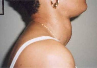 Substernal Goiter (Retrosternal Goiter). Patient with a goiter. Prominent side-view outline.
See 21 Hidden Clues to Diagnosing Nutritional Deficiencies, a Critical Images slideshow, to help identify clues to conditions associated with malnutrition.
Relevant Anatomy
Substernal goiters most commonly descend into the anterosuperior mediastinum. Recognize that the great vessels may be near the capsule of the gland or displaced anteriorly or inferiorly, although the predominant vascular supply is cervical. Typically, substernal goiters descend to one side of the trachea or the other, causing tracheoesophageal deviation to the contralateral side. Centrally located substernal goiters may compress the trachea between the sternum and spine, usually displacing the esophagus. When goiters are located in the anterosuperior mediastinum, the recurrent laryngeal nerve is not typically displaced from the tracheoesophageal groove.
Occasionally, substernal goiters may extend into the posterior mediastinum behind the trachea or even into the posterior thoracic cavity. These goiters are typically associated with greater anatomic considerations and complexity. Such goiters may extend between the trachea and esophagus, significantly displacing these structures or greatly distorting the normal relationship of the recurrent laryngeal nerve with the trachea. Goiters located in the posterior mediastinum or posterior thoracic cavity may also be complicated by a relatively normal location of the great vessels, making anterior exposure more difficult. Finally, note that goiters extending into the posterior thoracic cavity are extrapleural and behind the lung, which also makes anterior exposure more difficult.
Problem
Substernal goiters can remain asymptomatic for many years. Consequently, incidental discovery of these goiters is not uncommon, for example, on routine chest radiography. When symptoms arise, they are usually related to compression and compromise of adjacent structures from continued growth of the goiter within the bony confines of the thoracic cavity. [3] Symptoms of substernal goiter typically develop slowly and insidiously over long periods. Because such goiters can remain hidden within the thoracic cavity, they can occasionally become massive before discovery.
Conversely, while symptoms of substernal goiter can often develop slowly over many years, serious symptoms, such as airway compromise, can develop precipitously with little or no advance warning. Malignancy usually must be ruled out when substernal goiters are discovered in asymptomatic individuals, although fine-needle aspiration is often difficult or inadvisable in this location. For these reasons, many authors have advocated surgical removal of all substernal goiters, even when these goiters are asymptomatic.
Pathophysiology
The pathophysiology of goiters is well described in the Medscape Drugs and Diseases article Nontoxic Goiter. Substernal goiters are largely considered to result from the descent of a cervical goiter with the primary blood supply remaining in the neck, primarily from the inferior thyroid artery. Some theories postulate that small cervical thyroid nodules descend beneath the pretracheal fascia, possibly aided by the negative intrathoracic pressures that normally occur during respiration and swallowing. As the nodule gradually enlarges, it eventually becomes trapped below the thoracic inlet, where it continues to enlarge within the confines of the thoracic cavity, commonly hidden from view.
Substernal goiters originating from truly ectopic thyroid tissue located in the mediastinum are controversial. Arguments have been advanced that goiters, which are isolated within the mediastinum, may actually represent extensions of cervical goiters that have become sequestered (remaining connected only by fascial attachments), rather than arising truly de novo in the chest.
As substernal goiters enlarge within the bony thoracic cavity, vascular and visceral structures may slowly become compressed and compromised. Goiters often enlarge posteromedially to the great vessels and have the potential of compressing the vascular structures against the bone of the sternum or thoracic cage. Such compression can lead to superior vena cava syndrome. [4] Airway obstruction can occur because of compression or displacement of the trachea. Furthermore, over long periods, such compression can result in tracheomalacia. Dysphagia may originate because of compression of the esophagus. Dysphonia can occur because of compressive effects on the trachea or compromise of the recurrent laryngeal nerve.
Epidemiology
The incidence of substernal goiter is difficult to assess but reportedly ranges from 0.2% to 45% of patients undergoing thyroidectomy. [2] The wide range in reported incidence is largely due to variation in the definition of substernal goiter. Substernal goiters represent up to 7% of mediastinal tumors. [5]
In general, the diagnosis is made in individuals older than 50 years, and has a female predominance four-fold over males. [1]
Etiology
Most (85-95%) substernal thyroid masses represent benign goiter. Historically, goiters have generally occurred because of iodine deficiency, although this is now observed primarily in developing nations. Goiters can also be caused by ingestion of goitrogens (ie, substances that block formation of thyroid hormone), which can be found in turnip, cabbage, and kale, or they can occur as a familial form.
Malignancy occurs in 5-15% of substernal goiters.
Prognosis
No studies are available regarding the natural history of substernal goiter, nor those that compare expectant monitoring with surgery. [1]
Complications
Complications of untreated substernal goiters occur primarily because of compression of mediastinal structures. Tracheal compression can lead to tracheomalacia or respiratory compromise, which can be influenced by neck position. Dysphagia may occur due to esophageal compression. Compression of vascular structures can lead to the development of superior vena cava syndrome. Neurologic complications such as Horner syndrome, phrenic nerve palsy, or recurrent nerve palsies raise concern for malignant disease; however, these can occur in benign goiters as well because of compression.
-
Substernal Goiter (Retrosternal Goiter). Patient with a goiter. Prominent side-view outline.