Overview
Following the September 11, 2001, terrorist attacks, government and civilian experts recognized the need to enhance and update hospital-based emergency preparedness plans to deal with a potential radiologic attack or accident. [1] The main challenge was adapting the existing models, whose primary focus was containing a hazardous material release, to one that reflected the chaos of a large-scale disaster involving a large number of affected individuals. The best recommendations included detailed safety measures under the direction of sophisticated radiation safety experts, which proved difficult to adapt to emergency department settings. [2, 3]
Emergency department staff play a significant role in any public catastrophe because victims go directly to hospital emergency departments as the primary access to the healthcare system. However, few emergency departments feel prepared for a radiation disaster, and many personnel question the practicality and safety of current recommendations. [4] Likely the distinct scientific language and differing cognitive processes between front-line clinicians and radiation safety experts contribute, at least in part, to the discrepancy between what experts think should be done and what front-line providers think can be done. [5]
The purpose of this article is to help bridge this communication gap for emergency department personnel. It includes basic radiation reference material, suggestions for designing a preparedness strategy, and practical material that the reader may need in an actual event. [6]
If the reader's objective is education or preparation for an exercise, then reading this article in its entirety is recommended. However, this article may also be used for readers who may be seeking information during a true event to obtain essential points quickly without a great deal of time spent exploring the underlying concepts.
If you are in the midst of an actual event, go to the staff safety section first, then to instructions on setting up your receiving area and how to use a Geiger counter.
Terminology
Radiation
Radiation is energy transmitted in the form of electromagnetic waves or energetic particles. Well-known forms of electromagnetic radiation include the following:
-
Radiowaves
-
Microwaves
-
Infrared
-
Visible light
-
Ultraviolet light
-
X-rays
-
Gamma rays
Ionizing versus nonionizing radiation
Ionization occurs when sufficient energy is transferred to an atom to liberate an orbital electron generating an electrically charge ion pair. These ion pairs can then chemically react with other pathways within living cells and, if sufficient in number, will disrupt cellular function, including damage to DNA. On the electromagnetic spectrum, only x-rays and gamma rays contain sufficient energy to cause ionization. High-energy particles can also cause ionization.
Ionizing radiation: electromagnetic radiation
Energy can travel through space in the form of electromagnetic radiation. Electromagnetic radiation is composed of massless waves of oscillating electric and magnetic fields. In a vacuum, these waves move at a constant speed, the speed of light (3 X 10⁸ m/s). All electromagnetic waves propagate with characteristic wavelength and frequency, with the wave's energy being directly proportional to frequency and inversely proportional to wavelength. Within the electromagnetic spectrum, only x-rays and gamma rays have enough energy to produce ion pairs. The remaining waves within the spectrum, such as microwaves and radiowaves, are nonionizing.
Ionizing radiation: particulate radiation
Ionizing radiation can also be in the form of particulate radiation, which includes subatomic l charged or neutral particles traveling near the speed of light and therefore with high very high kinetic energy. Since particles have mass, they are less penetrating than electromagnetic waves and deposit their energy readily. This property makes them easier to shield but also more hazardous if ingested.
Alpha particles
Alpha particles are charged particles made up of 2 protons and 2 neutrons—essentially the nucleus of a helium atom. Because of their relatively large mass and positive charge, alpha particles are highly effective in transferring energy to tissue but are also easily blocked by a piece of paper or clothing. These particles are only a concern when alpha-emitting isotopes are ingested or inhaled.
Beta particles
Beta particles are high-energy electrons. As in alpha radiation, the main hazard with beta particles lies with internal exposures. With significant skin exposure, however, beta particles have sufficient energy to cause cutaneous burns, "beta burns."
Neutrons
Neutrons are electrically neutral subatomic particles that can be liberated in a particle accelerator, a nuclear reactor, or a nuclear weapon as part of the fission process. Neutron exposure is most consequential in a nuclear reactor criticality accident or during nuclear weapons detonation.
Protons
Protons are positively charged subatomic particles that make up a major component of cosmic radiation originating from the sun. All but a small amount of the sun's proton radiation is deflected by the earth's magnetic field.
Radioactive decay
Radioactive decay is the process in which unstable atomic nuclei assume a more stable configuration by emitting particles with kinetic energy (alpha or beta particles) or electromagnetic waves (gamma rays).
Half-life
Radiological half-life is a physical property of each radioisotope and reflects the stability of the nucleus. Measured half-lives can range from fractions of a second to billions of years.
Biologic half-life is the time required for the body to eliminate one half of the material by natural biological means.
Effective half-life is the time whereby half of the radioactivity of a particular radioisotope is eliminated as a result of the combined action of radioactive decay and biological elimination.
Irradiation, contamination, and incorporation
Irradiation is exposure to ionizing radiation. For example, patients who receive x-rays or CT scans become irradiated. Once the radiographic machinery is turned off, radiation is no longer produced. Because these individuals are only in the path of radiation energy, as opposed to carrying a radioactive source on their bodies, they pose no radiation exposure risk to others.
Contamination means radioactive material is physically present where it is unwanted or unexpected.
External contamination is when unwanted radioactive material is deposited on clothing, skin, hair, or any area where the material has not been taken inside of the body.
Internal contamination is when unwanted radioactive material is deposited within the body by absorption through skin, ingestion, inhalation, or through wounds.
Incorporation of radioactive material occurs when internal contamination enters metabolic pathways. Radioactive atoms are chemically indistinguishable from nonradioactive atoms. They participate in the same chemical pathways and are difficult to remove once incorporation occurs.
Decontamination is any steps taken to remove radioactive material from clothing and the external surfaces of the body or from rooms, building surfaces, equipment, or other items.
Decorporation is the processes by which radioactive materials are mobilized from tissues and organs and removed from the body by enhanced excretion.
Radiation dose
Gray and Rad
Radiation dose is the quantity of energy deposited in living tissue by an exposure to ionizing radiation. The Gray is the standard SI unit for dose and is expressed as J/kg. The Rad is the more historical unit and is commonly used. One gray is equal to 100 rad.
Sievert and Rem
The biological effect of different types of radiation vary significantly. For instance, alpha particles tend to deliver 20 times the biological effect of an equivalent dose of gamma radiation. The equivalent doses are expressed in Sieverts (Sv) in SI units and Rem in classical units.
Radiation exposure rates
Ionizing radiation interacts with air generating electrically charged ions in the air. Geiger counters are designed to detect this phenomenon, and exposure rates are typically expressed as Roentgens per hour (R).
Biologic Effects of Ionizing Radiation
The principal pathologic effect of ionizing radiation results from damage to DNA. Regardless of the form of ionizing radiation, the common pathway of injury is that the radiation deposits a relatively large amount of energy into the electron orbitals of atoms in the biologic medium. This energy transfer raises the energy level (excites) of the electron and, if sufficient, ejects the electron from the atom, resulting in a now positively charged atom (ionization).
These charged particles are chemically active, resulting in breakage of the chemical bonds within the DNA molecule. In addition to this direct injury, ionization radiation interacts with cellular water, forming free radicals, which can also damage the chemical bonds in the DNA strands. Regardless of the form of ionizing radiation (alpha, beta, gamma, x-ray, neutrons) or whether the damage is direct or indirect via free radical formation, the final pathologic injury is disruption of chemical bonds in the DNA strands. [7]
DNA damage is dose dependant, with the most common damage being single-strand breaks. As long as the number of breaks is not overwhelming and the complementary DNA template remains intact, these injuries are repaired with little biological consequence. In contrast, if the injury results in a double-strand break, the repair template is lost and can result in cell death, mutations, or carcinogenesis. [7]
Since DNA damage is the principal cause of the biologic effects of radiation, tissues with a high turnover rate are more sensitive to the toxic effects of radiation than cells that are more differentiated. Cells and tissues can repair a certain amount of this damage with no apparent clinical consequence; however, at higher doses, normal homeostatic mechanisms are overwhelmed. Physiologically, damage on the chemical level progresses to cellular dysfunction, which leads to tissue dysfunction, then organ failure and, ultimately, to death. [7]
Early versus late radiation effects
Early effects of radiation are seen after large doses of radiation are delivered over short periods and especially affect rapidly dividing, self-renewing organs including skin, bone marrow, and gut epithelium. Clinical syndromes reflect the failure of replenishment of tissues with rapid turn over and include acute radiation sickness and radiation burns. They are typically seen within a short period following a massive exposure.
Late toxicities, such as cataract formation and cancer, may appear months or years following an exposure. Other delayed effects are seen in organs with slowly dividing or quiescent, terminally differentiated cells such as the central nervous system, kidneys, and liver. Many of these late radiation effects are attributable to a combination of parenchymal cell death and microvascular disease.
The most well-known delayed complication of radiation exposure is malignancy. Exposures to the Chernobyl accident and atomic bomb testing in the Marshall Islands led to high incidences of thyroid malignancies. [8, 9, 10] Atomic bomb survivors have been shown to have an increased risk of leukemia, and young women with Hodgkin disease treated with radiation therapy have been shown to have an increased risk for breast cancer. [11, 12] The explosions at the Fukushima Daiichi Japanese nuclear power plant after the March 2011 massive earthquake increased fear of contamination from radiation and possible health risks. [13]
Biological Effect Relative to Exposure Dose
Exposure to ionizing radiation is an unavoidable fact of life on earth and has been a reality since the beginning of time. The various natural sources include cosmic rays from the sun and stars, radioactive elements in granite and other rocks, and radioactive elements that compose living tissue, predominantly potassium-40 and carbon-14. In addition, radioactivity exists in the environment from fallout from nuclear explosives testing, from both coal and nuclear power plants, and industrial accidents including Chernobyl in 1986 and the current nuclear reactor disaster in Fukashima, Japan. Finally, medical use of ionizing radiation is a growing source of exposure, although this exposure is generally limited to the individuals undergoing the various diagnostic studies and therapeutic interventions.
The National Council on Radiation Protection and Measurements publication No. 160 estimates the average effective dose of radiation to individuals in the United States to be 6.2 milliSieverts a year, with 3.0 milliSieverts coming from natural sources and an additional 3.0 from medical exposure, primarily from CT scanning and nuclear medicine procedures. [14]
The question of the relative benefit and the hazard of ionizing radiation is beyond the scope of this article, and current regulatory standards are used as a basis for operational practicalities with some extrapolation to address the gaps for the unknown. Regulatory agencies set standards for the lawful use of radioactive material to keep exposure doses as close to background as feasible and within specified maximum guidelines. [15] Such guidance highlights the principle that the risk of health effects of exposure to ionizing radiation must be balanced by the expected individual and societal benefit to justify exposure beyond natural background.
It is clear that the severity of toxicity caused by radiation is directly related to the quantity of energy deposited into the living organism and the subsequent disruption of metabolic and reproductive pathways. Exposure to high doses of ionizing radiation results is acute illness and potentially death. Low-level exposure may lead to relatively minor toxicity acutely and possible adverse health effects in the future.
Linear no-threshold model
The linear no-threshold model (LNT), which is used by most regulatory agencies, assumes a direct and proportional relationship between radiation exposure and cancer risk with all radiation doses. These risks are derived from the linear relationship between radiation doses and health effects (mostly cancer) in the atomic bomb survivor studies. However, at low exposure doses, the high natural incidence of the disease masks any additional effects that may have been caused by radiation. Because of this, the LNT assumes that cancer incidence as it relates to radiation dose behaves in the same way as at higher doses; that is, in a linear manner.
However, there is little conclusive direct evidence of adverse health effects at doses below 100 mSv, questioning the certainty to the LNT risk model. Some experts postulate that the actual hazard at low radiation doses is lower than those predicted by LNT. Whether the actual effect is linear or otherwise remains unknown, and the core principle of radiation safety is to ensure that radiation levels are kept as low as reasonably achievable (ALARA).
In light of this uncertainty and given the uncontrolled nature of a contamination radiological event, current guidance for immediate response to an emergency focuses first on prevention of very high-dose exposures; next, on promptly recognizing and controlling levels of contamination that may progress to acute health risks if not immediately controlled; and then, if feasible, on evaluating and managing lower levels of contamination and exposures near to background levels.
Clinical Syndromes
Localized cutaneous injury
Localized exposure to high doses of radiation may cause cutaneous injury similar to burns. Blistering, erythema, desquamation, and ulceration often present about 12-20 days after irradiation with the onset and severity related to the magnitude of exposure.
A local exposure of 3 Gy leads to second phase erythema and temporary epilation within 1-2 weeks. A local exposure of 7 Gy may cause immediate signs of burn and definitive epilation within 25-30 days. Greater exposures of 10-15 Gy lead to dry desquamation, and exposures of 15-25 Gy lead to wet desquamation (partial-thickness burn) within 2-3 weeks. For doses greater than 25 Gy, a cutaneous syndrome of necrosis and ulceration may occur from damage to endothelial cells and small blood vessels. [16] Vascular complications may present months to years after exposure.
Acute radiation syndrome
The acute radiation syndrome (ARS) occurs after whole-body exposure to a large dose of ionizing radiation. This syndrome includes a number of characteristic signs and symptoms whose severity depends on magnitude of dose and duration of exposure. ARS, by definition, does not occur at doses less than 1 Gy and is uniformly fatal at doses greater than 8 Gy. [16] The estimated LD50/60 (the dose at which 50% of those exposed die within 60 days) is 3.5 Gy for humans without medical treatment and roughly 7.0 Gy with treatment.
Stages of acute radiation syndrome
Acute radiation syndrome (ARS) has been described according to progression of illness through 4 stages: (1) prodrome, (2) clinical latency, (3) manifest illness, and (4) recovery or death. The prodromal symptoms occur shortly after irradiation, with the dose of exposure determining severity, duration, and onset. Common prodromal symptoms include nausea, vomiting, anorexia, fatigue, diarrhea, abdominal cramping, and dehydration. At doses of greater than 10 Gy, those exposed show symptoms within 5-15 minutes; at lower doses such as 2-3 Gy, symptoms can take up to 12 hours to present. Immediate diarrhea, hypotension, and fever indicate a supralethal exposure. Severe and early onset of prodromal symptoms indicates higher dosage of exposure and a poor prognosis. Progression through the other phases depends on dosage of exposure.
Classic acute radiation syndromes
Acute radiation syndrome (ARS) is further described by its 3 classic subsyndromes: the hematopoietic syndrome, the gastrointestinal syndrome, and the cerebrovascular syndrome. [17] The hematopoietic syndrome typically occurs after exposures of 2-5 Gy. At these doses, lymphocytes die from radiation-induced apoptosis, and precursor cells in the bone marrow are destroyed preventing new production of leukocytes and platelets. During the period of a few weeks (clinical latency), circulating cells die off with no replacements; it is at this nadir that the full syndrome becomes clinically apparent with development of infections and possible hemorrhage. Anemia from red cell depression usually does not occur alone in the absence of hemorrhage. Early supportive care, treatment and prevention of infections, and the consideration of cytokine therapy are all important aspects of care for this subsyndrome. However, even if the hematopoietic syndrome is treated, death commonly still occurs from multiorgan failure.
The gastrointestinal syndrome usually occurs after exposures of more than 5-12 Gy. Irradiation leads to death of intestinal mucosal stem cells in the crypts. After loss of mucosal cells at the villi through normal functioning, the stem cells are unable to produce new cells, leading to denudation of the GI tract. As the normal GI boundary is compromised, bacterial growth proliferates increasing the risk of sepsis. Common symptoms include anorexia, nausea, vomiting, prolonged bloody diarrhea, abdominal cramps, dehydration, and weight loss. Often the prodrome onset is rapid, followed by a latent period of roughly 1 week followed by the return of symptoms. The mainstays of treatment are maintenance of fluid and electrolyte balance and infection prevention. Nevertheless, despite treatment death often follows in 3-10 days.
The cerebrovascular syndrome occurs after exposures of very high doses (>30 Gy) and is uniformly fatal. At doses of greater than 100 Gy, death occurs within hours. Although the exact mechanism of death is not fully understood, vascular damage is thought to lead to significant cerebral edema, producing neurologic and cardiovascular collapse. Immediate symptoms include nausea, vomiting, hypotension, ataxia, and convulsions. Death follows within a few days.
Staff Safety
Perhaps the most important issue for emergency planners to address prior and during any disaster is protection of the staff members and first responders. Workers should be monitored for radiation exposure and use caution rendering care, but the risk of health effects must be kept in perspective to allow for optimal evaluation and care of victims. According to Waselenko et al, no healthcare workers who have adhered to appropriate guidelines have become contaminated or suffered from radiation injuries from handling a contaminated patient. [18]
That being said, the only way to respond safely and intelligently is with properly functioning radiation monitoring equipment. While all hospitals have some access to equipment, it is absolutely essential that appropriate equipment be immediately available in the emergency department and that staff are sufficiently trained to perform basic radiation surveys 24 hours a day, 7 days a week.
During the unknown early phase of a disaster, staff must observe an all-hazards approach and assume that biological, chemical, and radiological hazards may be present. Only after other hazards are excluded should staff limit their focus to radiological hazards. Protection should focus on minimizing external exposure and preventing inhalation, or inadvertent ingestion. In most cases, protection is accomplished by observing standard precautions, also referred to as level D personal protective equipment (PPE), generally observed in the hospital setting.
Basic radiation surveillance should focus on identifying the presence of radiation, protecting staff and patients, and controlling the spread of contamination. Once available, radiation specialists should be able to characterize the hazards more specifically.
Optimally, all personnel entering or working in a radiation control zone should wear personal dosimeters. In the early phases of a disaster, this may not be feasible, in which cases, dose estimates will need to be reconstructed. Under all circumstances, radiation exposure should be kept as low as reasonably achievable (ALARA) while preserving critical operations and resources.
General radiation protection principles:
-
Time - Minimize time spent near radioactive source
-
Distance - Maximize distance from source
-
Shielding - Place physical shields around source
Additionally, during an uncontrolled situation, providers should observe universal precautions, including splash and water protection. Furthermore, staff actively attending contaminated patients should wear personal monitors, such as real-time dosimeters and/or film badges, unless the level of exposure is known to be negligible.
Risks of radiation exposure
Radiation protection agencies assume that even the smallest exposure to ionizing radiation has an incremental albeit small probability of causing a long-term effect, such as cancer. For example, there is a hypothetical risk of cancer from receiving radiation from a single chest x-ray, the risk is so small that it impossible to statistically prove this.
The annual background radiation dose is equivalent to about 12-15 chest x-rays a year, and this is considered a normal yearly exposure. Other activities, like smoking or airline travel, can increase one’s annual dose of radiation. Medical tests, especially imaging using radiation, also increase the dose of radiation exposure. The hazard is thought to be very small relative to the expected benefit of the test.
On the other hand, very high radiation doses received in a short period may cause serious illness and even death. The dose range where acute effects are seen are equivalent to the dose one would receive from several thousand chest x-rays.
The radiation doses to staff members involved in an emergency response are expected to be well below these thresholds and well within the annual range of radiation exposures established by radiation protection agencies.
Prehospital Care
Emergency services personnel should initiate their incident command structure as soon as a radiation event is suspected. They should secure the scene, ensure self-protection, and attempt to rescue injured or ill victims while minimizing radiation exposure to all individuals, including the rescuers. Personnel should wear appropriate protective gear prior to arrival at the scene and follow the guidance of the radiation safety officer or operations commander. Clinical operations may be required in high exposure areas, but exposure limits should be strictly regulated by supervisors.
In the field, it is far more important to detect the presence of abnormally high radiation fields than to identify specific contaminants. Personal radiation monitoring instruments should be worn by all personnel with alarm points pre-set by radiation specialists. Reasonable set points for individuals not performing life-saving or critical interventions are >100 mR/h, with 500 millirem as the total cumulative dose.
Once the scene is assessed to be safe for entry, rescue personnel should assess and treat life-threatening injuries. Conventional injuries and illnesses present a much greater acute hazard to patients than radiation exposure and contamination; therefore, standard medical treatment should be initiated as soon as feasible and not delayed to assess contamination status. Contamination control can be accomplished by removing the victims clothing and wrapping in clean sheets, but this should not delay resuscitative care. In contrast, if a contaminated patient has a non–life-threatening or serious condition, they should be optimally decontaminated prior to definitive medical treatment.
In a mass casualty event, individuals who are neither contaminated nor ill or injured should go home and take a regular shower with soap and shampoo. They should bag and store their potentially contaminated clothing in a safe place.
Decontamination at or near the site of exposure would be useful if feasible. However, such interventions are not feasible when dealing with a large number of frightened individuals in the early moments of a catastrophe. Generally, individuals leave a hazard scene quickly and self-present to hospitals. Therefore, hospitals must be prepared to deal with large numbers of contaminated and potentially contaminated victims.
Emergency Department
Emergency departments should activate their institution's Hospital Incident Command Structure (HICS) as soon as they are notified of an event. The Joint Commission on Accreditation of Healthcare Organizations requires all hospitals to maintain a written protocol for receiving and treating victims of radiation disasters.
Triage
In general, treatment of conventional injuries and illness takes precedence over radiation concerns because the quantity of radioactive material that a contaminated individual carries on his or her body is unlikely to present a significant radiation risk to either the patient or to the healthcare staff attending to the patient. Healthcare workers have cared for contaminated individuals in the past, and there are no documented cases of healthcare personnel becoming acutely ill caring for contaminated or irradiated victims of radiation accidents. Therefore, critically ill and injured patients should be moved into critical care areas of the hospital and should be decontaminated during resuscitation.
Performing rapid medical triage
This initial sorting function should be completed at the initial point of clinical contact by an experienced individual who is capable of sorting patients into the three designated treatment areas on the basis of their clinical status as assessed by a brief inspection. It is critical that this step does not bog down from excessive deliberation or indecision. The three designation areas, and their traditional color coding, are as follows:
-
Medical emergency (red)
-
Medical urgency (yellow)
-
Ambulatory, with or without minor illness or injury (green)
The medical emergency (red) category comprises patients who are seriously ill or injured and require immediate attention. Initial management is to bypass the decontamination shower; remove the patient’s clothing and cover with a clean sheet while moving into the critical care area.
Medical urgency (yellow) patients should undergo rapid radiologic triage. They should be decontaminated as completely as feasible before entering the yellow area.
Ambulatory with or without minor illness or injury (green) patients should proceed directly to thorough radiologic survey if indicated.
Rapid radiologic triage - Prioritizing radiologic screening of victims
If the number of victims is small, a thorough evaluation using a hand-held survey meter should be performed on all individuals according to procedures suggested by REAC/TS. However, in a mass casualty incident, it is likely that too many patients will require screening in a short period of time to allow performance of a thorough screening, and a radiologic screening triage will be needed. If feasible, a portal monitoring system should be set up for screening large numbers of individuals with low probability of significant contamination.
Patients with serious injury or illness related to the incident
For an explosive incident, patients seriously injured were most likely closest to the blast, with the highest levels of radiation exposure. These patients will probably require hospital admission, which will allow clinicians ample time for patient observation and assessment of internal contamination and radiation-dose exposure analysis. Note: Open wounds must be rapidly assessed to exclude high-activity fragments.
Patients with moderate or minor injury or illness related to the incident
As in the case of the more seriously injured patient, these individuals are at a higher risk of radiation contamination due to their proximity to the incident. While these patients may not require hospital admission, they may require assessment for radiation contamination and exposure.
Persons with no physical injury but with facial and/or upper body contamination
Facial and upper body contamination may indicate that the patient inhaled radioactive contamination and may have internal contamination that requires treatment.
Children < 18 years old and pregnant women in proximity (500 m) to the event
Exposure to radioactivity poses greater risk for these patients (in the case of pregnant women, for the fetus). Decontamination therefore has heightened importance for them, and their care should be prioritized accordingly.
All other individuals without physical injuries
Direct these individuals to an alternative care area to obtain demographics, counseling, and assessment.
Workup
Time to emesis
Time to emesis (TE) correlates with exposure dose, decreasing as exposure dose increases. For TE less than 1 hour, whole-body dose estimates are greater than 4 Gy. For TE between 1 and 2 hours, whole-body dose is estimated to be greater than 3 Gy, and for TE greater than 4 hours, whole-body dose is estimated to be around 1 Gy.
Lymphocyte depletion
The most useful laboratory test in the setting of acute radiation exposure is the serial complete blood count with differential obtained every 6-12 hours. [19] Lymphocyte count serves as an indicator for prognosis and as an estimate for the dose of radiation received. Patients with a minimal lymphocyte count (MLC) of 1000-1499/mm3 have an approximate absorbed dose of 0.5-1.9 Gy. Although these patients may have clinically significant effects, their prognosis is good because the absorbed dose is usually nonlethal.
Patients with MLC of 500-999/mm3 have an approximate absorbed dose of 2.0-3.9 Gy with severe injuries and fair prognosis. An MLC of 100-499/mm3 coincides with an approximate absorbed dose of 4.0-7.9 Gy predicting severe injury and a poor prognosis, and those with MLC less than 100/mm3 have an estimated absorbed dose of greater than 8 Gy and have a high incidence of death despite bone marrow stimulation. Survival has not been documented for those exposed to greater than 10 Gy.
Other assays
Blood can also be drawn for cytogenetic evaluation. If dicentrics (chromosomes with 2 centromeres) are found, they can be used to indicate extent of radiation exposure. Cytogenetic studies are time-consuming processes that are currently not being used for mass screening strategies. Realistically, these studies may be more useful from an inpatient standpoint with expert guidance.
Imaging
Ultrasund and thermography have been used successfully to evaluate skin damage in real time. [20]
Further Inpatient Care
Cutaneous
Patients with localized irradiation who present with cutaneous injury should be treated in the same manner as those with thermal burns. Pain control and infection prophylaxis are the mainstays of treatment. In the cases of severe burns, vasodilator therapy, grafts, or amputations may be necessary.
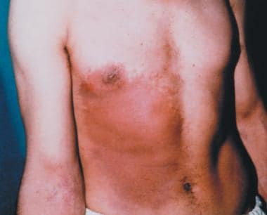
The images below show burns due to radiation exposure.
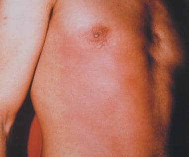 Early erythema in the frontal and antelateral right side of the chest 5 days after the exposure to an iridium-192 source (185 GBq, 5 Ci) mounted in a pen-size sourceholder for industrial radiography, which was placed to the pocket of the worker's overall and kept there for about 2 hours. Image courtesy of the WHO.
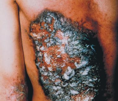 Desquamation and skin necrosis 21 days after exposure. Note: the white areas correspond to silver ointment. Image courtesy of the WHO.
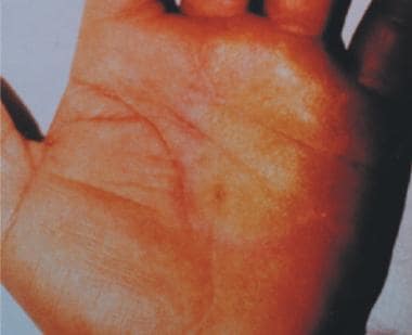 Tense painful bulla of the left palm on day 20 evolving from erythema with early blistering, which had appeared on day 10 after the initial contact for a few minutes with the iridium-192 source. Image courtesy of the WHO.
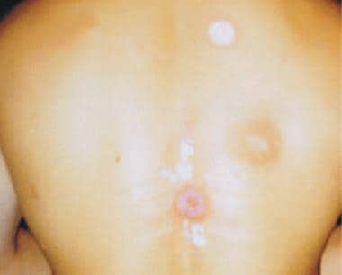 An ulcer in subacute stage and 5 ulcers following self-healing with depigmentation caused by being unawarely exposed 4-8 months earlier to the same164 GBq (4.4 Ci) cesium-137 source (placed in a pocket of a trench coat used as a blanket). Image courtesy of the WHO.
Internal contamination
In cases of internal ingestion or contamination of unknown radioactive materials, some measures (eg, lavage, charcoal) to decrease absorption may be effective. Similarly, specific therapies exist to remove some internally deposited radionuclide albeit with limited efficacy. These treatments are often in limited supply, complex in action, and associated with significant risks. Thoughtful and critical analysis should occur prior to administration.
Internal contamination by radioactive iodine can be treated with a saturated solution of potassium iodide (SSKI), a blocking agent that reduces uptake of radionuclide in the thyroid. SSKI is most effective when taken within a few hours of exposure. In reactor accidents involving radioactive iodine, massive quantities are released into the environment. Both exposed victims and rescue personnel should take SSKI to reduce thyroid uptake of radioiodine to reduce risk of future malignancies. Specific dosages for SSKI administration can be viewed at Guidance for Industry – KI in Radiation Emergencies—Questions and Answers.
Chelating agents, such as penicillamine, bind specific radioactive metals causing decreased tissue uptake and increased excretion. Exposure to isotopes of cesium can be treated with ferric hexacyanoferrate (Prussian blue) to decrease gastrointestinal absorption. Agents such as Ca-DTPA and Zn-DTPA can be administered after internal contamination with substances such as americium, curium, and plutonium. Administration of specific agents should be completed in consultation with professionals familiar with these agents.
Large-dose whole-body radiation exposure
Patients with intractable vomiting should be treated supportively with intravenous fluids, analgesics, and antinausea medication. This supportive treatment should continue until the initial illness has abated.
For exposures of around 5 Gy, immunosuppression and bowel dysfunction occur relatively quickly and patients will become ill in a few days. Supportive care includes, antibiotics, consideration of cytokine therapy, and transfusions as needed.
For exposures greater than 5 Gy, death from gastrointestinal syndrome is highly likely. Along with antibiotics and platelet transfusions, as needed, all measures to avoid to infection should be emphasized.
During periods of infection, antibiotics should be tailored toward the source of infection. If absolute neutrophil count (ANC; see the Absolute Neutrophil Count calculator) is less than 500 cells/mm3, most experts recommend prophylactic antibiotics, including an extended-spectrum quinolone, for coverage of gram-negative and gram-positive infections; an antiviral agent (acyclovir in those with a history of herpes simplex virus); and an antifungal agent. Once fever and infection occur, broader-spectrum antibiotics with additional antipseudomonal coverage should be initiated.
Hematopoietic growth factor treatment
In 2015, the US Food and Drug Administration (FDA) approved filgrastim (Neupogen) to increase survival in patients acutely exposed to myelosuppressive doses of radiation (hematopoietic syndrome of acute radiation syndrome [H-ARS]). Efficacy studies of filgrastim could not be conducted in humans with acute radiation syndrome for ethical and feasibility reasons. Approval was based on efficacy studies conducted in animals and data supporting the use of filgrastim for other approved indications.
Efficacy of filgrastim was demonstrated in a randomized, blinded, placebo-controlled study in a nonhuman primate model of radiation injury. The planned sample size was 62 animals, but the study was stopped at the interim analysis with 46 animals because efficacy was established.
In the study, rhesus macaques were randomized to a control (n = 22) or treated (n = 24) group. Animals were exposed to total body irradiation of 7.4 ± 0.15 Gy delivered at 0.8 ± 0.03 Gy/min, representing a dose that would be lethal in 50% of animals by 60 days of follow-up (LD50/60). Starting on day 1 after irradiation, animals received daily subcutaneous injections of placebo (5% dextrose in water) or filgrastim (10 mcg/kg/day).
Blinded treatment was stopped when one of the following criteria was met: ANC ≥1,000/mm3 for 3 consecutive days, or ANC ≥10,000/mm3 for more than 2 consecutive days within study day 1 to 5, or ANC ≥10,000/mm3 any time after study day 5. Animals received medical management consisting of IV fluids, antibiotics, blood transfusions, and other support as required.
Filgrastim significantly (at 0.023 level of significance) reduced 60-day mortality in the irradiated nonhuman primates. Mortality was 21% (5/24) in the filgrastim group compared with 59% (13/22) in the control group. [21]
Waselenko et al proposed recommendations for giving colony-stimulating factors (CSF) to victims of radiation exposure. [18] His team recommended giving granulocyte-macrophage colony-stimulating factor (GM-CSF) to those who receive more than 3 Gy of radiation and in those with multiple injuries who are exposed to more than 2 Gy. Recommended doses include initiating therapy with filgrastim at 5 mcg/kg/day or sargramostim at 250 mcg/m2/day subcutaneously immediately after exposure and continuing until absolute neutrophil count increases to greater than 1000 cells/mm3. Alternative treatment with subcutaneous pegfilgrastim weekly to those greater than 45 kg has also been suggested.
Pegfilgrastim (Neulasta, Udenyca) is also approved by the FDA for treatment of H-ARS adult and pediatric patients. It is a long-acting form of filgrastim that consists of filgrastim covalently bonded to polyethylene glycol (PEG).
Sargramostim (Leukine) is approved for treatment of H-ARS in adult and pediatric patients. Sargramostim is available in 2 forms for parenteral administration. The sterile ready-to-use solution may be administered without further dilution as a subcutaneous injection. Note that this preparation contains benzyl alcohol; therefore, it is not suitable for neonates or infants owing to the risk for gasping syndrome in this population with benzyl alcohol-containing products. In neonates and infants, use only the lyophilized powder that must be reconstituted with sterile water for injection without preservatives.
Obtain a baseline CBC with differential and then serial CBCs approximately every third day. The weight-based dose is administered once daily until the ANC remains >1000/mm3 for 3 consecutive CBCs.
Similarly to filgrastim, sargramostim studies were conducted in primates and showed an improved survival by 85% compared with placebo (78% vs 42%; p=0.0018) at day 60. [22]
PLX-R18
In May 2018, the FDA cleared emergency use of the investigational biologic PLX-R18 (Pluri Inc, Haifa, Israel) to treat acute radiation exposure in a nuclear event. Full FDA approval will depend on the results of a phase III clinical trial.
Emergency access was issued based on the completion of a midsize study of PLX-R18 in the treatment of acute radiation syndrome (ARS) in animals via the FDA’s “animal rule pathway” sponsored by the US National Institutes of Health’s National Institute of Allergy and Infectious Diseases. PLX-R18 will be provided up to 96 hours following radiation exposure. It is administered in two intramuscular doses 1 week apart. [23]
PLX-R18 consists of 3D-expanded placenta-derived cells. PLX-R18 elicits biological properties, including the ability to protect and regenerate bone marrow. The cells secrete a broad assortment of cytokines—including granulocyte colony-stimulating factor (G-CSF), interleukin-6, monocyte chemoattractant protein 1 (MCP-1), MCP-3, and growth-related oncogene (GRO)—that contribute to the rebuilding of the hematopoietic and immune systems. [24]
Procedures for Ambulatory Radiation Monitoring
Radiation portal monitor
Radiation portal monitors can be set up to screen large numbers of patients with low suspicion of contamination. Those patients with high risk of contamination should be decontaminated empirically or screened with a Geiger counter with special attention to detecting high-activity particles.
Radiation portal monitors can be used in a fashion similar to security screens performed in airports. Individuals should enter the portal frame and step on a footplate activating the monitor. Individuals should then turn sideways to enhance detection of very small sources. Portal monitors can generally detect as little as 1 microcurie of Cs-137, and the relevance of detecting such a low level of activity in a mass casualty situation is questionable.
Since the portal monitor’s main role will be to rapidly and reliably clear a large number of people as not contaminated, it is essential to avoid contaminating the portal monitor or the area immediately in the vicinity of the monitor. Anyone at high or intermediate risk of contamination should either undergo screening with a Geiger counter or be decontaminated empirically.
Survey using a typical Geiger-Mueller (GM) counter
Adapted from REAC/TS. [25]
Prepare the meter:
Position the Geiger counter with the meter away from you.
Locate and open the battery compartment.
Put the batteries in the meter using proper orientation (up/down).
Close and latch the battery compartment.
Check the batteries using the "range" switch or "bat" button; the method depends on the type of instrument. The meter needle should move to area on the scale marked battery; this indicates that the batteries are good. If the batteries are not good, find a flashlight or other source of 2 D-cells and place them in the meter; these batteries should be checked as well.
Turn the "F/S" switch to "S" (Slow).
Turn the "audio" switch to "ON."
Measure background radiation:
Ensure that the "F/S" switch is on "S" (Slow).
Adjust the range switch to the most sensitive position.
Remove the probe cover, as necessary.
Measure the background radiation for 60 seconds, and write down this reading. Background radiation varies with time; therefore, it may be necessary to take several count readings and average the results. Record this reading as well. A reading of 20-100 counts/min, or a reading of approximately 0.02 mR/hr (ie, 0.2 on the 0.1 range setting), or 0.2 micro Sv/hr, is expected in normal circumstances.
Record the background reading.
Conduct the survey:
- The person being surveyed should stand on a clean pad, with feet spread slightly and arms extended with the palms facing up and fingers pointing straight out.
- Move the "F/S" switch to "F" (fast response).
- Set the instrument selector switch to the most sensitive range of the instrument.
- Hold the probe approximately 1/2 to 1 inch from the person's skin and systematically survey the entire body from head to toe on all sides.
- Survey both hands and arms. Repeat the survey with the hands and arms turned over.
- Start at the top of the head and cover the entire body. Carefully monitor the forehead, nose, mouth, neckline, torso, knees, and ankles.
- Instruct the person to turn around. Repeat the survey on the back of the person's body.
- Monitor the soles of the feet.
Note the following:
-
Move the probe slowly (about 1 inch per second). The probe should not touch anything.
-
Maintain a constant distance from the person.
-
Pay particular attention to hands, face, and feet.
-
Some Geiger-Mueller instruments cannot detect alpha radiation and some low-energy beta radiation. Because alpha radiation is nonpenetrating, it cannot be detected through even a thin film of water, blood, dirt, clothing, or through probe cover.
-
The presence of radiation is indicated by an increase in count rate or exposure rate above background.
-
Locate the point that produces the most clicks. (Turn the "F/S" switch to "S" to take a reading at this location. Remember to reset it to "F" before continuing survey.)
-
Adjust the range of the instrument by moving the range selector switch as needed.
-
Document the time and the radiation measurements.
-
Generally, areas are considered contaminated if they register more than twice the previously determined background level. For incidents involving alpha emitters, if the reading is less than twice the background radiation level, the person is not contaminated to a medically significant degree. If the event circumstances indicate that an alpha emitter (eg, plutonium) or low energy beta emitter could be a contaminant, consult a health physicist.
End the radiation survey:
- Turn the meter off.
- Cover the meter probe with the cap.
- Remove the batteries.
- Insert the Geiger counter back in its case.
Procedures for Keeping the Facility Clean
In the early phases of a disaster, detecting the presence of radiation is more important than quantifying the readings precisely. Radiation specialists may not be immediately available, and front-line clinicians will be required to assume radiation safety roles early in a disaster. Later, when the full radiological support team arrives, more accurate measurements can be made. Until then, the number one priority for radiation safety is identifying, if present, high radiation areas or hot spots from relatively low radiation areas.
While it will be very challenging to determine specific radiation levels, efforts should be made to demarcate the radiation control zones that are consistent with safe radiation control practices.
Most importantly, it is essential to realize that homogeneous distributions of radiation will not occur. Rather, hot spots should be anticipated with attention to the possibility of highly radioactive fragments such as shrapnel imbedded into patients.
Procedures should be instituted to minimize radioactive contamination of the hospital, recognizing that some contamination may be unavoidable. As a general principle, only patients with serious illness or injury should enter the hospital prior to optimal decontamination. Those patients entering the hospital prior to decontamination must have their clinical care and radiation decontamination accomplished simultaneously when possible.
Other patients should be treated in other care areas within or near the hospital after optimal decontamination. Patients triaged to other areas of care should not be transferred to the ED unless their clinical condition deteriorates and the alternative care areas are incapable of providing adequate care.
General radiation control practices for treatment areas
Evacuation of the emergency department and preparation of the area for the arrival of new patients should follow established HICS guidelines. While treatment principles for a radiation contamination event prioritize clinical status over radiation concerns, steps to minimize contamination of the facility and promote safety practices include the following:
-
Secure entry points with hospital security.
-
Establish a control line at the entrance to the treatment and decontamination areas.
-
Clearly mark the area to differentiate the controlled (contaminated) from the nonradiation controlled (uncontaminated) side.
-
Set up controlled treatment areas large enough to hold the anticipated number of victims.
-
Cover traffic area floors and treatment areas. If time permits, securely tape durable nonslip covering to the floor. Placing floor covering should not delay urgent or emergent medical care.
-
Clearly mark the area to prevent unauthorized entry.
Restrict access to decontamination and treatment areas
Use strict isolation precautions, including protective clothing and double bagging discarded items, clothing, and waste. In addition, establish a buffer area or control line.
Monitor anyone or anything leaving the controlled areas. Frequently monitor/survey controlled treatment areas. Frequent environmental monitoring is recommended to ensure that high contamination or hot sources are found and limited. Results of surveys should be posted and visible to all staff.
Prepare survey instruments
-
Check survey instruments, including batteries. Surveyor should document background radiation levels.
-
Monitor anyone or anything potentially contaminated within the area and anyone or anything leaving a controlled area.
-
Cover equipment and stretchers. Cover all equipment in treatment areas. Protect stretchers with several layers of waterproof, disposable sheets.
Manage waste
Waste containment steps include:
-
Change instruments, outer gloves, drapes, etc, when they become contaminated.
-
Use waterproof materials to limit the spread of contaminated liquids.
-
Manage contaminated water spills.
-
Do not allow potentially contaminated water to collect in pools or to spread. Mops may simply spread the contamination, and contaminated mops should be impounded.
-
Absorbing small quantities into absorbent pads may be effective. A wet vacuum may be useful to remove pools as well.
-
Limit movements of patients and visitors in radiation control areas.
-
Restrict the movement of patients and visitors within and between controlled treatment areas to limit any unnecessary radiation contamination.
Conclusions
Emergency medicine providers will undoubtedly be involved in response to a contaminating radiological event, even if their fluency in the topic remains limited. Similarly, there will continue to be a discrepancy between the best practice perceived by the expert, fluent in all technical elements of a complex industry, and the value of these strategies as perceived by the front-line provider. More than a decade after September 11, 2001, this gap is significantly smaller.
Nevertheless, recognition of this limitation is critical to patient and staff safety. The likelihood that any particular neighbor will be affected is very small, but the risk that some neighborhood somewhere will be affected is quite substantial. Clinicians who find themselves in a radiation disaster should do the following:
-
Prioritize personnel safety.
-
Treat life-threatening illness and injury first.
-
Use radiation detection equipment.
-
Look specifically for high radiation sources (hot particles).
-
Observe universal precautions.
-
Expect early patients to arrive unannounced.
-
Prepare alternative care areas for ambulatory patients seeking radiation screening.
-
Undress and wash potentially contaminated patients.
-
Prioritize care to children and pregnant women.
-
Do not expect to keep all radiation out of the facility.
Resources
Management resources
Development of an Extended Framework for Emergency Response Criteria IAEA (2005)
Generic Procedures for Medical Response During a Nuclear or Radiological Emergency IAEA (2005b)
Management of Persons Contaminated with Radionuclides NCRP Publication 161 (2008)
Management of Terrorist Events Involving Radioactive Material NCRP Publication 138 (2001)
Manual for First Responders to a Radiological Emergency IAEA (2006)
Medical Management of Radiological Casualties Handbook AFRRI (2003)
NYC Hospital Guidance for Responding to a Contaminating Radiation Incident (2009)
Procedures for Medical Emergencies Involving Radiation HPS (2006)
Protecting People Against Radiation Exposure in the Event of a Radiological Attack ICRP (2005a)
Federal resources
Centers for Disease Control and Prevention, Radiation Emergencies
US Department of Health and Human Services, Radiation Event Medical Management Site
Department of Homeland Security
Environmental Protection Agency
Federal Emergency Management Agency (FEMA)
United States Armed Forces Radiobiology Research Institute
US Department of Labor: Occupational Safety and Health Administration
International resources
International Atomic Energy Agency
International Commission of Radiological Protection
Nongovernmental authorities
Council of Radiation Control Program Directors
Oak Ridge Institute for Science and Education, Radiation Emergency Assistance Center/Training Site
-
Early erythema in the frontal and antelateral right side of the chest 5 days after the exposure to an iridium-192 source (185 GBq, 5 Ci) mounted in a pen-size sourceholder for industrial radiography, which was placed to the pocket of the worker's overall and kept there for about 2 hours. Image courtesy of the WHO.
-
Early erythema 11 days after exposure. Image courtesy of the WHO.
-
Desquamation and skin necrosis 21 days after exposure. Note: the white areas correspond to silver ointment. Image courtesy of the WHO.
-
Tense painful bulla of the left palm on day 20 evolving from erythema with early blistering, which had appeared on day 10 after the initial contact for a few minutes with the iridium-192 source. Image courtesy of the WHO.
-
An ulcer in subacute stage and 5 ulcers following self-healing with depigmentation caused by being unawarely exposed 4-8 months earlier to the same164 GBq (4.4 Ci) cesium-137 source (placed in a pocket of a trench coat used as a blanket). Image courtesy of the WHO.
Tables

What would you like to print?
- Overview
- Terminology
- Biologic Effects of Ionizing Radiation
- Biological Effect Relative to Exposure Dose
- Clinical Syndromes
- Staff Safety
- Prehospital Care
- Emergency Department
- Workup
- Further Inpatient Care
- Procedures for Ambulatory Radiation Monitoring
- Procedures for Keeping the Facility Clean
- Conclusions
- Resources
- Show All
- Media Gallery
- References


