| Patient Education |
|
Click here for patient education.
|
|
You are in: eMedicine Specialties >
Emergency Medicine > NEUROLOGY
Vertebrobasilar Atherothrombotic Disease
Article Last Updated: Jun 3, 2008
AUTHOR AND EDITOR INFORMATION
Section 1 of 10
Author: Eddy Lang, MDCM, CCFP (EM), CSPQ, Assistant Professor, Department of Family Medicine, McGill University; Consulting Staff, Department of Emergency Medicine, The Sir Mortimer B Davis-Jewish General Hospital
Eddy Lang is a member of the following medical societies: American College of Emergency Physicians
Coauthor(s):
Marc Afilalo, MD, FACEP, FRCPC, MCFP (EM), CSPQ, Director, Emergency Department, Associate Professor, Faculty of Medicine, Section of Emergency Medicine, The Sir Mortimer B Davis-Jewish General Hospital
Editors: Richard S Krause, MD, Clinical Assistant Professor, Residency Program Director, Department of Emergency Medicine, State University of New York at Buffalo School of Medicine; Francisco Talavera, PharmD, PhD, Senior Pharmacy Editor, eMedicine; J Stephen Huff, MD, Associate Professor of Emergency Medicine and Neurology, Department of Emergency Medicine, University of Virginia Health Sciences Center; John D Halamka, MD, MS, Associate Professor of Medicine, Harvard Medical School, Beth Israel Deaconess Medical Center; Chief Information Officer, CareGroup Healthcare System and Harvard Medical School; Attending Physician, Division of Emergency Medicine, Beth Israel Deaconess Medical Center; Rick Kulkarni, MD, Medical Director, Assistant Professor of Surgery, Section of Emergency Medicine, Yale-New Haven Hospital
Author and Editor Disclosure
Synonyms and related keywords:
vertebrobasilar atherothrombotic disease, VBATD, vertebrobasilar insufficiency, basilar artery occlusion, lateral medullary infarction, Wallenberg syndrome, stroke, transient ischemic attacks, TIA, vertebrobasilar atherothrombotic disease, vertebrobasilar insufficiency, VBI, labyrinthitis, vestibular neuronitis, benign paroxysmal positional vertigo, brainstem infarction, atherosclerosis, syncope, medial medullary infarct, basilar artery syndrome, subclavian steal syndrome, Horner syndrome, hypertension, diabetes mellitus, smoking, fibromuscular dysplasia, rotational occlusion, Bow hunter's stroke, vertebral artery dissection, vertebrobasilar aneurysm
Background
Vertebrobasilar atherothrombotic disease (VBATD) describes a wide spectrum of clinical entities with a common pathophysiology. Transient ischemic attacks (TIAs) in this vascular territory are also referred to as vertebrobasilar insufficiency (VBI). This more commonly used term was developed in the 1950s after Fisher introduced the term carotid insufficiency to describe TIAs of the anterior circulation, which frequently serve as the prodrome to carotid branch infarcts. Although carotid insufficiency has been dropped from common medical jargon, VBI persists as the term that encompasses all TIA syndromes of the posterior circulation. In this article, VBATD describes both transient and permanent ischemic deficits as they affect posterior cerebral circulation. VBATD deserves special attention among emergency physicians because it is difficult to diagnose and important not to misdiagnose. Signs and symptoms of VBATD overlap those of other more common benign entities (eg, labyrinthitis, vestibular neuronitis, benign paroxysmal positional vertigo).
Pathophysiology
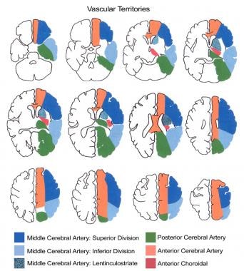
Vertebrobasilar (posterior) circulation constitutes the arterial supply to the brain stem, cerebellum, and occipital cortex. Any interruption in blood flow to these areas may manifest in a myriad of symptoms. These symptoms are determined by which particular branch or branches of the vertebrobasilar circulation have been compromised, extent of any collateral circulation, and degree of occlusion. The brain stem is a focal point of neurologic activity, housing cranial nerves, the reticular activating system, and a series of ascending and descending neurosensory tracts. When this compact area of neurologic activity malfunctions as a result of impaired blood flow, several different but overlapping clinical syndromes can result. Studies show that embolic phenomena cause infarction in vertebrobasilar territory in 9-40% of reported cases. The vertebrobasilar bed appears less susceptible than carotid circulation to embolic occlusion. The vertebral artery is often classified into intracranial and extracranial segments; the atherosclerotic process tends to affect vertebrobasilar circulation at specific intracranial and extracranial sites. The extracranial site is defined as the initial segment of the vertebral artery just proximal to its take-off from the subclavian. The intracranial site is defined as the proximal portion of the basilar artery, just after the joining of the 2 vertebral arteries or just distal to the pontomedullary junction.
Frequency
United States
Approximately one fourth of strokes and TIAs occur in the vertebrobasilar distribution.
- Brainstem infarctions have been reported in autopsy series at a rate of 2 per 1000 cases. One clinical study has suggested that the disease occurs 25% as frequently as occlusions of the carotid artery and its branches.
- Recent developments in neuroimaging provide new perspectives about the disease's prevalence. Some studies using MRI suggest that 40% of patients with vertebrobasilar TIAs have evidence of brainstem infarction.
Mortality/Morbidity
Vertebrobasilar ischemic disease encompasses a vast spectrum of clinical syndromes, extending from subclinical to lethal brainstem infarctions.
- Of patients who suffer infarctions in the vertebrobasilar territory, 50% report TIAs in the days or weeks (rarely months) prior to onset of the permanent deficit. A systematic review suggests that although overall mortality is no different than anterior territory ischemia, the early risk (within 7 d) of recurrent stroke or TIA progression to stroke is higher with VBATD.1
- Basilar artery syndrome, which presents as a locked-in state, is caused by complete occlusion of the intracranial portion of vertebrobasilar circulation. It is a devastating disease with a mortality rate of 75-85%.
Sex
As with atherosclerosis, this disease affects men twice as often as it does women.
Age
Vertebrobasilar ischemic disease occurs in the late decades of life (eg, 70s and 80s).
History
Vertebrobasilar TIAs typically have shorter duration than attacks involving the carotid territory, lasting 8 minutes on average compared with 14 minutes for carotid TIAs. Vertigo is the hallmark symptom of patients experiencing ischemia in the vertebrobasilar distribution. Many patients describe their vertigo as nonviolent or more of a swimming or swaying sensation. Exact incidence of vertigo is unknown, yet as many as one third of patients with VBI may experience vertigo as the sole manifestation of their illness. Visual disturbances (diplopia) as well as auditory phenomena have been described (sudden sensorineural hearing loss).
Other symptoms include the following: - Facial numbness or paresthesias
- Dysphagia, dysarthria, hoarseness
- Syncope (drop attacks)
- Hemisensory extremity symptoms (eg, contralateral to facial component)
- Lateral medullary infarct (Wallenberg syndrome): When VBI progresses to a complete brainstem infarction, a common syndrome is impaired neurologic functioning in the lateral aspect of the medulla, first described by Wallenberg. This is characterized by the following:
- Ipsilateral facial pain and numbness
- Ipsilateral ataxia (falling to side of lesion)
- Vertigo, nausea, vomiting
- Contralateral pain and thermal impairment over body and occasionally face
- Medial medullary infarct: Occlusion of a vertebral artery or branch of the lower basilar artery may produce the following symptoms:
- Contralateral arm and leg weakness (facial sparing)
- Diplopia
- Basilar artery syndrome: Caused by complete basilar artery occlusion, this is characterized by the following:
- Locked-in state (awake quadriplegia)
- Paralysis or weakness of all extremities
- Horizontal gaze paresis, stupor, coma
- Subclavian steal syndrome: This syndrome results from retrograde blood flow down the vertebral artery in response to increased demands from the left upper limb.
- One of the earliest descriptions of VBI was reported in patients who suffered from stenotic lesions of their left subclavian arteries, just proximal to the take-off of the vertebral artery. Half of these patients reported vertigo symptoms consistent with posterior circulation ischemia when exercising their left arms.
- Some series suggest that arm claudication and headache are the most prominent features in patients with symptomatic subclavian steal syndrome.
Physical
Most patients with early stage VBI have only transient episodes of neurologic dysfunction. As a result, most commonly cited physical symptoms may be minimal or nonexistent. Patients with ongoing symptoms, or those who already have incurred an ischemic deficit, demonstrate physical findings that reflect brainstem and cerebellar dysfunction. Crossed signs (eg, contralateral motor and sensory findings) are hallmarks of many types of brainstem strokes.
- Vertebrobasilar insufficiency
- Nystagmus
- Limb ataxia
- Truncal ataxia (falling to side of lesion)
- Contralateral deficit in pain and temperature perception
- Ipsilateral limb and trunk numbness
- Ipsilateral loss of taste
- Visual field defects
- Lateral medullary infarct (Wallenberg syndrome)
- Contralateral impairment of pain and thermal sensation to the extremities
- Nystagmus
- Ipsilateral Horner syndrome (eg, ptosis, miosis, anhydrosis)
- Medial medullary syndrome
- Ipsilateral paralysis and atrophy of the tongue
- Contralateral deficit in proprioception and fine touch (facial sparing)
- Internuclear ophthalmoplegia
- Basilar artery syndrome
- Bifacial and oropharyngeal palsy
- Horizontal gaze paresis
- Decreased level of consciousness
Causes
Atherosclerosis is by far the most common cause of VBI, making VBI most common among patients with cardiovascular risk factors such as age, hypertension, diabetes mellitus, smoking, and dyslipidemias. VBI may result from any disease process that has an impact on the arterial supply to the posterior fossa, including the following:
- Fibromuscular dysplasia
- Rotational occlusion (Bow hunter's stroke) - Mechanical occlusion or stenosis of the vertebral artery at the C1-C2 level caused by lateral flexion
- Vertebral artery dissection
- Vertebrobasilar aneurysms
- Dolichoectasia of basilar artery
Benign Positional Vertigo
Dissection, Vertebral Artery
Labyrinthitis
Multiple Sclerosis
Stroke, Hemorrhagic
Stroke, Ischemic
Transient Ischemic Attack
Vestibular Neuronitis
Other Problems to be Considered
Aneurysms, vertebrobasilar
Basilar artery dissection
Basilar artery migraine
Posterior fossa tumor
Transtentorial herniation
Vasculitis (involving vertebrobasilar circulation)
Lab Studies
Imaging studies are the primary tools used to confirm a vertebrobasilar atherothrombotic disease (VBATD) diagnosis. They also help exclude differential diagnoses that would preclude such therapies as anticoagulants. Ancillary evaluations, however, are important in the search for other conditions whose signs and symptoms overlap VBATD's complex clinical syndrome. These include the following:
- CBC count
- Electrolyte profile
- BUN level
- Serum glucose level
- Erythrocyte sedimentation rate (ESR)
- Urinalysis
- Thyroid function testing
- Venereal disease research labs (VDRL) test
- Coagulation profile
Imaging Studies
- CT scanning helps rule out CNS hemorrhage or mass effect secondary to cerebellar infarction. However, CT is not a good choice for detecting brainstem infarctions because of bony interference.
- MRI is far superior to CT for brainstem and posterior fossa imaging. MRI is more sensitive to small ischemic areas that characterize branch occlusion of the vertebrobasilar circulation. As a result MR is the imaging method of choice for patients with posterior circulation ischemia.
- Magnetic resonance angiography (MRA) may be as good as cerebral angiography for detecting occlusions and stenoses of the vertebrobasilar circulation, but it may not be as good for quantifying degree of stenosis.
- Doppler ultrasonography may complement MRA and provide important hemodynamic data on degree of vertebrobasilar stenosis.
- Transcranial Doppler helps assess and monitor vertebrobasilar patency in patients who have received intraarterial thrombolysis.
- Chest radiography may be indicated to rule out conditions whose signs and symptoms are similar to those of VBATD.
- A therapeutic gap is recognized.
- Diagnostic neuroimaging developments have surpassed therapeutic interventions available for patients with VBATD.
- Surgery is not an established treatment option for VBATD, in contrast to its value for treating carotid territory ischemia.
- Invasive techniques such as intraarterial thrombolysis and angioplasty generally are investigational at this time.
- Specialized neuroimaging (eg, MRI, angiography) has no impact on clinical decision making for the vast majority of patients with VBATD.
- For imaging patients with uncomplicated VBATD, the American Heart Association recommends CT scanning and duplex ultrasonography of the carotid vessels to search for surgically amenable stenoses.2
Other Tests
- ECG may be indicated to rule out conditions whose signs and symptoms are similar to those of VBATD.
Procedures
- Consider a lumbar puncture (LP) when differential diagnosis includes subarachnoid hemorrhage (negative CT scan) or meningoencephalitis.
Emergency Department Care
Vertebrobasilar atherothrombotic disease (VBATD) management in the emergency department (ED) varies on the basis of the patient's symptoms and condition.
- For patients with VBATD who have experienced ischemic infarcts, management falls into 2 major categories: supportive measures and interventions to reestablish patency in the infarct-related artery or to prevent occlusion of a vessel at risk for atherothrombotic or embolic occlusion.
- Airway issues must be addressed in patients with brainstem infarction resulting from VBATD.
- Compromise of ninth and tenth cranial nerves can blunt the gag reflex and inhibit even a conscious or awake patient from handling secretions effectively.
- Secure the airway of patient with an unstable course or severe deficits before starting prolonged diagnostic imaging studies.
- Patients who present to the ED with ischemic stroke are often hypertensive, even in the absence of premorbid blood pressure elevations.
- Given the autoregulatory curve's tendency to shift to the right during hypertension, most authors caution against lowering the blood pressure in the first 24-48 hours after onset of stroke.
- A precipitous drop in blood pressure can have a significant impact on cerebral perfusion pressure.
- Consider antihypertensive medication only in cases of concomitant hypertensive emergency (ongoing end-organ damage), mean arterial pressure (MAP) greater than 130 mm Hg, or systolic blood pressure greater than 220 mm Hg.
- Because most patients with significant neurologic symptoms are denied oral intake until swallowing mechanisms are evaluated, goals of intravenous fluid therapy are to provide isotonic hydration and to avoid hyperglycemia, which appears to exacerbate neuronal injury in stroke.
- Treat vomiting with antiemetics; vomiting may be severe in some brainstem infarctions.
- If a hemorrhagic lesion has been excluded, patients with VBATD are treated with antiplatelet agents or, in certain circumstances, an anticoagulant such as warfarin (see Medication).3 Reperfuse the infarct-related artery by intraarterial thrombolysis or percutaneous transluminal angioplasty (see Consultations).
Consultations
- Neurologist
- Neurosurgeon: Consultation with a neurosurgeon is indicated for surgical evacuation of cerebellar hemorrhages and to manage cerebellar infarction complicated by hydrocephalus.
- Interventional neuroradiologist
- Intraarterial thrombolysis: The high mortality rate associated with basilar artery occlusion and resulting brainstem infarction has prompted research into reperfusion therapy via intraarterial infusion of thrombolytic agents (see Medication). Several case series and small randomized controlled trials have shown promise with regards to recanalization and improved clinical outcomes in basilar artery occlusion and vertebrobasilar stroke.4
- Percutaneous transluminal cerebral angioplasty: Increasingly, investigators have described successful dilation of high-grade vertebral artery stenoses in patients with VBATD who did not respond to medical therapy. Although this approach is not without risk (rate of stroke as high as 40% in some series), other studies have described 80% success rates in restoring flow and eliminating symptoms.
Antiplatelet medications constitute first-line treatment for patients with vertebrobasilar atherothrombotic disease (VBATD). This approach is supported by a large body of clinical research in the secondary prevention of strokes, but its application to posterior circulation events is not well established. Despite this, important inferences can be drawn from the European Stroke Prevention Study, which examined the efficacy of a daily regimen of 225 mg of dipyridamole and 990 mg of aspirin in 2500 patients randomized to receive drug therapy or placebo.5 All participants had a history of TIA or stroke. Fully one third had diseases classified as vertebrobasilar. During a 2-year follow-up study, combined incidences of stroke or death in patients with VBATD were 14% among those receiving treatment and 14% among those receiving placebos. No randomized clinical trials have been conducted to determine antiplatelet therapy's efficacy in treating VBATD. Antiplatelet therapy's widely perceived benefits for cerebrovascular disease may prevent an ethically acceptable trial with a placebo arm. Data from the International Stroke Trial (IST) revealed a small but real clinical benefit of antiplatelet therapy in patients who experienced a completed stroke.6 The IST results suggest that only 1% of patients may benefit from aspirin therapy. Arguments for anticoagulant therapy in VBATD are much more tenuous. A nonrandomized, concurrent, cohort study suggested that anticoagulation provided superior stroke protection for patients with vertebrobasilar TIAs than for patients with carotid TIAs.7 No randomized clinical trials involving patients with vertebrobasilar TIAs have compared anticoagulants to antiplatelet therapy or to placebos. A strong argument favoring use of anticoagulants in VBATD includes settings in which the embolic source of thrombi is known or suspected (eg, atrial fibrillation). Use of low-molecular-weight heparins has shown no significant improvement in outcome over conventional treatments. No clinical evidence supports using intravenous administered thrombolytics to patients with posterior circulation infarcts, let alone VBI. No published subgroup analyses of trials studying thrombolysis in the general stroke population suggest a beneficial role for thrombolytics in treating brainstem or cerebellar infarction. All thrombolytics are plasminogen activators and act either directly (urokinase, alteplase) or indirectly (streptokinase). The scientific literature describes 5 case series totalling 150 patients with acute brainstem infarction who received intra-arterial thrombolysis. Recanalization of the infarcted artery was achieved in only 50-75% of patients in this limited sample; however, patients who experienced reperfusion had a mortality rate of 40-50%, which compares favorably with the 75-85% mortality rate associated with historical controls. The incidence of intracerebral hemorrhage as a complication of treatment was apparently 10%, similar to rates seen in stroke trials using systemic thrombolysis.
Drug Category: Anticoagulants
These agents prevent recurrent or ongoing thromboembolic occlusion of the vertebrobasilar circulation.
| Drug Name | Heparin |
| Description | Augments activity of antithrombin III and prevents conversion of fibrinogen to fibrin. Does not actively lyse yet is able to inhibit further thrombogenesis. Prevents reaccumulation of clots after spontaneous fibrinolysis. |
| Adult Dose | Loading dose: 40-170 U/kg IV Maintenance infusion: 18 U/kg/h IV Alternatively, start with 50 U/kg/h IV, followed by continuous infusion of 15-25 U/kg/h IV; increase dosage by 5 U/kg/h q4h prn using aPTT results |
| Pediatric Dose | Loading dose: 50 U/kg/h IV Maintenance infusion: 15-25 U/kg/h IV Increase dosage by 2-4 U/kg/h IV q6-8h prn using aPTT results |
| Contraindications | Documented hypersensitivity; subacute bacterial endocarditis; active bleeding; history of heparin-induced thrombocytopenia |
| Interactions | Digoxin, nicotine, tetracycline, and antihistamines may decrease effects; conversely, NSAIDs, aspirin, dextran, dipyridamole, and hydroxychloroquine may increase heparin toxicity |
| Pregnancy | C - Fetal risk revealed in studies in animals but not established or not studied in humans; may use if benefits outweigh risk to fetus
|
| Precautions | In neonates, preservative-free heparin recommended to avoid possible toxicity (ie, gasping syndrome) by benzyl alcohol, which is used as preservative; caution in severe hypotension and shock |
| Drug Name | Warfarin (Coumadin) |
| Description | Interferes with hepatic synthesis of vitamin K-dependent coagulation factors. Used for prophylaxis and treatment of venous thrombosis, pulmonary embolism, and thromboembolic disorders. Tailor dose to maintain INR in range of 2-3. |
| Adult Dose | 5-15 mg/d PO qd for 2-5 d; adjust dose according to desired INR |
| Pediatric Dose | 0.05-0.34 mg/kg/d PO; adjust dose according to desired INR (infants may require doses at or near high end of this range) |
| Contraindications | Documented hypersensitivity; severe liver or kidney disease; open wounds; GI ulcers |
| Interactions | Drugs that may decrease anticoagulant effects include griseofulvin, carbamazepine, glutethimide, estrogens, nafcillin, phenytoin, rifampin, barbiturates, cholestyramine, colestipol, vitamin K, spironolactone, oral contraceptives, and sucralfate Medications that may increase anticoagulant effects include oral antibiotics, phenylbutazone, salicylates, sulfonamides, chloral hydrate, clofibrate, diazoxide, anabolic steroids, ketoconazole, ethacrynic acid, miconazole, nalidixic acid, sulfonylureas, allopurinol, chloramphenicol, cimetidine, disulfiram, metronidazole, phenylbutazone, phenytoin, propoxyphene, sulfonamides, gemfibrozil, acetaminophen, and sulindac |
| Pregnancy | D - Fetal risk shown in humans; use only if benefits outweigh risk to fetus
|
| Precautions | Do not switch brands after achieving therapeutic response; caution in active tuberculosis or diabetes mellitus; patients with protein C or S deficiency are at risk of developing skin necrosis |
Drug Category: Antiplatelet agents
These agents inhibit the cyclooxygenase system, decreasing the level of thromboxane A2, which is a potent platelet activator.
| Drug Name | Aspirin (Anacin, Ascriptin, Bayer aspirin) |
| Description | Inhibits prostaglandin synthesis, which prevents formation of platelet-aggregating thromboxane A2. Studies report 300 mg/d dose as effective as larger dose and may be associated with fewer adverse effects. |
| Adult Dose | 650 mg PO divided bid/qid; not to exceed 1.3 g/d |
| Pediatric Dose | Not established |
| Contraindications | Documented hypersensitivity; liver damage; hypoprothrombinemia; vitamin K deficiency; bleeding disorders; asthma Because of association with Reye syndrome, do not use in children (<16 y) with flu |
| Interactions | Antacids and urinary alkalinizers may decrease effects; corticosteroids decrease serum levels; anticoagulants may cause additive hypoprothrombinemic effects and increase bleeding time; may antagonize uricosuric effects of probenecid and increase toxicity of phenytoin and valproic acid; doses >2 g/d may potentiate glucose-lowering effect of sulfonylurea drugs |
| Pregnancy | D - Fetal risk shown in humans; use only if benefits outweigh risk to fetus
|
| Precautions | May cause transient decrease in renal function and aggravate chronic kidney disease; avoid use in patients with severe anemia, with history of blood coagulation defects, or taking anticoagulants |
| Drug Name | Ticlopidine (Ticlid) |
| Description | Second-line antiplatelet therapy for patients who cannot tolerate aspirin or in whom aspirin is ineffective. |
| Adult Dose | 250 mg PO bid |
| Pediatric Dose | Not established |
| Contraindications | Documented hypersensitivity; neutropenia or thrombocytopenia; liver damage; active bleeding disorders |
| Interactions | Corticosteroids and antacids may decrease effects; theophylline, cimetidine, aspirin, and NSAIDs increase toxicity |
| Pregnancy | B - Fetal risk not confirmed in studies in humans but has been shown in some studies in animals
|
| Precautions | Discontinue if absolute neutrophil count decreases to <1.2 x 10⁹/L or if platelet count falls to <80 x 109/L |
| Drug Name | Clopidogrel (Plavix) |
| Description | Inhibits platelet aggregation by inhibiting binding of ADP to platelet receptor and subsequent ADP-mediated activation of glycoprotein GPIIb/IIIa complex. Rapidly absorbed from GI tract. Used as second-line therapy for patients with TIA crescendo symptoms who are already taking aspirin. |
| Adult Dose | 75 mg PO qd with or without food |
| Pediatric Dose | Not recommended |
| Contraindications | Documented hypersensitivity; lactation; active bleeding from peptic ulcer; intracranial hemorrhage |
| Interactions | Evening primrose oil, garlic, ginger, ginkgo biloba, grapeseed extract increase effects; warfarin increases risk of bleeding |
| Pregnancy | B - Fetal risk not confirmed in studies in humans but has been shown in some studies in animals
|
| Precautions | Caution in patients at increased risk of bleeding from trauma, surgery, or other pathologic conditions; caution in patients with lesions with propensity to bleed (eg, ulcers) |
Drug Category: Thrombolytics
These agents restore perfusion in the infarct-related artery.
| Drug Name | Alteplase (Activase) |
| Description | Tissue plasminogen activator exerts effect on fibrinolytic system to convert plasminogen to plasmin. Plasmin degrades fibrin, fibrinogen, and procoagulant factors V and VIII. Serum half-life is 4-6 min but half-life lengthened when bound to fibrin in clot. Used in management of acute myocardial infarction (MI), acute ischemic stroke, and pulmonary embolism (PE). Heparin and aspirin are not given for 24 h after tPA. Must be given within 3 h of stroke onset. Exclude hemorrhage by CT scan. If hypertensive, lower BP with labetalol, 10 mg IV. Safety and efficacy of concomitant administration with aspirin and heparin during first 24 h after onset of symptoms have not been investigated. |
| Adult Dose | 0.9 mg/kg IV over 60 min; not to exceed 90 mg; 10% of total dose administered as initial IV bolus over 1 min; administer only within 3 h of onset of stroke symptoms |
| Pediatric Dose | Not established |
| Contraindications | Documented hypersensitivity; active internal bleeding, cerebrovascular accident or stroke within last 2 mo, intracranial or intraspinal surgery or trauma, intracranial hemorrhage on pretreatment evaluation, suspicion of subarachnoid hemorrhage, intracranial neoplasm, arteriovenous malformation or aneurysm, bleeding diathesis, or severe uncontrolled hypertension |
| Interactions | Anticoagulants and antiplatelets may increase risk of bleeding; may give heparin with and after alteplase infusions to reduce risk of rethrombosis; either heparin or alteplase may cause bleeding complications |
| Pregnancy | C - Fetal risk revealed in studies in animals but not established or not studied in humans; may use if benefits outweigh risk to fetus
|
| Precautions | Monitor for bleeding, especially at arterial puncture sites, with coadministration of vitamin K antagonists; control and monitor blood pressure frequently during and following alteplase administration (when managing acute ischemic stroke); do not use >0.9 mg/kg to manage acute ischemic stroke; doses >0.9 mg/kg may cause ICH |
Complications
- Basilar artery occlusion is a rare but devastating complication of vertebrobasilar atherothrombotic disease (VBATD).
- It is associated with a 75-85% mortality rate and high rate of neurovegetative states in survivors.
Prognosis
- Vertebrobasilar TIAs (ie, VBI) generally may have a more favorable prognosis than carotid territory TIAs because the risk of developing a completed stroke is less. Collateral circulation may account for improved outcome in these patients.
- Lateral medullary infarction (Wallenberg syndrome) is characterized by persistent symptoms that last for years.
Medical/Legal Pitfalls
- Failure to recognize the etiology of vertigo in the elderly
- Deciphering the cause of vertigo in older patients can be challenging.
- Patients with VBI are generally believed to manifest neighboring signs of brainstem dysfunction that would alert clinicians to the central etiology of patient's vertigo (as opposed to peripheral). However, some studies suggest that as many as one third of patients with VBI manifest vertigo as the only symptom of their condition
- Failure to avoid excluding VBATD on the basis of a negative CT scan: CT scanning fails to detect patients with vertebrobasilar TIAs and may not reveal small brainstem infarctions.
- Failure to initiate therapy after hemorrhage has been reliably excluded
- Failure to initiate antiplatelet therapy when diagnosis of brainstem infarction is initially suspected: This may increase the likelihood of permanent ischemic deficits; in a small but definite subsegment of patients, VBI eventually progresses to brainstem infarction.
- Failure to take precautions when feeding patient with brainstem infarction
- Aspiration pneumonitis is the leading cause of death in patients with vertebrobasilar strokes.
- Even alert patients with good extremity strength may have compromised swallowing mechanisms.
- Failure to identify candidates who might be eligible for thrombolytic therapy either intravenously or via an intra-arterial mode of administration
- Flossmann E, Rothwell PM. Prognosis of vertebrobasilar transient ischaemic attack and minor stroke. Brain. Sep 2003;126(Pt 9):1940-54. [Medline].
- Feinberg WM, Albers GW, Barnett HJ. Guidelines for the management of transient ischemic attacks. From the Ad Hoc Committee on Guidelines for the Management of Transient Ischemic Attacks of the Stroke Council of the American Heart Association. Circulation. Jun 1994;89(6):2950-65. [Medline].
- Coull BM, Williams LB, Goldstein LS. Anticoagulants and antiplatelet agents in acute ischemic stroke. Report of the Joint Stroke Guideline Development Committee of the American Academy of Neurology and the American Stroke Association (a Division of the American Heart Association). Neurology. 2002;59 (1):13-22. [Medline].
- Brandt T, von Kummer R, Muller-Kuppers M. Thrombolytic therapy of acute basilar artery occlusion. Variables affecting recanalization and outcome. Stroke. May 1996;27(5):875-81. [Medline].
- Sivenius J, Riekkinen PJ, Smets P. The European Stroke Prevention Study (ESPS): results by arterial distribution. Ann Neurol. Jun 1991;29(6):596-600. [Medline].
- International Stroke Trial Collaborative Group. The International Stroke Trial (IST): a randomised trial of aspirin, subcutaneous heparin, both, or neither among 19435 patients with acute ischaemic stroke. Lancet. May 31 1997;349(9065):1569-81. [Medline].
- Whisnant JP, Cartlidge NE, Elveback LR. Carotid and vertebral-basilar transient ischemic attacks: effect of anticoagulants, hypertension, and cardiac disorders on survival and stroke occurrence--a population study. Ann Neurol. Feb 1978;3(2):107-15. [Medline].
- Becker KJ, Purcell LL, Hacke W. Vertebrobasilar thrombosis: diagnosis, management, and the use of intra- arterial thrombolytics. Crit Care Med. Oct 1996;24(10):1729-42. [Medline].
- Caplan LR. Vertebrobasilar occlusive disease. In: Stroke: Pathophysiology, Diagnosis and Management. Vol 1. 1992:549-619.
- Culebras A, Kase CS, Masdeu JC. Practice guidelines for the use of imaging in transient ischemic attacks and acute stroke. A report of the Stroke Council, American Heart Association. Stroke. Jul 1997;28(7):1480-97. [Medline].
- Delaney KA. Bedside diagnosis of vertigo: value of the history and neurological examination. Acad Emerg Med. Dec 2003;10(12):1388-95. [Medline].
- Froehling DA, Silverstein MD, Mohr DN. The rational clinical examination. Does this dizzy patient have a serious form of vertigo?. JAMA. Feb 2 1994;271(5):385-8. [Medline].
- Libman RB, Kwiatkowski TG, Hansen MD. Differences between anterior and posterior circulation stroke in TOAST. Cerebrovasc Dis. 2001;11(4):311-6. [Medline].
- Lipinski CA, Swanson ER. Vertebrobasilar distribution stroke mimicking transtentorial herniation. Ann Emerg Med. May 1998;31(5):640-2. [Medline].
- Macleod MR, Davis SM, Mitchell PJ. Results of a multicentre, randomised controlled trial of intra-arterial urokinase in the treatment of acute posterior circulation ischaemic stroke. Cerebrovasc Dis. 2005;20(1):12-7. [Medline].
- Ois A, Gomis M, Rodriguez-Campello A, et al. Factors associated with a high risk of recurrence in patients with transient ischemic attack or minor stroke. Stroke. Jun 2008;39(6):1717-21. [Medline]. [Full Text].
- Phan TG, Wijdicks EF. Intra-arterial thrombolysis for vertebrobasilar circulation ischemia. Crit Care Clin. Oct 1999;15(4):719-42, vi. [Medline].
- Piechowski-Jozwiak B, Bogousslavsky J. Basilar occlusive disease: the descent of the feared foe?. Arch Neurol. Apr 2004;61(4):471-2. [Medline].
- Sauvaget E, Kici S, Petelle B. Vertebrobasilar occlusive disorders presenting as sudden sensorineural hearing loss. Laryngoscope. Feb 2004;114(2):327-32. [Medline].
- Terada T, Higashida RT, Halbach VV. Transluminal angioplasty for arteriosclerotic disease of the distal vertebral and basilar arteries. J Neurol Neurosurg Psychiatry. Apr 1996;60(4):377-81. [Medline].
- Voetsch B, DeWitt LD, Pessin MS. Basilar artery occlusive disease in the New England Medical Center Posterior Circulation Registry. Arch Neurol. Apr 2004;61(4):496-504. [Medline].
Vertebrobasilar Atherothrombotic Disease excerpt Article Last Updated: Jun 3, 2008
|