Continually Updated Clinical Reference
 
 
  All Sources     eMedicine     Medscape     Drug Reference     MEDLINE
 
eMedicine - Ultraviolet Keratitis : Article by

Quick Find
Authors & Editors
Introduction
Clinical
Differentials
Workup
Treatment
Medication
Follow-up
Miscellaneous
Acknowledgments
Multimedia
References

Related Articles
Conjunctivitis

Corneal Ulceration and Ulcerative Keratitis

Iritis and Uveitis


Patient Education
Eye and Vision Center

Corneal Flash Burns Overview

Corneal Flash Burns Causes

Corneal Flash Burns Symptoms

Corneal Flash Burns Treatment


Author: Reed Brozen, MD, Director of Air Transport, Associate Professor, Department of Emergency Medicine, Dartmouth Medical School, Dartmouth-Hitchcock Medical Center

Reed Brozen is a member of the following medical societies: American Academy of Emergency Medicine, American College of Emergency Physicians, New Hampshire Medical Society, and Society for Academic Emergency Medicine

Coauthor(s): Christian Fromm, MD, Director of Research, Department of Emergency Medicine, Maimonides Medical Center, Assistant Professor of Emergency Medicine, Mt Sinai School of Medicine

Editors: Eric Kardon, MD, FACEP, Associate Staff, Division of Emergency Medicine, Athens Regional Medical Center; Francisco Talavera, PharmD, PhD, Senior Pharmacy Editor, eMedicine; James S Walker, DO, Program Coordinator, Associate Professor, Department of Emergency Medicine, University of Oklahoma Health Sciences Center; John D Halamka, MD, MS, Associate Professor of Medicine, Harvard Medical School, Beth Israel Deaconess Medical Center; Chief Information Officer, CareGroup Healthcare System and Harvard Medical School; Attending Physician, Division of Emergency Medicine, Beth Israel Deaconess Medical Center; Rick Kulkarni, MD, Medical Director, Assistant Professor of Surgery, Section of Emergency Medicine, Yale-New Haven Hospital

Author and Editor Disclosure

Synonyms and related keywords: actinic keratitis, snow blindness, flash burn, welder's flash, arc eye, ultraviolet keratitis, UV keratitis, UV radiation injury, corneal damage, corneal abrasion, corneal ulceration, corneal laceration, superficial punctate keratitis, SPK, UV keratoconjunctivitis, ultraviolet keratoconjunctivitis 

Background

UV light is the most common cause of radiation injury to the eye. The cornea absorbs most UV radiation. UV radiation damage to the corneal epithelium is cumulative, similar to the effects with dermal epithelium (sunburn). Ozone in the atmosphere effectively filters most of the harmful UV radiation of wavelengths shorter than 290 nm; natural UV sources, such as the sun, rarely cause injury after short exposures. However, unprotected exposures to the sun or solar eclipses or exposure to the sun on highly reflective snow fields at high elevation can lead to direct corneal epithelial injury. The latter clinical scenario is known as snow blindness.

Artificial sources of UV radiation also cause corneal damage. Injury from a welder's arc commonly is known as flash burn, welder's flash, or arc eye. Other sources of UV radiation injury include suntanning beds, carbon arcs, photographic flood lamps, lightning, electric sparks, and halogen desk lamps.

Prolonged exposures to UV radiation can lead to chronic solar toxicity, which is associated with several ocular surface disorders, eg, pinguecula, pterygium, climatic droplet keratopathy, and even squamous metaplasia and carcinoma. The only ocular cancer associated with UV radiation is epidermoid carcinoma of the bulbar conjunctiva, which occurs with increased frequency in the tropics and subtropics and which has been experimentally replicated in animal models using UV radiation. Rarely, retinal absorption of visible to near-infrared (400-1400 nm) radiation from welding arcs can lead to permanent, sight-threatening injury.

Pathophysiology

UV rays irritate the superficial corneal epithelium, causing inhibition of mitosis, production of nuclear fragmentation, and loosening of the epithelial layer. Under experimental conditions in animals, phototoxic effects have been demonstrated at all levels of the cornea, including the stroma and endothelium.

An inflammatory response occurs, which includes edema and congestion of the conjunctiva and a stippling of the corneal epithelium known as superficial punctate keratitis (SPK). SPK is a nonspecific corneal condition associated with multiple ocular disorders. It is characterized by small pinpoint defects in the superficial corneal epithelium, which stain with fluorescein. If SPK is severe, it may be followed by total epithelial desquamation, with conjunctival chemosis, lacrimation, and blepharospasm. Reepithelialization usually occurs within 36-72 hours, and long-term sequelae are rare. This SPK contrasts with the more severe effects frequently encountered with corneal damage caused by alkaline or strongly acidic chemicals.

In general, ocular pain and decreased visual acuity occurs 6-12 hours after the injury. This lag time involves an unexplained pattern of corneal sensory loss and return and is thought to indicate a probable photochemical injury rather than a thermal injury to the cornea.

Frequency

United States

UV keratitis and UV keratoconjunctivitis are the only radiant exposure conditions of the cornea that occur with any significant frequency in the United States.

Mortality/Morbidity

No reported mortality exists.

  • Morbidity results from UV radiation damage to the superficial corneal epithelium, which usually heals spontaneously within 48 hours of the exposure.
  • Long-term sequelae, which may result from superinfection, are rare.

Sex

No difference in incidence exists between males and females.


History

  • Document information regarding the nature and duration of the exposure.
  • Patients experience the onset of a foreign-body sensation, irritation, pain, photophobia, tearing, blepharospasm, and decreased visual acuity 6-12 hours after the exposure.
  • Obtain the pertinent history, including contact lens use, past ocular trauma or surgery, current medications, and allergies to medications.

Physical

Prior to examination or treatment, assess visual acuity, with corrective lenses if relevant. Perform a full examination of the eyes, including inspection of all external structures together with funduscopic and slit lamp examinations.

  • Examination of the lids and conjunctiva may reveal varying lid edema and conjunctival hyperemia.
  • A diffuse corneal haze may be seen in severe cases.
  • Perform a standard slit lamp examination with prior application of proparacaine.
    • Fluorescein staining reveals superficial punctate epithelial surface irregularities, which usually cover the entire surface of the cornea. This condition usually is referred to as SPK.
    • If the patient's eyelid was partially closed during the exposure, a well-demarcated line separates normal from damaged corneal epithelium.
    • Involvement of the lens is rare and occurs only after intense exposure.

Causes

Radiation injury to the eye may be caused by unprotected or long exposures to the sun, particularly at high altitude; exposure to UV radiation reflected off snow, ice, or water; and viewing of solar eclipses. In addition to the sun, sources of UV radiation include the following:

  • Welder's arcs
  • Carbon arcs
  • Suntanning beds
  • Photographic flood lamps
  • Lightning
  • Electric sparks
  • Halogen desk lamps


Conjunctivitis
Corneal Ulceration and Ulcerative Keratitis
Iritis and Uveitis

Other Problems to be Considered

Dry eye syndrome - Poor tear lake or a decreased tear break-up time
Blepharitis - Erythema, telangiectasias, and crusting of the eyelid margins
Trauma - Can occur from relatively mild trauma, such as chronic eye rubbing
Exposure keratopathy - Poor eyelid closure with failure to cover the entire globe
Topical drug toxicity - Neomycin, tobramycin, or drops with preservatives, including artificial tears
Contact lens–related disorder - Chemical toxicity, tight-lens syndrome, contact lens overwearing syndrome, or giant papillary conjunctivitis
Thygeson SPK - Bilateral, recurrent SPK without conjunctival injection
Foreign body under the upper eyelid - Typically linear SPK with fine scratches arranged vertically
Trichiasis and/or distichiasis - Eyelashes rubbing on the cornea
Entropion or ectropion - superior or inferior SPK
Floppy lid syndrome - Extremely loose lids that pull away from the eye very easily
Associated retinopathy - Sun-gazer retinopathy associated with UV keratitis, seen particularly in psychiatric patients who stare directly into the sun


Procedures

  • Perform standard slit lamp examination with fluorescein.


Prehospital Care

  • Flush eyes for several minutes with water or saline solution.

Emergency Department Care

  • Administer a short-acting cycloplegic drop (eg, cyclopentolate 1%) to relieve the pain of reflex ciliary spasm.
  • Administer a topical antibiotic ointment or drops, such as erythromycin or gentamicin. If an eye patch is used with an antibiotic ointment, the patient should use antibiotic drops after the eye patch has been removed.
  • Application of a semipressure dressing with the eye well closed underneath was once the standard of care, but today this method of treatment is controversial. While these dressings may offer good pain relief, they may also delay reepithelialization. Moreover, some patients find the loss of sight and depth perception (in the case of single-eye patching) more traumatic than the pain itself.
    • When used, the dressing is to be left on for 24 hours.
    • The dressing should not be replaced by the patient without professional assistance because excessive pressure can cause further damage to the eye.
  • Because of the severely painful nature of this type of injury, patients should be treated with nonsteroidal anti-inflammatory drugs and oral narcotic analgesics.
    • All patients without contraindications should take ibuprofen 600 mg qid and oxycodone with acetaminophen for breakthrough pain.
    • Topical anesthetic may be administered only in the ED; frequent use retards epithelial healing and may lead to corneal ulcer formation. The relief provided by topical anesthetics is so immediate and dramatic that patients are highly motivated to take a bottle home with them. Be careful not to allow the patient to take the bottle surreptitiously.
  • Topical NSAIDs are not approved for this indication but are used for many other painful and allergic eye conditions. Topical NSAIDs for UV keratitis and corneal abrasions is an off-label use but has become common practice in many places.

Consultations

Ophthalmologic consultation usually is not necessary for this condition but may be obtained at the discretion of the emergency physician in the ED or if substantial healing has not occurred within 24-48 hours.


The goal of therapy is to treat the pain associated with damage in the corneal epithelium resulting from UV light exposure and to prevent infection while the cornea heals. Some medications include ophthalmic antibiotics, topical cycloplegics, ophthalmic anesthetics, NSAIDs (both ophthalmic and parenteral), and other analgesics.

Drug Category: Ophthalmic anesthetics

These agents are indicated for pain relief. Local anesthetics stabilize the neuronal membrane and prevent the initiation and transmission of nerve impulses, thereby producing the local anesthetic action.

Drug NameProparacaine 0.5% (Alcaine, Ophthetic)
DescriptionHas rapid onset of anesthesia that begins 13-30 sec after instillation. However, has short duration of action of about 15-20 min. Since prolonged eye anesthesia can eliminate patient's awareness of mechanical damage to the cornea, drug should not be used outside the ED. Frequent use of anesthetics may retard healing. Least irritating of all topical anesthetics. Prevents initiation and transmission of impulse at nerve cell membrane by stabilizing and decreasing ion permeability.
Adult Dose2-3 gtt in affected eye q15-20min during ED examination
Pediatric DoseAdminister as in adults
ContraindicationsDocumented hypersensitivity; prolonged use
InteractionsIncreases effects of phenylephrine and tropicamide
PregnancyC - Fetal risk revealed in studies in animals but not established or not studied in humans; may use if benefits outweigh risk to fetus
PrecautionsCaution in cardiac disease or hyperthyroidism and with abnormal or reduced levels of plasma esterases

Drug Category: Cycloplegics

These agents relax ciliary muscle spasm that can cause a deep aching pain and photophobia. Cycloplegics are used to facilitate eye examination and provide relief of symptoms in patients with moderate-to-severe eye injury. Cycloplegic agents also are mydriatics, thus before using them it is important to ensure that the patient does not have glaucoma. This medication could provoke an acute angle-closure glaucoma attack in a susceptible patient.

Drug NameCyclopentolate 0.5-1% (Cyclogyl)
DescriptionPrevents muscle of ciliary body, and sphincter muscle of iris from responding to cholinergic stimulation. Induces mydriasis in 30-60 min and cycloplegia in 25-75 min.
Adult Dose1 gtt of 1% solution to induce cycloplegia; repeat in 5-10 min prn
Pediatric Dose<1 year: 1 gtt of 0.5% into each eye 5-10 min before examination
>1 year: 1 gtt of 0.5%, 1%, or 2% solution; repeat in 5-10 min prn
ContraindicationsDocumented hypersensitivity; narrow-angle glaucoma
InteractionsDecreases effects of carbachol and cholinesterase inhibitors
PregnancyC - Fetal risk revealed in studies in animals but not established or not studied in humans; may use if benefits outweigh risk to fetus
PrecautionsCaution in patients (eg, elderly patients) in whom increased intraocular pressure may be present; can cause toxic anticholinergic systemic adverse effects, but incidence is rare when used sparingly (more common in children, especially infants); compressing the lacrimal sac with digital pressure for 1-3 min following application may minimize systemic absorption

Drug Category: Antibiotics, ophthalmic

The routine use of topical antibiotics remains controversial. Many emergency physicians have stopped its use for minor injuries, although others continue routine treatment with a broad-spectrum antibiotic ointment for lubrication and infection prophylaxis. This treatment persists despite its unproven efficacy, discomfort, and evidence that ointments may retard corneal epithelial healing.

Drug NameErythromycin 0.5% ointment (Ilotycin, AK-Mycin)
DescriptionIndicated for treatment of infections caused by susceptible strains of microorganisms and for prevention of corneal and conjunctival infections.
Adult DoseApply 0.5-inch (1.25-cm) ribbon 2-8 times/d, depending on severity of the infection
Pediatric DoseAdminister as in adults
ContraindicationsDocumented hypersensitivity; viral, mycobacterial, and fungal infections of the eye; patients using steroid combinations after the uncomplicated removal of a foreign body from the cornea should avoid using this product
InteractionsNone reported
PregnancyB - Fetal risk not confirmed in studies in humans but has been shown in some studies in animals
PrecautionsNot for use to treat ocular infections that are likely to become systemic; prolonged or repeated therapy may result in bacterial or fungal overgrowth of nonsusceptible organisms and may lead to a secondary infection; take appropriate measures if superinfection occurs

Drug NameGentamicin (Genoptic, Garamycin)
DescriptionAminoglycoside antibiotic used for gram-negative bacterial coverage.
Adult DoseOintment: Apply 0.5-inch (1.25-cm) ribbon bid/tid to the affected eye q3-4h
Solution: 1-2 gtt in affected eye q2-4h, as often as q1h for severe infections
Pediatric DoseAdminister as in adults
ContraindicationsDocumented hypersensitivity; mycobacterial, viral, and fungal infections of the eye; patients taking steroid combinations after uncomplicated removal of a foreign body from cornea should also avoid using this product
InteractionsNone reported
PregnancyC - Fetal risk revealed in studies in animals but not established or not studied in humans; may use if benefits outweigh risk to fetus
PrecautionsDo not use to treat ocular infections that may become systemic; prolonged or repeated antibiotic therapy may result in bacterial or fungal overgrowth of nonsusceptible organisms that may lead to a secondary infection; take appropriate measures if superinfection occurs

Drug Category: Analgesics

Although most NSAIDs are used primarily for their anti-inflammatory effects, they are effective analgesics and are useful for the relief of mild to moderate pain. Pain control is essential to quality patient care. Although oral narcotics may be sedating they should be prescribed and are nearly always needed to gain adequate pain control. Prescribing adequate pain medications on the first visit is essential to prevent a revisit solely for pain control. Only a small quantity is needed since this is a self-limited problem with short duration.

Drug NameIbuprofen (Motrin, Advil, Nuprin, Rufen)
DescriptionUsually the DOC for mild to moderate pain, if no contraindications exist; inhibits inflammatory reactions and pain, probably by decreasing cyclooxygenase activity, which results in the inhibition of prostaglandin synthesis
Adult Dose200-800 mg PO q4-6h, while symptoms persist; not to exceed 3.2 g/d
Pediatric Dose10-70 mg/kg/d PO divided tid/qid
ContraindicationsDocumented hypersensitivity; because of potential cross-sensitivity to other NSAIDs, do not give these agents to patients in whom aspirin, iodides, or other NSAIDs induce hypersensitivity; peptic ulcer disease; recent GI bleeding or perforation; renal insufficiency; high risk of bleeding
InteractionsProbenecid may increase the concentrations and possibly the toxicity; may decrease the effect of loop diuretics when administered concurrently; PT may increase when administered concurrently with anticoagulants (monitor PT closely, and instruct patients to watch for signs and symptoms of bleeding); may increase serum lithium levels and risk of methotrexate toxicity
PregnancyB - Fetal risk not confirmed in studies in humans but has been shown in some studies in animals
D - Fetal risk shown in humans; use only if benefits outweigh risk to fetus
PrecautionsCaution in congestive heart failure, hypertension, and decreased renal and hepatic function

Drug NameOxycodone and acetaminophen (Percocet, Tylox, Roxicet)
DescriptionDrug combination indicated for the relief of moderate to severe pain.
Adult Dose1-2 tab or cap PO q4-6h prn
Pediatric Dose0.05-0.15 mg/kg/dose PO q4-6h prn; not to exceed 5 mg/dose of oxycodone
ContraindicationsDocumented hypersensitivity
InteractionsPhenothiazines may decrease analgesic effects; toxicity increases when administered concurrently with CNS depressants or tricyclic antidepressants
PregnancyC - Fetal risk revealed in studies in animals but not established or not studied in humans; may use if benefits outweigh risk to fetus
PrecautionsDuration of action may increase in elderly patients; be aware of the total daily dose of acetaminophen; maximum dose of acetaminophen is 4,000 mg/d, higher doses may cause liver toxicity

Drug Category: Ophthalmic analgesic

Some ophthalmologists are advocating that diclofenac (Voltaren) or ketorolac (Acular) drops be used despite lack of official indications. These topical agents have been shown to relieve pain in multiple situations including corneal abrasions, allergies, and postsurgical pain.

Drug NameKetorolac tromethamine 0.5% (Acular)
DescriptionInhibits prostaglandin synthesis by decreasing activity of the enzyme, cyclooxygenase, which results in decreased formation of prostaglandin precursors, which, in turn, results in reduced inflammation.
Adult Dose1 gtt into each affected eye qid, continue for a maximum of 2 wk
Pediatric Dose<12 years: Not established
>12 years: Administer as in adults
ContraindicationsDocumented hypersensitivity
InteractionsNone reported
PregnancyC - Fetal risk revealed in studies in animals but not established or not studied in humans; may use if benefits outweigh risk to fetus
PrecautionsPerform ophthalmologic studies in patients who develop eye complaints during therapy; discontinue therapy if changes are noted; changes may include blurred or diminished vision, corneal deposits and retinal disturbances, scotomata, changes in color vision, and macula degeneration

Drug NameDiclofenac (Voltaren)
DescriptionInhibits prostaglandin synthesis by decreasing the activity of the enzyme cyclooxygenase, which results in decreased formation of prostaglandin precursors.
Adult Dose1 gtt into affected eye qid, continue for a maximum of 2 wk
Pediatric Dose<12 years: Not established
>12 years: Administer as in adults
ContraindicationsDocumented hypersensitivity
InteractionsNone reported
PregnancyC - Fetal risk revealed in studies in animals but not established or not studied in humans; may use if benefits outweigh risk to fetus
PrecautionsCorneal thinning may occur


Further Outpatient Care

  • Follow-up care with an ophthalmologist usually is not necessary except in cases of extensive corneal damage or in patients with preexisting serious eye conditions.

Complications

  • Superinfection, rarely
  • Vision loss, rarely

Prognosis

  • Excellent for full recovery in 24-76 hours

Patient Education

  • Educate patients about proper eye precautions, such as the use of UV-filtering lenses or limiting exposure to the sun.
  • For excellent patient education resources, visit eMedicine's Eye and Vision Center. Also, see eMedicine's patient education article Corneal Flash Burns.


Medical/Legal Pitfalls

  • Do not discharge the patient with UV keratitis from the ED with anesthetized eye drops for pain control. The anesthetic eye is vulnerable to subsequent ulcer formation and secondary traumatic injuries, eg, unnoticed foreign bodies or vigorous eye rubbing. Repeated use of anesthetic drops may retard epithelial healing.
  • Document whether contact lenses were worn at the time of injury.
  • Document whether protective eyewear was worn at the time of injury. Many workmen's compensation forms require this documentation, and the information often is important for workplace safety education programs.
  • Always look for and document that you assessed the presence of foreign bodies in the eye. The diagnosis of UV keratitis does not preclude a comorbid ocular condition such as the presence of a foreign body.
  • Cases of UV keratitis that involve extensive corneal injury merit follow-up care with an ophthalmologist in 24-48 hours. Follow-up care also is recommended for patients who have persistent pain or vision deficits 48 hours after injury.
  • Never treat UV keratitis with steroids; the outcome may be disastrous if a concomitant herpetic ocular lesion is present.


The authors and editors of eMedicine gratefully acknowledge the contributions of previous editor, Craig Feied, MD, to the development and writing of this article.


Media file 1:  Ultraviolet keratitis. 
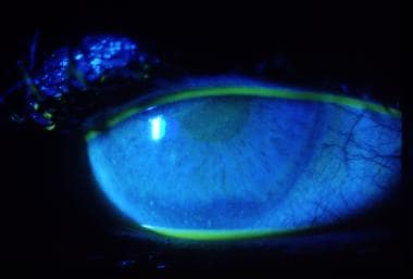
Diffuse uptake of fluorescein stain as seen in ultraviolet keratitis.
Click to see larger pictureClick to see detailView Full Size Image
Media type:  Photo


  • Belin MW, Catalano RA, Scott JL. Burns of the eye. In: Catalano RA, ed. Ocular Emergencies. Philadelphia, Pa: WB Saunders Co; 1992:192-4.
  • Brown MD, Cordell WH, Gee AS. Do ophthalmic nonsteroidal anti-inflammatory drugs reduce the pain associated with simple corneal abrasion without delaying healing?. Ann Emerg Med. Oct 1999;34(4 Pt 1):526-34. [Medline].
  • Brunette DD, Ghezzi K, Renner GS. Ophthalmologic disorders. In: Rosen P, Barkin R, eds. Emergency Medicine: Concepts and Clinical Practice. 4th ed. St. Louis, Mo: Mosby Year Book; 1998:2704.
  • Crumpton KL, Shockley LW. Ocular trauma: a quick illustrated guide to treatment, triage, and medicolegal implications. Emerg Med Rep. 1997;18:223-34.
  • Daxecker F, Blumthaler M, Ambach W. Ultraviolet exposure of cornea from sunbeds. Lancet. Sep 24 1994;344(8926):886. [Medline].
  • Schein OD. Phototoxicity and the cornea. J Natl Med Assoc. Jul 1992;84(7):579-83. [Medline].
  • Spencer WH. The Cornea. Ophthalmic Pathology. 4th ed. Philadelphia, Pa: WB Saunders Co; 1996:233-35.
  • Weaver CS, Terrell KM. Evidence-based emergency medicine. Update: do ophthalmic nonsteroidal anti-inflammatory drugs reduce the pain associated with simple corneal abrasion without delaying healing?. Ann Emerg Med. Jan 2003;41(1):134-40. [Medline].
  • Wittenberg S. Solar radiation and the eye: a review of knowledge relevant to eye care. Am J Optom Physiol Opt. Aug 1986;63(8):676-89. [Medline].
  • Yen YL, Lin HL, Lin HJ, et al. Photokeratoconjunctivitis caused by different light sources. Am J Emerg Med. Nov 2004;22(7):511-5. [Medline].

Ultraviolet Keratitis excerpt

Article Last Updated: Feb 4, 2008