Practice Essentials
Peripheral vascular injuries may result from penetrating trauma or blunt trauma to the extremities. If not recognized and treated rapidly, injuries to major arteries, veins, and nerves may have disastrous consequences, resulting in the loss of life and limb. Deciding whether the injury requires surgical intervention is a major priority of initial management. The pitfall to avoid in the emergency department is a missed diagnosis. In the patient without 'hard' signs (see explanation of hard and soft signs below) of a peripheral vascular injury, a careful history of bleeding, physical examination that includes an ankle-brachial index (ABI) or brachial-brachial index (BBI), and CT arteriography (depending on an ABI < 0.9) should determine the diagnosis of arterial injury. [1, 2, 3, 4, 5]
Hard and soft signs
The presence of "hard" signs of vascular injury has a 92-95% sensitivity for injuries requiring intervention. The vast majority of patients exhibiting the following "hard" signs require intervention, with a positive predictive value greater than 95% [6] :
-
Bruit or thrill: These are present in only 45% of patients with an arteriovenous fistula
-
Active or pulsatile hemorrhage
-
Pulsatile or expanding hematoma
-
Signs of limb ischemia and elevated compartment pressure including the 5 "P's: pallor, paresthesias, pulse deficit, paralysis, and pain on passive extension of the compartment. Pain on passive extension is the earliest and most sensitive physical finding.
-
Diminished or absent pulses: This is not a sensitive prognostic finding, as up to 25% of patients with major vascular injuries requiring repair have normal pulses distal to the injury.
The following "soft" signs are much less useful in predicting or excluding major vascular injuries that require intervention. The positive predictive value of "soft" signs indicating abnormal findings on an arteriogram is only 35%. The vast majority of these lesions do not require emergent repair.
-
Hypotension or shock
-
Neurologic deficit due to primary nerve injury occurs immediately after injury. In contrast, ischemic neuropathy is delayed in onset, developing within minutes to hours after injury.
-
Stable, nonpulsatile or small hematoma
-
Proximity of the wound to major vascular structures
Location of injury
In the upper extremity, the areas of greatest concern include the axilla and the area from the deltopectoral groove distally across the elbow to the proximal forearm. The axilla, medial and anterior upper arm, and antecubital fossa particularly are considered high-risk areas because of the superficial location of the axillary and brachial arteries in these regions. [7, 8, 9]
Wounds distal to the bifurcation of the brachial artery are less likely to result in serious limb ischemia, as long as either the ulnar or radial artery remains intact. Injuries to a single distal artery can often be managed by ligation alone if the palmar arches are complete and no prior injury is present. This is the case in 95% of these patients.
In the lower extremity, the area of greatest concern extends from the top of the leg marked by the inguinal ligament anteriorly and by the inferior gluteal fold posteriorly, across the knee inferiorly to the level of the mid calf. The inguinal region, medial thigh, and popliteal fossa particularly are considered high-risk locations. [10, 11]
Below the knee, the popliteal artery trifurcates to form the anterior and posterior tibial arteries and the peroneal artery. Arterial wounds affecting a single vessel distal to the trifurcation are unlikely to produce serious limb ischemia. If distal collateralization is adequate, injuries to a single branch may therefore be managed by ligation.
Risk factors
The highest risk of serious vascular injury is associated with high-energy gunshot wounds, such as those produced by military rifles and shotguns. Explosives are a frequent cause of vascular injury in military combat. The rate of vascular injury in modern combat (ie, the wars in Iraq and Afghanistan) is 5 times greater than in the past. [12] Blunt and penetrating trauma injuries resulting in extremity fractures also have a high incidence of concomitant vascular injuries, even in the absence of clinical signs. The likelihood of serious vascular injury is lower in patients who sustain low-energy wounds, such as those produced by handguns and knives.
Patients with "soft" signs of injury should preferentially be further evaluated by MDCT angiography or, alternatively by duplex ultrasonography. Certain high-risk injuries, such as shotgun wounds and major vessel proximity injuries, may undergo MDCT or conventional arteriography despite the absence of "hard" or "soft" signs. Low-risk injuries without "hard" and "soft" signs should be observed for possible progression of injury either in the hospital or on an outpatient basis. Major venous injuries of the lower extremities are typically repaired because this improves wound healing and decreases the incidence of compartment syndrome, venous thrombosis, and chronic edema.
Delayed diagnosis and treatment may result in thrombosis, embolization, or rupture with exsanguinating hemorrhage.
Risk factors for amputation include elevated compartment pressure, arterial transection, concomitant open fractures, and the combination of injuries above and below the elbow or knee in the same extremity. [13]
Nonocclusive injury
Nonocclusive injuries do not disrupt flow and include the following:
-
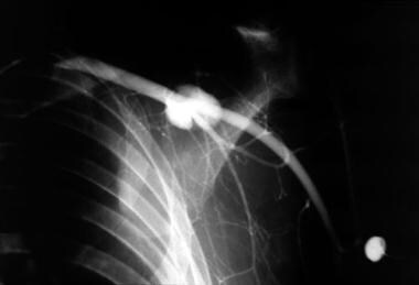
Pseudoaneurysms may resolve completely or grow over time presenting months to years later. They may cause neuropathy due to compression or embolization and can present as a growing pulsatile mass. (See the image below.)
-
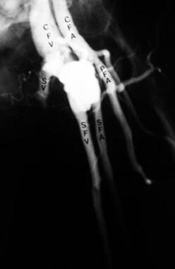
Arteriovenous fistulae typically take months to mature and often require surgical repair. (See the image below)
-
Intimal tears and flaps generally heal spontaneously over time.
-
Segmental narrowing can cause diminished flow, but pulses may remain intact. This injury may resolve spontaneously with fluids and rest and rarely require surgical intervention.
Management
Perform the following for peripheral vascular injuries:
-
Stabilize the extremity in the anatomic position.
-
Control hemorrhage with direct pressure.
-
Apply a tourniquet proximal to the injury if direct pressure is not effective in controlling hemorrhage.
Immediately reduce displaced or angulated fractures if any evidence or suspicion of vascular compromise exists. Promptly reduce dislocations of the elbow and knee to prevent further injury to neurovascular structures.
External hemorrhage usually can be controlled with direct pressure, but a blood pressure cuff or tourniquet should be applied proximally to the injury if compression fails or is not possible.
Once the patient has been stabilized, identify peripheral vascular injuries and restore normal circulation as rapidly as possible.
Do not apply clamps or hemostats to vascular structures, since this may make definitive repair more difficult and damage surrounding tissues.
A vascular surgeon must be consulted whenever a major vascular injury is a concern.
Surgical exploration and repair is performed as soon as possible for patients with "hard" signs of vascular injury, refractory hypotension, and obvious limb ischemia. Conventional arteriography to further define the injury may be performed preoperatively at the discretion of the vascular surgeon. Endovascular repair with stent placement is now used with increased frequency. [16]
Most non-occlusive injuries presenting without "hard" signs resolve over time. [17] Long-term follow-up with scheduled, repeat physical examinations is a safe and effective approach.
Patients must be given explicit instructions to present for neurovascular checks of the extremities on a scheduled basis. Instruct patients to return to the ED if they experience increased pain, edema, or active bleeding from the wound or if any weakness, numbness, or paresthesias develops in the injured extremity.
Epidemiology
Peripheral injuries account for 80% of all cases of vascular trauma. The lower extremities are involved in two thirds of all patients with vascular injuries.
Penetrating trauma accounts for 70-90% of vascular injuries. In the past, iatrogenic injuries related to endovascular procedures accounted for less than 10% of all cases. This percentage is increasing due to the growing use of endovascular procedures for diagnostic and therapeutic purposes.
Death due solely to peripheral vascular injuries is uncommon, but it does occur due to exsanguination or development of a necrotizing myofascial infection. Major venous injuries accompany 13-51% of significant arterial injuries.
Compartment syndrome may result from ischemia of a muscle compartment. Limb survival is threatened by delays in diagnosis and treatment, particularly when limb perfusion is compromised for more than 6 hours at body temperature ("warm" ischemia).
Extensive concurrent musculoskeletal, nerve, and skin injuries indicate a poor prognosis. Concomitant peripheral nerve injuries may be missed and can lead to long-term disability and deformity. [18]
Crush injuries associated with open tibial fractures are particularly likely to result in loss of the lower leg and amputation.
Ninety percent of patients with peripheral vascular injuries are male.
Vascular injuries most often occur in patients aged 20-40 years.
-
Pseudoaneurysm of the axillary artery.
-
Arteriovenous fistula between common femoral artery and vein.