Practice Essentials
Diagnosis of ectopic pregnancy has been greatly improved by the advent of rapid serum beta human chorionic gonadotropin (β-hCG) tests in the early 1980s and then the widespread adoption of transvaginal pelvic ultrasonography (TVUS) in the late 1980s. [1] Starting in the mid-1990s, the rise of bedside ultrasonography performed by emergency physicians has brought further improvements in time to diagnosis and treatment.
Currently, the approach focuses on diagnosing ruptured or suspected ectopic pregnancy versus normal (or failing) intrauterine pregnancy (IUP) by using early TVUS in the emergency department (ED). The process is as follows: first the physician must suspect the potential for a possible ectopic pregnancy diagnosis by obtaining a b-hCG measurement. Next determine the location of the pregnancy utilizing US. Ninety-eight percent of all ectopic pregnancies are implanted in the fallopian tube. [2] Finally, appropriate consultation and/or follow-up is then based on whether there are indeterminate results on ultrasonography such as pregnancies of unknown location (PULs) in relation to the discriminatory zone of serum b-hCG measurements.
Diagnoses of ectopic pregnancy in the ED may be rising in the United States. From 2006 to 2013, the overall ratio of ED visits with an ectopic pregnancy diagnosis increased from 11.0 per 1000 live births to 13.7 per 1000 live births. [3]
Go to Ectopic Pregnancy, Ultrasonographic Imaging in the Diagnosis of Ectopic Pregnancy, and Surgical Management of Ectopic Pregnancy for complete information on these topics.
Ultrasonography
The most critical step in beginning the workup is to have a high clinical suspicion for ectopic pregnancy (eg, in any woman of childbearing age). After a positive urine pregnancy test, any necessary initial resuscitation, and physical examination (including pelvic examination to rule out an open cervical os or completed abortion), ultrasonography should be performed. [4] This initial sonogram should be obtained at the bedside by an emergency physician, where feasible.
Bedside pelvic sonography is the imaging test of choice to investigate early pregnancy complaints in the emergency department (ED). It is noninvasive, portable, repeatable, does not involve contrast or ionizing radiation, and can be performed concurrently with resuscitation of an unstable patient. [5]
The goals of bedside pelvic ultrasonography are to find a definitive intrauterine pregnancy (IUP), a definitive or suspected ectopic pregnancy, and findings indicative of failed IUP. Because ultrasonographic findings of early normal IUP development (< 7 wk) are well correlated with beta human chorionic gonadotropin (β-hCG) levels, the absence of a normal IUP on the sonogram together with a β-hCG level above the discriminatory zone virtually rules out a normal IUP.
Pelvic sonography is usually conducted first with the transabdominal approach (which can reliably identify IUP at a β-hCG level above 6500 mIU/mL) and then with the transvaginal approach (which can extend the discriminatory zone down to 1500 mIU/mL). M-mode imaging is useful for measuring the fetal heart rate. Color Doppler ultrasonography can help identify some ectopic pregnancies by identifying a placental blood flow pattern in the adnexa.
A protocol using bedside emergency physician–performed transvaginal ultrasonography (TVUS) showed a large reduction in the incidence of discharged patients who later had ruptured ectopic pregnancies. [6] Emergency physician–performed ultrasonography has been shown to speed time to diagnosis compared to ultrasonography performed by a consulting obstetrician/gynecologist or by the radiology department. [7]
Experienced emergency physicians are sometimes able to correctly diagnose ectopic pregnancies initially missed by consulting obstetrician/gynecologists. [8] The only lawsuit found in a search of the WESTLAW nationwide litigation database concerning emergency physicians and ultrasonography was filed for failure to perform ED ultrasonography in an ectopic pregnancy that ruptured several days later. [9]
Hemodynamically unstable patients should first be scanned in the right upper quadrant of the abdomen; the finding of free fluid in Morison’s pouch in the right clinical setting by the emergency physician has been shown to decrease time to the operating room (OR). [10] Attention should be paid to the adnexa, even when an intrauterine pregnancy (IUP) is visualized, to rule out the rare heterotopic pregnancy, especially in patients with a history of assisted reproduction.
Ultrasonographic findings suggestive of ectopic pregnancy (empty uterus with a tubal ring, complex adnexal mass, or a moderate-to-large amount of free fluid) or a definite extrauterine pregnancy warrant an immediate gynecologic consult for medical or surgical treatment. Patients with evidence of a failed IUP should be followed up in consultation with gynecology for either repeat ultrasonography and serial β-hCG, dilation and curettage (D&C), or expectant management.
Patients with indeterminate ultrasonographic findings (empty uterus, gestational sac < 8 mm without a yolk sac) should have a β-hCG level drawn and should be followed up closely with gynecology to monitor serial β-hCG levels and ultrasonography. Although a certain percentage of pregnancies of unknown location (PULs) will eventually be diagnosed with an ectopic pregnancy (15% in 1 study), these are much less likely to require surgical treatment than those diagnosed on the initial sonogram.
Patients with a live IUP on a sonogram are essentially ruled out for ectopic pregnancy, have a low risk of eventually aborting (about 9% in 1 study, higher if associated with vaginal bleeding [11] ), and can be discharged from the ED after routine further care.
A spectrum of ultrasonographic findings exists and can be categorized according to a variety of different schemes, depending on the experience of the sonographer and the institutional setting.
Go to Ultrasonographic Imaging in the Diagnosis of Ectopic Pregnancy for complete information on this topic.
Intrauterine gestational sac containing yolk sac or fetal pole
A definitive IUP virtually rules out ectopic pregnancy (aside from heterotopic pregnancies). Keep in mind that interstitial or cornual pregnancies can appear intrauterine; however, they will be located eccentrically in the uterus, with a myometrial mantle (distance from the outer edge of the bright decidual layer to the outside border of the uterus) of less than 5 mm. [12]
Differentiating a cervical ectopic pregnancy from an impending spontaneous abortion can be difficult: the presence of fetal heart motion, a small hourglass-shaped uterus, and the absence of the “sliding sign” (in which the gestational sac slides against the cervix with gentle pressure from the ultrasound probe) are particularly helpful. [13]
Intrauterine gestational sac >16 mm without fetal pole or >8 mm without yolk sac, or intrauterine fetal pole >5 mm without heart motion
An intrauterine gestational sac larger than 16 mm without a fetal pole or larger than 8 mm without a yolk sac or an intrauterine fetal pole larger than 5 mm without heart motion is indicative of failed IUP. Although some patients with these findings are later diagnosed with live IUPs, they are also essentially ruled out for ectopic pregnancy.
Other findings suggestive of impending failure are a live IUP with a large amount of subchorionic hemorrhage (appearing as fluid separating the chorion from the endometrial lining) or a fetal heart rate of < 100 bpm. A gestational sac with a mean sac diameter less than 5 mm greater than the crown-rump length has an 80% risk of pregnancy loss. [14]
Extrauterine sac containing yolk sac or fetal pole, with or without heart motion
Although an extrauterine sac containing a yolk sac or a fetal pole, with or without heart motion, is definitive for ectopic pregnancy (see the image below), only 16-32% of ectopic pregnancies have this finding on TVUS. [15]
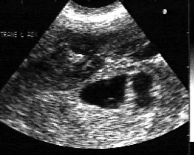 Endovaginal sonogram reveals complex mass outside uterus with small yolk sac present within. Mass is more echogenic than uterus above it and represents ectopic pregnancy.
Tubal ring
A tubal ring is a thick-walled cystic structure in the adnexa, independent of the ovary and uterus, and is highly predictive of ectopic pregnancy. [16] It can sometimes be confused with a corpus luteum cyst when the ovary is not well visualized. The corpus luteum cyst wall tends to be thinner and less echogenic than the endometrium, and the cyst tends to contain clear fluid. [17] When surrounded by free fluid, it can sometimes be confused with a hemorrhagic ovarian cyst. [18]
Complex adnexal mass
A complex adnexal mass is the sign most frequently found in ectopic pregnancies. [19] It can be somewhat cystic-appearing or entirely solid in nature, surrounded by free fluid, and ill-defined. If it cannot be moved independently of the ovary, it is unlikely to be an ectopic pregnancy. [20]
Moderate amount of anechoic free fluid (or any echogenic free fluid)
The cul-de-sac (pouch of Douglas) must be assessed when a definitive IUP is absent. A small amount of free fluid is physiologic. A moderate amount of anechoic free fluid (tracking more than one third of the way up the posterior wall of the uterus), or any echogenic free fluid, has a higher chance of being ultimately diagnosed as an ectopic pregnancy. [21]
Double decidual sac sign (gestational sac < 8 mm without yolk sac or fetal pole)
Although the double decidual sac sign is considered diagnostic of IUP by experienced sonographers, it can easily be confused with the pseudogestational sac found in ectopic pregnancy (caused by breakdown of stimulated endometrial lining) and lead to incorrect ruling out of ectopic pregnancy. [15] The pseudogestational sac (seen in 10-20% of ectopic pregnancies [22] ) can be differentiated by its central location in the uterus, oval shape, thin echogenic rim, and lack of double decidual sac sign (see the image below). [14]
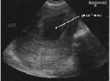 Pseudogestational sac of ectopic pregnancy can be confused with embryonic demise. Pseudogestational sac is produced when ectopic pregnancy stimulates endometrium, with degeneration of central decidual reaction.
Empty uterus without any of adnexal findings above
An empty uterus lacking any of the above adnexal findings may indicate an early IUP, completed abortion, or an ectopic pregnancy. In this case, a β-hCG above the discriminatory zone essentially rules out an early IUP, though it does not help diagnose or rule out ectopic pregnancy. A thin (< 8 mm) endometrial stripe appears somewhat predictive of eventual diagnosis of ectopic pregnancy in patients with a β-hCG below 1000 mIU/mL, [23] but there is sufficient overlap with eventual failed IUPs and normal IUPs to make this finding is a poor diagnostic test. [24]
Other findings
Molar pregnancy, retained products of conception (as in an incomplete abortion), or a corpus luteum cyst (which can be difficult to differentiate from an ovarian ectopic pregnancy, but is far more common) may be found.
Other Diagnostic Imaging Modalities
Magnetic resonance imaging
Although most emergency departments (EDs) have more ready access to ultrasonography than to magnetic resonance imaging (MRI), the latter’s lack of ionizing radiation may give it a role to play in the broader workup of pregnant women with abdominal pain (eg, ruling out appendicitis). MRI could also be indicated in those rare cases where ultrasonography results are inconclusive as to whether a visualized pregnancy is intrauterine or interstitial. [25]
Computed tomography
Computed tomography (CT) scanning involves the use of a substantial amount of ionizing radiation. Accordingly, it is, at best, a third-line choice of diagnostic study for pregnant patients.
Laboratory Studies
Serum and urine beta human chorionic gonadotropin
The first laboratory test to obtain is a qualitative urine beta human chorionic gonadotropin (β-hCG) test, which can be performed rapidly at the bedside. The qualitative urine β-hCG value can be unreliable at low quantitative serum β-hCG levels (ie, < 75 mIU/mL); therefore, in cases of high clinical suspicion, a serum quantitative β-hCG level should be obtained. [26]
Serum β-hCG levels can definitively rule out pregnancy if negative, though there have been case reports of pathology-proven ruptured ectopic pregnancy and hemorrhagic shock despite an undetectable serum β-hCG. [27] In a series of symptomatic patients presenting to an urban emergency department (ED), the odds of having an ectopic pregnancy were more than twice as high in those with an initial serum β-hCG below 1500 mIU/mL than in those with an abnormal intrauterine gestation. [28]
Additionally, an upper cutoff value of 40,000 mIU/mL is 99% sensitive for ectopic pregnancy. [29] Because ruptured ectopic pregnancies have been reported at a wide range of β-hCG levels, the β-hCG level should not be a factor in determining whether or not transvaginal ultrasonography (TVUS) should be performed.
In the early stages of a normal intrauterine pregnancy (IUP), the serum β-hCG rises along a well-defined curve; therefore, serial β-hCG tests can be useful for determining the ultimate location of a pregnancy of unknown location (PUL). The lower limit of the normal rise in β-hCG is 53% in 2 days. [30] A lower threshold, 35% rise over 2 days, would minimize the risk of intervening on a normal IUP. Unfortunately, 15% of PULs that ultimately turn out to be ectopic pregnancies will also meet this threshold. [31]
Although failed IUPs (spontaneous abortions) do not show a decrease in β-hCG along any well-defined curve, patients with a β-hCG level that falls more than 50% in 2 days are at low risk of having an ectopic pregnancy. [32]
Serum progesterone
Unlike β-hCG levels, serum progesterone levels tend to be stable over time during the first trimester, and concentrations are higher in a normal IUP. A single serum progesterone level has been used alone to discriminate between normal and failing IUPs, but it cannot accurately discriminate between IUPs and ectopic pregnancies. [33] It also cannot reliably diagnose ectopic pregnancy in conjunction with an indeterminate sonogram. [34]
Other laboratory tests
Research is ongoing concerning CA 125, pregnancy-associated plasma protein-A, vascular endothelial growth factor, and creatine kinase. None of them have yet shown superiority to serial β-hCG measurements in distinguishing between IUP and ectopic pregnancy. [35]
Other laboratory studies that should be obtained include the following:
-
Complete blood count, if significant hemorrhage is suspected
-
Metabolic panel to rule out electrolyte imbalances and also to rule out hepatic or renal abnormalities in case methotrexate therapy is being considered
-
Serum lactate level in cases of suspected shock
-
Urinalysis to eliminate urinary tract infection as a cause of pelvic pain
-
Blood type and Rh factor, if transfusion is required or if RhoGAM will be provided for Rh-negative patients with vaginal bleeding
Other Tests
Dilation and curettage
Dilation and curettage (D&C) can be used to rule out ectopic pregnancy by determining the presence of chorionic villi. The obvious drawback to its frequent use is that a certain number of normal intrauterine pregnancies (IUPs) will be aborted. It may be an option in the further workup of PUL when the pregnancy is undesired.
Culdocentesis
Culdocentesis was previously used to diagnose a ruptured ectopic pregnancy by the presence of free fluid in the pouch of Douglas. However, ultrasonography is noninvasive and has largely replaced culdocentesis where available.
Diagnostic laparoscopy
Diagnostic laparoscopy formerly played an important role in ultimately diagnosing patients with pregnancy of unknown location (PUL). Today, with the rise of serial beta human chorionic gonadotropin (β-hCG) measurements and ultrasonography, it is rarely performed. A recent case series of women with PUL found that only 1 of the 363 patients required diagnostic laparoscopy. [36]
Treatment & Management
Patients in shock require prehospital care to treat hypotension. Patients who have ultrasonographic findings suggestive of ectopic pregnancy or those who are clinically unstable at a location where pelvic ultrasonography is unavailable should be transferred to a facility that provides a higher level of care.
When an ectopic pregnancy is identified, medical or surgical treatment is provided as indicated. Those with ectopic pregnancy who require admission or surgery should be admitted to an obstetrics/gynecology service.
Pharmacologic treatment
The current standard medical treatment of unruptured ectopic pregnancy consists of the administration of methotrexate (MTX). [37, 38, 39] This decision to initiate MTX therapy should be made in conjunction with, if not by, the consulting obstetrician/gynecologist. The ideal candidate for medical treatment should have the following characteristics:
-
Hemodynamic stability
-
No severe or persisting abdominal pain
-
The ability to follow up multiple times
-
Normal baseline liver and renal function test results
Absolute contraindications include existence of intrauterine pregnancy (IUP), immunodeficiency, moderate-to-severe anemia, leucopenia, or thrombocytopenia, sensitivity to MTX, active pulmonary or peptic ulcer disease, clinically important hepatic or renal dysfunction, or breastfeeding.
Sonogram findings of an ectopic gestational sac greater than 4 cm in size, (or 3.5 cm if the ectopic pregnancy has fetal heart motion) on a sonogram, an initial beta human chorionic gonadotropin (β-hCG) concentration higher than 5000 mIU/mL, and the presence of significant free fluid are indicators of likely failure of MTX therapy and therefore constitute relative contraindications.
The multiple-dose MTX regimen consists of daily 1 mg/kg doses, given intramuscularly (IM) on alternating days with leucovorin (folinic acid, which reduces side effects), until there is a 15% decline in β-hCG over 2 days. The single-dose MTX regimen consists of a 50 mg/m2 dose, followed by a repeat β-hCG on day 4; if the β-hCG level has declined by less than 15% between days 4 and 7, another 50 mg/m2 dose of MTX is given.
Both treatment regimens show an efficacy similar to surgical management for unruptured ectopic pregnancies in the ideal patient population. Common side effects include increase in abdominal girth, vaginal bleeding or spotting, abdominal pain, gastrointestinal symptoms, stomatitis, and dizziness. Rare side effects include severe neutropenia, reversible alopecia, and pneumonitis. [37]
Surgical treatment
Surgical approaches to managing ectopic pregnancy when medical management is not possible or is ruled out by other associated medical conditions are described elsewhere (see Surgical Management of Ectopic Pregnancy).
Consultations
Obstetrics/gynecology should be consulted as needed for ectopic pregnancies and for follow-up care of patients with failing or failed intrauterine pregnancies (IUPs) or pregnancies of unknown location (PULs). Any patient who is clinically unstable should have the consultation in the emergency department (ED).
Obstetrics/gynecology or radiology should also be consulted for transvaginal ultrasonography (TVUS) as needed, according to institutional policy.
Outpatient monitoring
Patients with PULs should follow up with obstetrics/gynecology in 2 days for repeat β-hCG and ultrasonography. Patients with failing or failed IUPs should arrange for follow-up with obstetrics/gynecology for dilation and curettage (D&C) or expectant management. Patients receiving MTX in the ED should follow up with obstetrics/gynecology according to protocol.
In a retrospective study (1998-2006) of 161 women with successfully expectantly managed nonviable tubal ectopic pregnancies, median serum β-hCG clearance time was 19 days. [40] Although not statistically significant, women who had an initially plateauing β-hCG levels in the declining phase appeared to take longer to clear β-hCG. These findings may have clinical implications regarding follow-up duration and expectant management in the setting of tubal ectopic pregnancies. [40]
-
Endovaginal sonogram reveals complex mass outside uterus with small yolk sac present within. Mass is more echogenic than uterus above it and represents ectopic pregnancy.
-
Pseudogestational sac of ectopic pregnancy can be confused with embryonic demise. Pseudogestational sac is produced when ectopic pregnancy stimulates endometrium, with degeneration of central decidual reaction.


