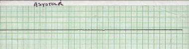
Continually Updated Clinical Reference
 
 
  All Sources     eMedicine     Medscape     Drug Reference     MEDLINE
 
eMedicine - Asystole : Article by

Quick Find
Authors & Editors
Introduction
Clinical
Differentials
Workup
Treatment
Medication
Follow-up
Miscellaneous
Multimedia
References

Related Articles
Ventricular Fibrillation


Patient Education
Public Health Center

Cardiopulmonary Resuscitation (CPR)


Author: Richard M Caggiano, MD, FACEP, Adjunct Faculty, Department of Medicine, University of Washington, School of Medicine; Director of Emergency Services, Medical Director of Employee Health, Assistant Director of Trauma Services, Pullman Regional Hospital

Richard M Caggiano is a member of the following medical societies: American College of Emergency Physicians

Editors: Edward Bessman, MD, Chairman, Department of Emergency Medicine, John Hopkins Bayview Medical Center; Assistant Professor, Department of Emergency Medicine, Johns Hopkins University; Francisco Talavera, PharmD, PhD, Senior Pharmacy Editor, eMedicine; Gary Setnik, MD, Chair, Department of Emergency Medicine, Mount Auburn Hospital; Assistant Professor, Division of Emergency Medicine, Harvard Medical School; John D Halamka, MD, MS, Associate Professor of Medicine, Harvard Medical School, Beth Israel Deaconess Medical Center; Chief Information Officer, CareGroup Healthcare System and Harvard Medical School; Attending Physician, Division of Emergency Medicine, Beth Israel Deaconess Medical Center; Jonathan Adler, MD, Attending Physician, Department of Emergency Medicine, Massachusetts General Hospital; Division of Emergency Medicine, Harvard Medical School

Author and Editor Disclosure

Synonyms and related keywords: flat line, asystole, cardiac standstill, pulseless electrical activity, PEA, primary asystole, secondary asystole, bradyasystolic rhythm, bradydysrhythmia, bradyasystole, asystolic cardiopulmonary arrest, asystolic cardiac arrest

Background

Asystole is cardiac standstill with no cardiac output and no ventricular depolarization; it eventually occurs in all dying patients.

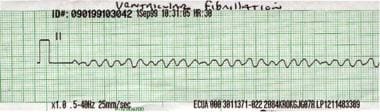
Pulseless electrical activity (PEA) is the term applied to a heterogenous group of dysrhythmias unaccompanied by a detectable pulse. Bradyasystolic rhythms are slow rhythms; they can have a wide or narrow complex, with or without a pulse, and often are interspersed with periods of asystole. When discussing PEA, ventricular fibrillation (VF) and ventricular tachycardia (VT) are excluded.

Pathophysiology

Asystole can be primary or secondary. Primary asystole occurs when the heart's electrical system intrinsically fails to generate a ventricular depolarization. This may result from ischemia or from degeneration (ie, sclerosis) of the sinoatrial (SA) node or atrioventricular (AV) conducting system. Primary asystole usually is preceded by a bradydysrhythmia due to sinus node block-arrest, complete heart block, or both.

Reflex bradyasystole/asystole can result from ocular surgery, retrobulbar block, eye trauma, direct pressure on the globe, maxillofacial surgery, hypersensitive carotid sinus syndrome, or glossopharyngeal neuralgia. Episodes of asystole and bradycardia have been documented as manifestations of left temporal lobe complex partial seizures. These patients experienced either dizziness or syncope. No sudden deaths were reported, but the possibility exists if asystole were to persist. The longest interval was 26 seconds.

Secondary asystole occurs when factors outside of the heart's electrical conduction system result in a failure to generate any electrical depolarization. In this case, the final common pathway is usually severe tissue hypoxia with metabolic acidosis. Asystole or bradyasystole follows untreated VF and commonly occurs after unsuccessful attempts at defibrillation. This forebodes a dismal outcome.

Frequency

United States

The number of adults in cardiopulmonary arrest who had bradyasystole as the initial arrest rhythm is difficult to measure accurately. Reports vary and may be skewed by the patient population studied and/or by the method of reporting the initial rhythm. In a 1991 study of 185 patients in cardiopulmonary arrest at the time of arrival to the emergency department, 9% had survived to hospital admission and none were discharged alive (Gray, 1991). This study was not limited to patients with asystole.

International

In one study from Goteborg, Sweden, asystole was the presenting rhythm in the field in 35% of patients with cardiac arrest (Engdahl, 2000).

When the incidence of coronary artery disease in the population of a country is relatively low, asystole is relatively more common as a manifestation of cardiopulmonary arrests. This is because cardiac ischemia more frequently results in VF. Asystole is most likely to be found in cardiopulmonary arrests occurring in children; this is usually secondary to another noncardiac event (ie, respiratory arrest due to sudden infant death syndrome, infection, choking, drowning, or poisoning).

Mortality/Morbidity

Asystole is associated with a poor outcome regardless of its initial cause. In the Goteborg study, 10% of 1,635 asystolic patients survived to hospital admission and 2% survived to hospital discharge.

Resuscitation is likely to be successful only if it is secondary to an event that can be corrected immediately, such as a cardiac arrest due to choking on food (a cafe coronary), and only if an airway can be established and the patient may be rapidly reoxygenated. Occasionally, primary asystole can be reversed if it is due to pacemaker failure, which could be either intrinsic or extrinsic, and this is corrected immediately by external pacing.

Sex

Frequency of asystole, as a percentage of all cardiopulmonary arrests, is higher in women than in men; however, the frequency of cardiac arrest in general is proportional to the underlying incidence of heart disease, which is more common in males until around 75 years of age.

Age

Prevalence of asystole as the presenting cardiac rhythm is lower in adults (25-56%) than in children (90-95%).


History

Immediate diagnosis of asystole requires the recognition of a full cardiac arrest and a confirmed flat-line rhythm in 2 perpendicular leads. Lightheadedness or syncope may precede asystole when it follows a bradyasystolic rhythm.

Physical

If the rhythm is truly asystole and has been present for more than several seconds, the patient will be unconscious and unresponsive. A few agonal (final gasping) breaths may be noted, but detectable heart sounds and palpable peripheral pulses are absent.

Causes

  • Examples of common conditions that can result in secondary asystole include suffocation, near drowning, stroke, massive pulmonary embolus, hyperkalemia, hypothermia, MI complicated by VF or VT that deteriorates to asystole, defibrillation, and sedative-hypnotic or narcotic overdoses leading to respiratory failure.
  • Hypothermia is a special circumstance, since asystole can be tolerated for a longer period of time under such conditions and can be reversed with rapid rewarming while cardiopulmonary resuscitation (CPR) is being performed. If available, institute cardiopulmonary bypass immediately, because it can accomplish both of these goals. Most survivors have received cardiopulmonary bypass.
  • Primary asystole develops when cellular metabolic functions are no longer intact and an electrical impulse cannot be generated. With severe ischemia, pacemaker cells cannot transport the ions necessary to effect the transmembrane action potential. Implantable pacemaker failure may also be a cause of primary asystole.
    • Proximal occlusion of the right coronary artery can cause ischemia or infarction of both the SA and the AV nodes.
    • Extensive infarction can cause bilateral bundle-branch block (ie, infranodal complete heart block).
    • Idiopathic degeneration of the SA or AV node can result in sinus arrest-block and/or AV heart block, respectively. This process is slow and progressive, but the symptoms may be acute and asystole may result. An implantable pacemaker is usually required for these conditions.
    • Occasionally, asystolic sudden death occurs from congenital heart block, local tumor, or cardiac trauma.
    • Asystole can occur following an indirect lightning strike (ie, direct current [DC]) that depolarizes all the cardiac pacemakers. A rhythm may return spontaneously or shortly after CPR is initiated. These patients may survive intact if given immediate attention. Alternating current (AC) from man-made sources of electrical current usually results in VF.


Ventricular Fibrillation

Other Problems to be Considered

ECG lead misplacement


Lab Studies

  • A potassium level may be useful if deemed appropriate, and results are immediately available.
  • Arterial blood gas (ABG) analysis may be used to obtain rapid reporting of potassium level in many institution's laboratories. ABG level may also help to evaluate the ventilatory and acid-base status of the patient as well as hemoglobin level. If the patient is in full arrest, a blood gas level does not accurately reflect the pH status of the tissues.

Imaging Studies

  • Asystole is generally a diagnosis made via ongoing cardiac monitoring or ECG and physical examination with pulselessness. Bedside ultrasonography may be useful to confirm cardiac standstill. The heart may be viewed via either a subxiphoid view or an intercostal view and wall motion, or lack thereof, observed.
  • For documentation purposes, in addition to including rhythm strips from 2 separate leads identifying asystole, an image of 2-dimensional echocardiography taken over time in M-mode may be taken, saved, and included in the medical record. This will further document lack of heart wall motion.

Other Tests

  • Continuous cardiac monitoring is useful during attempts at resuscitation to determine rhythm and effects of intervention.
    • Asystole is best determined in 2 separate cardiac leads. This helps protect against reading a flat line due to lead malfunction incorrectly as asystole, as well as in differentiating fine VF, particularly in an isoelectric lead, from asystole.
    • Eliminate a possible diagnosis of VF, which may masquerade as asystole, by checking 2 leads perpendicular to each other. In spite of this caveat, using a flat-line protocol based on a 3-lead check, occult VF was documented in only 3 (2.5%) of 118 asystolic patients, indicating that VF masquerading as asystole is uncommon. Technical problems were much more common, observed in 10 patients (8%).
    • Exclude the possibility of lead misplacement by always checking for the presence of a pulse.
  • Spurious asystole can be seen when using manual defibrillator paddles to monitor the rhythm. This is usually very brief, but nevertheless, can be avoided by switching to monitoring leads, especially after several successive shocks.
  • Pulse oximetry may be used during resuscitation to monitor for effectiveness of forward flow or CPR; however, usually flow is too inadequate for the sensor to register any results.

Procedures

  • Endotracheal intubation is indicated with attempts at resuscitation.
  • Central venous access or intraosseous access may be needed for vascular access.


Prehospital Care

  • The only 3 drugs recommended or acceptable by the American Heart Association (AHA) for adults in asystole are epinephrine, vasopressin, and atropine. In spite of full vagolytic doses of atropine (0.03 mg/kg) and high-dose epinephrine (0.20 mg/kg), or the use of vasopressin 40 units (U), few patients survive to leave the hospital neurologically intact. Atropine is no longer recommended in young children and infants in asystole but can be considered in adults with slow PEA rhythms.
  • If spontaneous circulation has not been restored, administering vasopressin 40 U IV for the first 2 doses or followed by epinephrine given at the physician's discretion has recently showed superior results. A recent study showed that more patients survived to hospital discharge, although the neurologic status of the patients at discharge was not clearly stated. The number of patients with asystole in the study was 528. Of those, 12 patients in the vasopressin group survived to discharge compared with 4 in the standard therapy group. In this study, the odds ratio stated may not be statistically significant. The study also showed a nonstatistically significant trend toward worse results for VF and PEA (Wenzel, 2004). The advanced cardiac life support (ACLS) 2005 guidelines allow vasopressin 40 IU IV as a one-time dose treatment option in VF and asystole. It can be given either before epinephrine or after the first dose of epinephrine.
  • Transcutaneous pacing (TCP), even when used immediately, has not altered meaningful survival (ie, functional lifestyle) significantly. When no metabolic deficit exists, such as in a cardiac arrest preceded by a conduction or impulse generation disorder (ie, primary asystole), immediate use of TCP may be lifesaving.

Emergency Department Care

Mainstays of ED treatment are providing oxygenation and ventilation via endotracheal intubation and circulation via CPR, attempts at transcutaneous or transvenous pacing (that have some small potential to be fruitful in primary asystole that has just occurred), and administration of pharmacologic agents.

  • A case has been reported of a long but successful resuscitation from asystole of a patient who was hyperkalemic secondary to renal failure. The treatment included calcium chloride to reverse the physiologic effects of hyperkalemia and insulin and glucose to lower serum potassium levels. This cannot be recommended routinely for all cases of asystolic arrest.
  • Electrical defibrillation should not be applied indiscriminately to the patient in asystole. This is not only fruitless, but also detrimental, eliminating any possibility of recovering a rhythm. Asystole following electrical defibrillation has an even worse outcome than that in a patient whose first documented rhythm was asystole. One caution is that following defibrillation, a brief spurious asystole can occur using manual monitoring through the defibrillator paddles. This does not occur with the rhythm monitoring leads or hands-off monitor pads. If not taken into consideration, it could lead to a delay in defibrillation, when indeed VF is present.


Parasympathetic influences during cardiopulmonary arrest have not been elucidated fully, and the clinical benefits of atropine have yet to be confirmed. Atropine can be used for asystole, but the AHA now states that its use should be considered. High-dose epinephrine (0.20 mg/kg) may improve the hemodynamics of CPR, thereby increasing the rate of return to spontaneous circulation; however, it has not been demonstrated to influence the final clinical outcome. Therefore, these doses no longer are recommended for children or adults. Adenosine antagonists, such as aminophylline, have been investigated but have not been shown to be clinically useful.

Drug Category: Anticholinergic agents

The goal in using these agents is to enhance sinoatrial activity and to improve conduction through the SA or AV node by reducing vagal tone via muscarinic receptor blockade. This is effective only if the site of block is within the SA or AV node. For patients with infranodal block, this therapy is ineffective. It may increase a Mobitz II second-degree block to a higher degree of block or a third-degree block.

Drug NameAtropine (Atropair, Isopto, Atropisol)
DescriptionParasympatholytic agent used to eliminate vagal influence on SA and AV nodes. Not effective for infranodal third-degree heart block.
Adult Dose1 mg IV/IO, may repeat dose up to 3 mg total, or 0.03 mg/kg, which is completely vagolytic
If no IV/IO access is available, administer 2 or 2.5 mg via an endotracheal tube (ET); this is a less reliable method and should only be used as a last resort; dose should be followed (a "flush") with 5 mL of normal saline flush and 5 ventilations should be provided; a minimum dose of 0.1 mg IV/IO should be given to avoid centrally mediated paradoxical parasympathomimetic effect
Pediatric DosePediatric bradyasystolic arrest: Not recommended.
Symptomatic bradycardia with a pulse unresponsive to oxygen and fluids: 0.02 mg/kg IV/IO
Children: 0.1 mg IV minimum dose; not to exceed 0.5 mg; maximum total dose 1 mg
Adolescents: 0.1 mg IV minimum dose; not to exceed 1 mg; maximum total dose 2 mg
If no IV or IO access exists, give 0.03 mg/kg ET; the same holds for this route as stated above when applied to pediatrics
ContraindicationsNone when indicated for symptomatic sinus bradycardia or Mobitz type I second-degree heart block; contraindicated in Mobitz type II second-degree heart block; generally not effective for infranodal third-degree heart block
InteractionsNone for this indication
PregnancyC - Safety for use during pregnancy has not been established.
PrecautionsAdds to tachydysrhythmia if a rhythm resumes, especially when used with sympathomimetic agents; may potentially lower the VF threshold (This is theoretical and only an issue if perfusing rhythm present.)

Drug Category: Adrenergic agonists

These agents can produce constriction of skeletal and vascular muscle.

Drug NameEpinephrine (Adrenaline)
DescriptionConsidered the single most useful drug in cardiac arrest. Used to increase coronary and cerebral blood flow during CPR. May enhance automaticity during asystole. Can be used for bradycardia in adult and pediatric patients.
Adult Dose1 mg IV/IO; 2-2.5 mg ET if not given IV/IO; range was 0.01-0.20 mg/kg depending on standard-dose or high-dose epinephrine protocols
(High doses are no longer recommended by AHA.)
Pediatric Dose0.01 mg/kg IV/IO; alternative, 0.10 mg/kg ET if no IV/IO access
(High-dose epinephrine [0.20 mg/kg] is no longer recommended by AHA.)
ContraindicationsNone for this indication
InteractionsNone for this indication
PregnancyC - Safety for use during pregnancy has not been established.
PrecautionsMay result in tachydysrhythmia if rhythm resumes; this is additive to effects of atropine or other sympathomimetic agents

Drug NameVasopressin (Pitressin)
DescriptionHas vasopressor and ADH activity. Increases water resorption at distal renal tubular epithelium (ADH effect) and promotes smooth muscle contraction throughout vascular bed (vasopressor effects). Vasoconstriction is increased in splanchnic, portal, coronary, cerebral, peripheral, pulmonary, and intrahepatic vessels.
Adult Dose40 IU IV once, although some studies suggest a repeat dose of 40 IU (not an AHA recommendation)
Pediatric DoseNot established
ContraindicationsNone for this indication
InteractionsNone for this indication
PregnancyC - Safety for use during pregnancy has not been established.
PrecautionsMay result in tachydysrhythmia if rhythm resumes


Further Inpatient Care

  • The intensive care unit is the appropriate disposition for the occasional patient who survives asystolic cardiopulmonary arrest and requires further treatment and diagnostic evaluation.

Deterrence/Prevention

  • Primary asystole may be prevented by the appropriate use of a permanent pacemaker in those patients who have high-grade heart block or sinus arrest.
  • Preventing secondary asystole requires early recognition and treatment of the preceding event.

Complications

  • Complications include permanent neurologic impairment and complications from CPR or invasive procedures (eg, liver laceration, fractured ribs, pneumothorax, hemothorax, air embolus, aspiration, gastric/esophageal rupture).
  • Death often occurs.

Prognosis

  • Prognosis depends on the etiology of the asystolic rhythm, timing of interventions, and success or failure of advanced cardiac life support. Generally, the prognosis is dismal and postcountershock asystole in particular has an even worse survival rate.

Patient Education

  • Advice about electrical storm safety and prevention of hypothermia is appropriate for those likely to be exposed to these conditions.
  • For excellent patient education resources, visit eMedicine's Public Health Center. Also, see eMedicine's patient education article Cardiopulmonary Resuscitation (CPR).


Medical/Legal Pitfalls

  • Failure to anticipate impending asystolic arrest
  • Failure to recognize esophageal intubation
  • Withholding specific therapy for patients with hypothermia


Media file 1:  Rhythm strip showing asystole.
Click to see larger pictureClick to see detailView Full Size Image
Media type:  Rhythm Strip

Media file 2:  Rhythm strip showing ventricular fibrillation.
Click to see larger pictureClick to see detailView Full Size Image
Media type:  Rhythm Strip


  • American Heart Association. Guidelines for cardiopulmonary resuscitation and emergency cardiac care. Emergency Cardiac Care Committee and Subcommittees, American Heart Association. Part I. Introduction. JAMA. Oct 28 1992;268(16):2171-83. [Medline].
  • Barthell E, Troiano P, Olson D, et al. Prehospital external cardiac pacing: a prospective, controlled clinical trial. Ann Emerg Med. Nov 1988;17(11):1221-6. [Medline].
  • Bayes de Luna A, Coumel P, Leclercq JF. Ambulatory sudden cardiac death: mechanisms of production of fatal arrhythmia on the basis of data from 157 cases. Am Heart J. Jan 1989;117(1):151-9. [Medline].
  • Billi JE, Zideman DA, Eigel B, et al. Conflict of interest management before, during, and after the 2005 International Consensus Conference on cardiopulmonary resuscitation and emergency cardiovascular care science with treatment recommendations. Resuscitation. Nov-Dec 2005;67(2-3):171-3. [Medline].
  • Bognolo DA, Rabow FI, Vijayanagar RR, Eckstein PF. Traumatic sinus node dysfunction. Ann Emerg Med. Jun 1982;11(6):319-21. [Medline].
  • Comess KA, DeRook FA, Russell ML, et al. The incidence of pulmonary embolism in unexplained sudden cardiac arrest with pulseless electrical activity. Am J Med. Oct 1 2000;109(5):351-6. [Medline].
  • ECC Committee, Subcommittees and Task Forces of the American Heart Association. 2005 American Heart Association Guidelines for Cardiopulmonary Resuscitation and Emergency Cardiovascular Care. Circulation. Dec 13 2005;112(24 Suppl):IV1-203. [Medline].
  • Engdahl J, Bang A, Lindqvist J, Herlitz J. Can we define patients with no and those with some chance of survival when found in asystole out of hospital?. Am J Cardiol. Sep 15 2000;86(6):610-4. [Medline].
  • Gold RS, Pollard Z, Buchwald IP. Asystole due to the oculocardiac reflex during strabismus surgery: a report of two cases. Ann Ophthalmol. Dec 1988;20(12):473-5, 477. [Medline].
  • Gray WA, Capone RJ, Most AS. Unsuccessful emergency medical resuscitation--are continued efforts in the emergency department justified?. N Engl J Med. Nov 14 1991;325(20):1393-8. [Medline].
  • Herlitz J, Ekstrom L, Wennerblom B, et al. Effect of bystander initiated cardiopulmonary resuscitation on ventricular fibrillation and survival after witnessed cardiac arrest outside hospital. Br Heart J. Nov 1994;72(5):408-12. [Medline].
  • Lindholm DJ, Campbell JP. Predicting survival from out-of-hospital cardiac arrest. Prehospital Disaster Med. Apr-Dec 1998;13(2-4):51-4. [Medline].
  • Locatelli ER, Varghese JP, Shuaib A, Potolicchio SJ. Cardiac asystole and bradycardia as a manifestation of left temporal lobe complex partial seizure. Ann Intern Med. Apr 6 1999;130(7):581-3. [Medline].
  • Mader TJ, Smithline HA, Gibson P. Aminophylline in undifferentiated out-of-hospital asystolic cardiac arrest. Resuscitation. Jun 1999;41(1):39-45. [Medline].
  • Marill KA, Williamson S. Cardiac arrest: current concepts, controversies, and evidence. Emergency Medicine Practice. Nov 2002;4(11):1-24.
  • Moonie GT, Rees DL, Elton D. The oculocardiac reflex during strabismus surgery. Can Anaesth Soc J. Nov 1964;11:621-32. [Medline].
  • Niemann JT, Stratton SJ, Cruz B, Lewis RJ. Outcome of out-of-hospital postcountershock asystole and pulseless electrical activity versus primary asystole and pulseless electrical activity. Crit Care Med. Dec 2001;29(12):2366-70. [Medline].
  • Niemann JT, Cairns CB. Hyperkalemia and ionized hypocalcemia during cardiac arrest and resuscitation: possible culprits for postcountershock arrhythmias?. Ann Emerg Med. Jul 1999;34(1):1-7. [Medline].
  • Ornato JP, Peberdy MA. The mystery of bradyasystole during cardiac arrest. Ann Emerg Med. May 1996;27(5):576-87. [Medline].
  • Perers E, Abrahamsson P, Bang A, et al. There is a difference in characteristics and outcome between women and men who suffer out of hospital cardiac arrest. Resuscitation. Apr-May 1999;40(3):133-40. [Medline].
  • Pitetti R, Glustein JZ, Bhende MS. Prehospital care and outcome of pediatric out-of-hospital cardiac arrest. Prehosp Emerg Care. Jul-Sep 2002;6(3):283-90. [Medline].
  • Resuscitation Council [UK]. Spurious asystole when using manual defibrillators and monitoring through the defibrillator paddled. Resusitation Council [UK]. 2002;1-2.
  • Richman PB, Nashed AH. The etiology of cardiac arrest in children and young adults: special considerations for ED management. Am J Emerg Med. May 1999;17(3):264-70. [Medline].
  • Savage DD, Castelli WP, Anderson SJ, Kannel WB. Sudden unexpected death during ambulatory electrocardiographic monitoring. The Framingham Study. Am J Med. Jan 1983;74(1):148-52. [Medline].
  • Sayegh AJ, Swor R, Chu KH, et al. Does race or socioeconomic status predict adverse outcome after out of hospital cardiac arrest: a multi-center study. Resuscitation. Apr-May 1999;40(3):141-6. [Medline].
  • Starfield B. Continuous confusion?. Am J Public Health. Feb 1980;70(2):117-9. [Medline].
  • Stiell IG, Wells GA, Field B, et al. Advanced cardiac life support in out-of-hospital cardiac arrest. N Engl J Med. Aug 12 2004;351(7):647-56. [Medline].
  • Vukov LF, White RD. External transcutaneous pacemakers in prehospital cardiac arrest. Ann Emerg Med. May 1988;17(5):554-5. [Medline].
  • Wenzel V, Krismer AC, Arntz HR, et al. A comparison of vasopressin and epinephrine for out-of-hospital cardiopulmonary resuscitation. N Engl J Med. Jan 8 2004;350(2):105-13. [Medline].
  • Young KD, Gausche-Hill M, McClung CD, Lewis RJ. A prospective, population-based study of the epidemiology and outcome of out-of-hospital pediatric cardiopulmonary arrest. Pediatrics. Jul 2004;114(1):157-64. [Medline].

Asystole excerpt

Article Last Updated: Feb 8, 2007