Practice Essentials
Nail bed (nailbed) injuries are common, with fingertip injuries being the most commonly seen hand injuries. The fingertip is frequently injured because it is the point of interaction between the body and one's surroundings in the majority of activities performed on a daily basis, and it is the most distal portion of the upper extremities. In addition to long-term cosmetic consequences, injuries to the nail can affect daily living. The nail provides protection for the fingertip, offers the ability to pick up small objects, and plays a role in tactile sensation. It serves as a counter force when the finger pad touches an object; two-point discrimination distance widens substantially with removal of a nail. [1, 2, 3, 4, 5, 6]
Most injuries of the nail bed are due to crushing injuries, such as with a hammer. [1] Twenty-five percent of nail bed injuries involve the finger being crushed in a doorway, most commonly car doors. Crush injuries squeeze the soft tissue of the nail bed between the nail and the distal phalanx. This may result in a simple subungual hematoma or a simple or stellate laceration. Saws, knives, drills, moving belts, and lawnmowers are also common causes of nail bed injuries. Crush and avulsion injuries, as well as injuries associated with distal phalanx fractures, have a worse prognosis.
Treatment
Blunt trauma to the fingertip and nail bed requires adequate treatment to prevent secondary deformities and reduce the need for subsequent reconstruction. [7] Delayed or inadequate treatment can result in negative functional and cosmetic outcomes. Peak incidence of fingertip and nail bed injuries is from 4 to 30 years of age. According to Chang et al, 10% of such accidents are treated in the emergency department. In the case of fingertip injuries, the nail bed is injured in 15-24% of cases. [8]
The injured finger can usually be examined without anesthesia, although children or those in severe pain may require a digital block first. A complete examination of sensation (performed prior to a digital block), motor function, and vascular supply is necessary.
A digital block of 1% lidocaine hydrochloride without epinephrine provides anesthesia of sufficient duration for most repairs. Bupivacaine extends anesthesia time 4-8 hours for longer procedures. Children may require procedural sedation and analgesia.
Observe the posture of the fingers, and look for any presence of deformities signifying fracture, dislocation, or tendon avulsion, and the presence of glass, wood, metal, or other foreign body fragments.
Depending on the extent of injury, radiologic evaluation with anteroposterior, lateral, and oblique views of the injured finger(s) may be useful to rule out foreign bodies and fractures or dislocations of the distal finger. [6]
The prophylactic use of antibiotics is indicated, depending on mechanism and extent of injury, such as for crush injuries and human bites or animal bites. Many clinicians prescribe a first-generation cephalosporin when bone or joint is exposed below a nail bed injury.
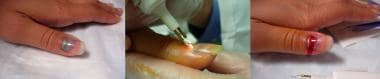
Small (less than 25% of the nail bed) and painless subungual hematomas require no intervention, as the hematoma will eventually reabsorb. If the subungual hematoma covers more than 25% of the nai lbed or is causing pain, the patient should be offered evacuation via trephination or nail removal.
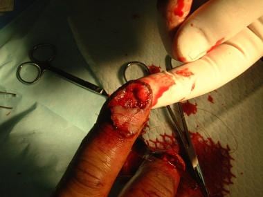
Lacerations to the nail bed should be repaired using 6-0 or smaller absorbable sutures. Minimal to no debridement should be performed because aggressive debridement may cause undue tension on the repair and results in scarring.
When repairing avulsed nails and nail beds, if the nail is detached proximally, it must be removed to inspect for any damage to the nail bed.
(See the treatment images below.)
Prognosis
Complications of nail bed injuries may include scarring, loss or obstruction of the nail fold, destruction of nail with lack of new nail growth, abnormal nail growth or disrupted nail growth, and infection.
Nail bed injuries generally heal well with appropriate treatment, although it may take months to years for the nail to grow back into the proper shape. Crush and avulsion injuries, as well as injuries associated with distal phalanx fractures, have a worse prognosis. Injuries that span the entire nail bed or most of the bed and fold also fare worse than those that are isolated to one to two thirds of the nail bed or only to the nail fold and germinal matrix.
All patients should be advised that a deformed nail is a possibility. New nail growth may take from 3 to 12 months, and even then it may be misshapen for a longer time. If problems with new nail growth exist at 6 or 12 months, patients may want to follow up with a hand surgeon for possible scar excision or nail bed revisions.
Pathophysiology
To fully appreciate the consequences and treatment of nail bed (nailbed) injuries, reviewing the anatomy of the nail bed and the surrounding tissues is useful. [1, 9, 10, 11]
-
Nail - Hard structure composed of desiccated, keratinized squamous cells
-
Perionychium - Composed of the nai lbed and paronychia
-
Nail bed - Soft tissue below the nail that is bound to the underlying periosteum of the distal phalanx and consists of the germinal and sterile matrix [11]
-
Paronychia - Lateral nail folds
-
Hyponychium - Junction between the nail bed and fingertip skin that contains large numbers of polymorphonuclear leukocytes and lymphocytes, which protect the subungual tissue from infection
-
Nail fold - Holds the proximal nail
-
Eponychium - Commonly known as the cuticle, or the distal portion of the nail fold where it attaches to the dorsum of the nail
-
Lunula - White opacity distal to the eponychium, caused by the presence of nail cell nuclei in the germinal matrix as they stream upward and distally to create nail
Nail formation is a collective production by 3 areas of the perionychium, as follows:
-
The germinal (intermediate) matrix, covering the ventral floor of the proximal volar nail fold to the lunula, produces 90% of nail volume. It is immediately distal to the extensor tendon attachment to the distal phalanx. As the cells are produced, they force cells ahead to flatten and stream distally into the nail because of the confining boundaries of the nail fold. The nuclei of the cells disintegrate as they grow beyond the lunula, giving the nail its clear appearance.
-
The sterile (ventral) matrix begins as the lunula ends and extends out to the hyponychium. It is closely adherent to the dorsal periosteum of the distal phalanx. It contributes a small amount to the nail but mostly provides adherence between the nail and the nail bed.
-
The proximal half of the dorsal roof of the nail fold produces cells that give the nail its shine.
Longitudinal nail growth takes between 70 and 160 days to cover the entire length of the nail. In general, a normal fingernail grows out completely in about 6 months, whereas toenails grow one-third to half the rate of fingernails; thus, toenails take 12-18 months to grow out entirely. The nail growth rate is less than normal in people who are immunocompromised, immobilized or paralyzed, malnourished, suffering from acute infection, or undergoing antimitotic drug therapy. [12]
After an injury, nail growth is stunted or absent for up to 21 days. The nail then grows rapidly for approximately the next 50 days and then slows again before a normal and sustained growth rate resumes. These relative accelerations and slowdowns in nail growth create the characteristic lump that is often observed on most nails that regrow after trauma.
As a result of scar tissue being unable to produce nail material, damage to specific components of the perionychium will lead to characteristic defects during regrowth of the posttraumatic nail. A scar of the dorsal roof of the nail fold creates a dull streak on the nail surface, while a scar of the germinal matrix may cause a split or absent nail, and a scar in the sterile matrix results in a split or nonadherent nail beyond the scar.
The nail bed is supplied by 2 volar arterial arches that are anastomoses between digital arteries of the finger or toe, just above the periosteum of the distal phalanx. Venous drainage coalesces in the proximal nail bed and proximal to the nail fold and drains over the dorsum of the finger. Abundant lymphatic vessels are present in the nail bed. The perionychium is innervated by the dorsal branches of the paired digital nerves, one to the nail fold, one to the fingertip, and one to the pulp.
Epidemiology
The hand is involved in 11-14% of on-the-job injuries and 10% of all accident cases in US emergency departments. However, the exact prevalence of nail bed (nailbed) injuries is unknown, since many patients with nail bed injuries do not bother to seek a physician's care for what they perceive as a minor trauma.
Complications of nail bed injuries include nail loss, abnormal growth, nonadherence of new nail, splitting of the nail, soft tissue infection, and osteomyelitis of the underlying distal tuft.
There is a 3:1 male-to-female predominance of injury.
Nail bed injuries occur in people of all ages; however, the most common age group is between 4 and 30 years. Fingertip injuries account for two thirds of hand injuries in children, and damage to the nail bed occurs in 15-24% of these injuries. [2, 3]
In a retrospective study of 457 children with finger nail bed injuries, most repairs were found to have been done by the pediatric emergency physician (72.2%). Predictors of complications were type of injury (stellate laceration and severe crushed nail bed injuries) and fracture of the distal phalanx, and predictors of use of antibiotics were mechanism of injury (crushed in door, sports injury, and road traffic accident) and fracture of the distal phalanx. [13]
-
Trephination of a subungual hematoma.
-
Nailbed repair.


