Overview
The most important aspect of emergent management of myasthenia gravis is the detection and treatment of the myasthenic crisis. Myasthenia gravis is a relatively rare autoimmune disorder of peripheral nerves in which antibodies form against acetylcholine (ACh) nicotinic postsynaptic receptors at the myoneural junction. A reduction in the number of ACh receptors results in a characteristic pattern of progressively reduced muscle strength with repeated use of the muscle and recovery of muscle strength following a period of rest. The bulbar muscles are affected most commonly and most severely, but most patients also develop some degree of fluctuating generalized weakness. [1]
Epidemiology
In the United States in 2021, the overall incidence of myasthenia gravis (MG) was 3.2 per 100,000 with similar estimates for males and females (3.2 vs. 3.1 per 100,000, respectively). [2] Total prevalence was estimated to be 37.0 per 100,000 with sex-specific estimates being comparable at 37.3 and 36.7 per 100,000 for males and females, respectively. [2] These figures suggest that cases of MG are increasing, likely due to increased awareness and diagnosis of the disease.
In older age groups, men are affected more often and the disease is often misdiagnosed. [3] As a result, there is a bimodal distribution with a female predominance in the 2nd to 3rd decade of life and male predominance in the 6th to 8th decades. [1, 3, 4] Ocular complaints are more common in the first year and are the presenting symptom in 50% of cases. Often within 1 year, patients have generalized symptoms such as weakness or fatigue and one third of patients develop respiratory weakness, requiring mechanical ventilation. [5] Myasthenic crisis occurs in about 20% of patients with generalized myasthenia gravis. [3] Over the years, due to changes in treatment, prognosis and mortality have changed. Mortality in the last 4 decades has seen a dramatic decrease from 75% to 4.5%. [4]
Patient History
Most patients who present to the emergency department (ED) have an established diagnosis of myasthenia gravis and are already taking appropriate medications. The activity of the disease fluctuates, and adjustments in medication dosages must be made accordingly. Noncompliance with medications, infection, and other physiologic stressors may result in a fulminant exacerbation of the disease. The most common cause of myasthenic crisis often is infection, although idiopathic causes are also common. [6]
Many other factors influence cholinergic transmission, including drugs, temperature, and emotional state. The adverse effects of many medications may provoke exacerbations; therefore, carefully obtaining a medication history is important. Medications reported to cause exacerbations of myasthenia gravis include the following:
-
Antibiotics - Macrolides, fluoroquinolones, aminoglycosides, tetracycline, and chloroquine
-
Antidysrhythmic agents - Beta blockers, calcium channel blockers, quinidine, lidocaine, procainamide, and trimethaphan
-
Antipsychotics - Phenothiazines, sulpride, atypicals [7]
-
Cardiovascular- Propanolol, quinidine, verapamil, bretylium, statins [7]
Thyroid disorders may be seen in as many as 10% of patients with myasthenia gravis, and symptoms of hyperthyroidism or hypothyroidism may be present.
Rarely does a patient present with undiagnosed myasthenia gravis. However, if this situation does occur, typical complaints are of generalized weakness and reduced exercise tolerance that improves with rest. [6] Patients with myasthenia gravis do not present with primary complaints of sleepiness or muscle pain. The patient may also complain of a specific weakness of certain muscle groups (eg, those used when climbing stairs).
The distribution of muscle weakness follows a characteristic pattern; initially 85% of patients have involvement of the eyelids and extraocular muscles, resulting in ptosis and/or diplopia. [1] The involvement of the facial muscles results in changes in expression and speech, whereas involvement of the pharyngeal muscles results in progressive difficulty with mastication and deglutition.
In 15-20% of patients, myasthenia gravis affects the bulbar muscles alone. The other patients progress to generalized myasthenia gravis. [1]
Neck and proximal limb weakness may occur. Eighty percent of patients with bulbar weakness go on to develop generalized weakness involving the limbs. [3] Respiratory weakness may be present. Respiratory failure occurs in 1% of patients.
Physical Examination
Severe exacerbations
Severe exacerbations of myasthenia gravis may present dramatically and should be considered a true neurological emergency. [7] Findings can include the following:
-
Facial muscles may be slack, and the face may be expressionless
-
The patient may be unable to support his or her head, which will fall onto the chest while the patient is seated
-
Jaw is slack
-
Voice has a nasal quality
-
Body is limp
-
Gag reflex is often absent, and such patients are at risk for aspiration of oral secretions [10]
-
Respiratory distress
The patient's ability to generate adequate ventilation and to clear bronchial secretions is of utmost concern with severe exacerbations of myasthenia gravis.
Inability to cough leads to an accumulation of secretions; therefore, rales, rhonchi, and wheezes may be auscultated locally or diffusely. The patient may have evidence of pneumonia (ie, fever, cough, dyspnea, consolidation).
The patient may appear anxious, with rapid and shallow breathing. Paradoxical chest movements due to diaphragmatic weakness may be present. [6]
Cholinergic crisis
One of the confusing factors in treating patients with myasthenia gravis is that insufficient medication (ie, myasthenic crisis) and excessive medication (ie, cholinergic crisis) can present in similar ways.
Cholinergic crisis results from an excess of cholinesterase inhibitors (ie, neostigmine, pyridostigmine) and resembles organophosphate poisoning. In this case, excessive ACh stimulation of striated muscle at nicotinic junctions produces flaccid muscle paralysis that is clinically indistinguishable from weakness due to myasthenia gravis. Despite muscle weakness, deep tendon reflexes are preserved.
Both myasthenic crisis and cholinergic crisis may cause bronchospasm with wheezing, bronchorrhea, respiratory failure, diaphoresis, and cyanosis. [10]
Miosis and the SLUDGE syndrome (ie, salivation, lacrimation, urinary incontinence, diarrhea, gastrointestinal [GI] upset and hypermotility, emesis) also may mark cholinergic crisis. However, these findings are not inevitably present.
Neonatal myasthenia gravis
Although most of these cases are apparent within 48 hours, the presentation may be delayed as long as 10 days after delivery. This delayed presentation should be kept in mind when evaluating newborn infants in the ED for weakness or poor feeding.
Imaging Tests
Chest radiography
Chest radiography is indicated to determine the presence of aspiration or other pneumonias, which commonly occur in patients with myasthenia gravis.
CT scanning and MRI
Computed tomography (CT) scanning and magnetic resonance imaging (MRI) of the chest are each highly accurate in detecting thymomas. Every patient with myasthenia gravis should be screened for these neoplasms. Chest radiography is relatively insensitive in screening for thymomas, as it does not detect them in up to 30% of cases.
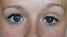
Ice Pack Test
Cooling may improve neuromuscular transmission. In a patient with myasthenia gravis who has ptosis, placing ice over an eyelid will lead to cooling of the lid, which leads to improvement of the ptosis.
Lightly placing ice that is in a surgical glove or that is wrapped in a towel over the eyelid will cool it within 2 minutes. [4] A positive test is clear resolution of the ptosis. [11, 3]
This test has a pooled sensitivity and specificity of 82% and 96%, respectively. However, the literature generally overestimates the test’s usefulness, since most of the studies were case-control designs. [12]
Additional Testing
Additional tests (eg, standard electromyography, single-fiber electromyography, repetitive nerve stimulation, assays for ACh receptor antibody [ARA]) are used to confirm the diagnosis of myasthenia gravis, but these tests usually are not available on an emergent basis. [11] The ARA test offers the highest specificity of these additional tests, since these other tests only reveal a disorder of neuromuscular conduction. [12]
Patients with respiratory distress should have an evaluation of pulmonary function, providing that the patient is not in obvious respiratory failure. This evaluation includes pulse oximetry, a measure of pulmonary function (ie, peak expiratory flow, forced expiratory volume in 1 second [FEV1]), and arterial blood gas (ABG) sampling to determine partial pressure of carbon dioxide (PCO2). [10] A negative inspiratory force (NIF) of 30 cm H2O or less or a forced vital capacity (FVC) of 20ml/Kg are indicative of a myathenic crisis and airway compromise. [7]
Evidence of hypoxemia, poor respiratory effort, or CO2 retention is an indication for intubation and mechanical ventilation. [10]
Patients can also undergo other testing, which, while useful for neurologists for outpatient care, does not apply in the ED. Examples of these tests are the Oculobulbar Facial Respiratory score and the MG-Activities of Daily Life (MG-ADL) score, which yield data on bulbar function but are most useful in long-term or serial assessment of patient function and therefore do not have a significant role in the ED. [13] A small retrospective study has shown that the MG-ADL score was higher on admission in patients requiring a stay in the ICU. [14]
Prehospital Care
Field personnel should recognize generalized muscle weakness of any etiology as a potential cause of respiratory failure. Patients with generalized weakness require transport to the hospital, and provisions for active airway intervention should be made en route.
Patients in frank respiratory arrest should be intubated and ventilated prior to transport, if possible. Suctioning of pulmonary secretions may be required to adequately ventilate the patient. Supplemental oxygen is indicated in all cases, and intravenous access is desirable prior to initiating transport.
Emergency Department Care
Patients with myasthenia gravis who are in respiratory distress may be experiencing a myasthenic crisis or a cholinergic crisis. Before these possibilities can be differentiated, ensuring adequate ventilation and oxygenation is important. [15] Patients with myasthenic crisis can develop apnea very suddenly, and they must be observed closely. Evidence of respiratory failure may be noted through ABG determination, pulmonary function tests, or pulse oximetry.
Airway maneuvers
Open the airway by suctioning secretions after positioning the jaw and tongue. Administer high-flow oxygen, and measure oxygen saturation by pulse oximetry. If respirations remain inadequate, ventilate by bag-valve mask while preparing to intubate. In the patient without an intact gag reflex, an oral airway may be placed.
Endotracheal intubation
Rapid sequence intubation should be modified, because depolarizing paralytic agents (eg, succinylcholine) have less predictable results in patients with myasthenia gravis. The relative lack of ACh receptors makes these patients relatively resistant to succinylcholine; therefore, higher doses must be used to induce paralysis. Once paralysis is achieved, it may be prolonged. [10]
A rapid-onset, nondepolarizing agent (ie, rocuronium, vecuronium) is the preferred paralytic agent for these patients. Although nondepolarizing agents delay the onset of paralysis, compared with succinylcholine, these medications do not result in unwanted prolonged paralysis. Following paralysis, intubation is accomplished as usual. ABG sampling guides ventilator settings.
Preliminary studies suggest that bilevel positive airway pressure (BiPAP) can prevent intubation in patients with myasthenic crisis without overt hypercapnia and should be considered in the patient who can be closely monitored. [10, 16] Hypercapnia present at the time of BiPAP initiation can predict failure and the need to proceed to endotracheal intubation. [17, 7]
Investigation and treatment
Once the airway is secured, investigation into the cause of the exacerbation of myasthenia gravis may proceed, with the most common reason for an exacerbation being infection, followed by inadequate treatment with cholinesterase inhibitors. However, up to 30% of patients will not have an identified cause of their exacerbation. [6] Differentiation from cholinergic crisis can proceed as described above.
In less severely ill patients, oral pyridostigmine can be administered until clinical improvement is seen. The patient should be closely observed and monitored during this trial. Other reasons for the exacerbation can then be investigated.
Although patients with myasthenia gravis can develop any common infection that can result in decompensation, the most likely source of infection is pulmonary. Cultures of blood, sputum, and urine may be indicated on an individual basis. Chest radiography is important in detecting pneumonia. Appropriate broad-spectrum antibiotics are indicated for sepsis and pneumonia. It is important to consider that fluoroquinolones and antibiotics may adversely affect cholinergic transmission in patients with myasthenia gravis, and these antibiotics should be avoided if possible.
Patients with myasthenia gravis are sensitive to high temperatures (core or ambient), and their muscle strength can improve when temperature is lowered with cooling measures or antipyretics.
Inpatient Care
Patients who present to the ED with myasthenic or cholinergic crisis will often require admission to an intensive care unit; [7] while patients with increasing muscle weakness of a less severe degree require admission to a monitored setting, because their course is unpredictable. [18] Patients with complications of the disease or treatment are admitted to a level of care corresponding to the nature and severity of the complication.
Patients with pneumonia should be admitted because they often are taking immunosuppressant medications and are at a high risk for aspiration pneumonia. [19]
Plasmapheresis has been found to be an effective short-term treatment of acute exacerbations of myasthenia gravis. Plasmapheresis removes circulating antibodies, including the autoimmune antibodies responsible for the disease. Clinical improvement takes several days to occur and lasts up to 3 weeks. [20] Because of the delayed onset of beneficial effects, plasmapheresis has limited utility in the ED setting, but often is used in the ICU setting. Some disagreement exists between various national societies on the amount of evidence supporting plasma exchange. The American Society for Apheresis considers therapeutic plasma exchange a first-line therapy for myasthenia gravis, whereas the American Academy of Neurology (AAN) states that the current evidence does not support or refute a benefit. [5, 21, 22]
Multiple observational and case series studies have shown a short-term benefit from plasma exchange, especially in myasthenic crisis. However, there is only 1 randomized clinical trial that showed no difference between the 2 treatment arms of plasma exchange versus intravenous immunoglobulin (IVIG). [23] Additionally the plasma exchange group had a higher mortality rate. [5]
Immunotherapy with intravenous gamma globulin appears to diminish the activity of the disease for unknown reasons. [24] The benefit begins within 2 weeks and may last for several months. Approximately 65% of patients with myasthenia gravis respond to intravenous gamma globulin. [25, 26] The AAN considers IVIG an effective therapy for moderate-to-severe cases of myasthenia gravis, as per their 2012 guidelines. [27]
Thymectomy is associated with clinical improvement in 85% of cases, and 35% of patients appear to have complete remission. [1] Patients past the age of puberty and younger than 50 years should have elective thymectomy as part of their treatment. [28]
The need for anticholinesterase medication fluctuates significantly in the postoperative period but overall is less than it was prior to thymectomy. [8]
Transfer, Consultations, and Monitoring
Patients with severe exacerbations of myasthenia gravis or cholinergic crisis should be transferred only after they have been stabilized and the airway has been secured. Persistent hypoxemia, hypercarbia, dysrhythmias, or unstable vital signs make transfer unwise, unless appropriate care cannot be delivered at the original facility.
Emergent consultation with a neurologist is indicated. Patients with severe exacerbations requiring intubation and mechanical ventilation are managed in an intensive care setting with appropriate consultation. [6]
With regard to patient monitoring, all patients with myasthenia gravis should be referred to a neurologist for ongoing care.
Questions & Answers
Overview
What is the prevalence of myasthenia gravis?
What causes myasthenic crisis in patients with myasthenia gravis?
Which medications may exacerbate myasthenia gravis?
Which clinical history findings are characteristic of myasthenia gravis?
Which physical findings are characteristic of severe myasthenia gravis?
How is a cholinergic crisis differentiated from a myasthenic crisis in myasthenia gravis?
Which physical findings are characteristic of neonatal myasthenia gravis?
What is the role of chest radiography in the emergency department evaluation of myasthenia gravis?
How are patients with myasthenia gravis screened for thymomas?
How is ptosis diagnosed in patients with myasthenia gravis?
How is a diagnosis of myasthenia gravis confirmed?
When is pulmonary function testing indicated in the evaluation of myasthenia gravis?
How is bulbar function assessed in myasthenia gravis?
What is included in prehospital care for myasthenia gravis?
What is the initial emergency department (ED) treatment for myasthenia gravis?
What is included in the ED investigation and treatment of exacerbation of myasthenia gravis?
What are the indications for inpatient care for myasthenia gravis?
When is patient transfer indicated for the treatment of myasthenia gravis?