Practice Essentials
Sodium levels are tightly controlled in a healthy individual by regulation of urine concentration and an intact thirst mechanism. Hypernatremia (defined as a serum sodium level >145 mEq/L) is rare in patients with preserved thirst mechanism. When hypernatremia is discovered in a patient, obtain urine osmolality and sodium levels. Check serum glucose level to ensure that osmotic diuresis has not occurred. The emergency department management of hypernatremia revolves around two tasks: restoration of normal serum tonicity and diagnosis and treatment of the underlying etiology. [1]
Hypernatremia is associated with a high mortality rate (>50% in most studies). Consequently, the emergency physician must be able to recognize and treat this condition. This article discusses the patients in whom hypernatremia should be suspected and how to initiate workup and administer appropriate treatment.
In general, hypernatremia can be caused by derangement of the thirst response or altered behavioral response thereto (primarily psychiatric patients, and elderly patients who are institutionalized), impaired renal concentrating mechanism (diabetes insipidus [DI]) secondary to kidney pathology (nephrogenic DI) or difficulty with the neurohormonal control of this concentrating mechanism (central DI), or by losses of free water from other sources.
Assessment and treatment of a hypernatremic patient focuses on two important questions:
-
What is the patient's volume status?
-
Is the problem acute or chronic?
Signs and symptoms of hypernatremia
Symptoms of hypernatremia tend to be nonspecific. Anorexia, restlessness, nausea, and vomiting occur early. These symptoms are followed by altered mental status, lethargy or irritability, and, eventually, stupor or coma. Musculoskeletal symptoms may include twitching, hyperreflexia, ataxia, or tremor. Neurologic symptoms are generally nonfocal (eg, mental status changes, ataxia, seizure), but focal deficits such as hemiparesis have been reported.
Workup in hypernatremia
When hypernatremia is discovered in a patient, obtain urine osmolality and sodium levels. Check the serum glucose level to ensure that osmotic diuresis has not occurred.
Serum sodium level indications include the following:
-
Serum sodium levels of more than 190 mEq/L usually indicate long-term salt ingestion
-
Serum sodium levels of more than 170 mEq/L usually indicate diabetes insipidus (DI)
-
Serum sodium levels of 150-170 mEq/L usually indicate dehydration
Head computed tomography (CT) scanning or magnetic resonance imaging (MRI) is suggested in all patients with severe hypernatremia. Traction on dural bridging veins and sinuses caused by movement of water from the brain and brain shrinkage can lead to intracranial hemorrhage, most often in the subdural space. Hemoconcentration from total body water loss may lead to dural sinus thrombosis. Imaging studies may indicate a central cause for hypernatremia.
Emergency department management of hypernatremia
The emergency department management of hypernatremia revolves around two tasks: restoration of normal serum tonicity and diagnosis and treatment of the underlying etiology. When possible, providing free water to a patient orally is preferred. Hypernatremia should not be corrected at a rate greater than 1 mEq/L per hour.
Using isotonic sodium chloride solution, stabilize hypovolemic patients who have unstable vital signs before correcting free water deficits, because hypotonic fluids quickly leave the intravascular space and do not help to correct hemodynamics. Once stabilization has occurred, free water deficits can be replaced either orally or intravenously.
Euvolemic patients can be treated with hypotonic fluids, either orally or intravenously (ie, dextrose 5% in water solution [D5W], quarter or half isotonic sodium chloride solution), to correct free fluid deficits.
Hypervolemic patients require removal of excess sodium, which can be accomplished by a combination of diuretics and D5W infusion. Patients with acute renal failure may require dialysis.
Traditionally, correction of hypernatremia begins with the following calculation of fluid deficit:
Free Water Deficit = Body Weight (kg) X Percentage of Total Body Water (TBW) X ([Serum Na /140] - 1)
Pathophysiology
Water homeostasis is maintained by a balance between water intake and the combined water loss from renal excretion, respiratory, skin, and GI sources. Under normal conditions, water intake and losses are matched. To maintain salt homeostasis, the kidneys similarly adjust urine concentration to match salt intake and loss. See the image below.
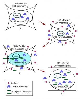 Figure A: Normal cell. Figure B: Cell initially responds to extracellular hypertonicity through passive osmosis of water extracellularly, resulting in cell shrinkage. Figure C: Cell actively responds to extracellular hypertonicity and cell shrinkage in order to limit water loss through transport of organic osmolytes across the cell membrane, as well as through intracellular production of these osmolytes. Figure D: Rapid correction of extracellular hypertonicity results in passive movement of water molecules into the relatively hypertonic intracellular space, causing cellular swelling, damage, and ultimately death.
Hypernatremia results from disequilibrium of one or both of these balances. Most commonly, the disorder is caused by a relative free water loss, although it can be caused by salt loading. The various ways in which these equilibria can be disturbed are discussed in Causes.
When hypernatremia (of any etiology) occurs, cells become dehydrated. Either the osmotic load of the increased sodium acts to extract water from the cells or a portion of the burden of the body's free water deficit is borne by the cell. (Sodium, primarily an extracellular ion, is actively pumped out of most cells and is the primary determinant of serum osmolarity.) Dehydrated cells shrink from water extraction.
Cells immediately respond to combat this shrinkage and osmotic force by transporting electrolytes across the cell membrane, thus altering rest potentials of electrically active membranes. After an hour of hypernatremia, intracellular organic solutes are generated in an effort to restore cell volume and to avoid structural damage. This protective mechanism is important to remember when treating a patient with hypernatremia. Cerebral edema ensues if water replacement proceeds at a rate that does not allow for excretion or metabolism of accumulated solutes.
The effects of cellular dehydration are seen principally in the CNS, where stretching of shrunken neurons and alteration of membrane potentials from electrolyte flux lead to ineffective functioning. If shrinkage is severe enough, stretching and rupture of bridging veins may cause intracranial hemorrhage.
Epidemiology
Frequency
United States
Hypernatremia occurs in approximately 1% of hospitalized patients. The condition usually develops after hospital admission. An incidence closer to 2% has been reported in debilitated elderly persons and in breastfed infants. [2, 3]
A retrospective study by Otterness et al in a single emergency department found that out of more than 57,400 adult patients in whom sodium was measured, mild, moderate, and severe hypernatremia were found in 1%, 0.2%, and less than 0.1% of patients, respectively. [4]
International
Pediatric patients in developing nations may be at increased risk for hypernatremia because infant feeding may be complicated by poor maternal milk production (secondary to nutritional status) and errors in reconstitution of powdered formula.
An Italian study, by Giordano et al, found that hypernatremia accounted for just 4.4% of all cases of electrolyte imbalance in the study’s emergency department (compared with 44% for hyponatremia). [5]
Mortality/Morbidity
The mortality rate from hypernatremia is high, especially among elderly patients. Mortality rates of 42-75% have been reported for acutely evolving hypernatremia and 10-60% for chronic hypernatremia.
The aforementioned study by Otterness and colleagues of adult patients in a single emergency department found the adjusted odds ratios (ORs) for hospital mortality in persons with mild, moderate, or severe hypernatremia were 3.65, 8.58, and 55.75, respectively. The risk of death for patients with hypernatremia was greater than that for those with hyponatremia. [4]
Because patients with hypernatremia often have other serious comorbidities, precisely evaluating the degree of mortality directly due to hypernatremia is difficult. Morbidity in survivors is high, with many patients experiencing permanent neurologic deficits.
Most deaths are due to an underlying disease process, rather than the hypernatremia itself. Delay in treatment (or inadequate treatment) of hypernatremia increase mortality.
A study by Vedantam et al reported that in patients with severe traumatic brain injury (TBI), an independent association exists between the development of hypernatremia after hospital admission, whether mild, moderate, or severe, and an increased likelihood of early mortality. The investigators cited mortality hazard ratios for mild, moderate, and severe hypernatremia of 3.4, 4.4, and 8.4, respectively, in severe TBI. [6]
A study by Huang et al indicated that in patients with chronic kidney disease, hypernatremia is associated with an increased risk for all-cause mortality and for deaths unrelated to cardiovascular problems or malignancy. Hyponatremia was found to be associated with an increased risk for the same, as well as for cardiovascular- and malignancy-related mortality. The study included 45,333 patients with stage 3 or 4 chronic kidney disease, 9.2% of whom had dysnatremia. [7]
A Turkish study, by Ates et al, indicated that in patients presenting to emergency departments with severe hypernatremia, independent risk factors for mortality included low systolic blood pressure, low pH, Na+ level over 166 mmol/L, increased plasma osmolarity, a mean sodium reduction rate of -0.134 mmol/L/h or less, dehydration, and, pneumonia. The retrospective study included 256 patients. [8]
A study by Castello et al indicated that in patients with sepsis, the presence of hypernatremia or moderate to severe hyponatremia, at presentation to the emergency department, is an independent risk factor for mortality. For hypernatremia, the hazard ratios for 7- and 30-day mortality were 3.52 and 2.14, while for mild to moderate hyponatremia they were 4.89 and 1.79. [9]
In hospitalized patients, persistent hypernatremia and protracted hypotension have been associated with a very poor prognosis. A study by Jung et al indicated that in patients with community-acquired hypernatremia, an independent association exists between admission to the hospital from the emergency department and hospital mortality, with the same being true for oral intake restriction, mean arterial pressure, and respiratory rate. Also with regard to hospital mortality, multivariate analysis revealed a peak sodium level in the moderate or severe range to be an independent risk factor. [10]
The aforementioned study by Giordano et al stated that the great majority of electrolyte imbalances encountered in the report were associated with other systemic diseases. Dividing the study population into young, middle aged, and elderly, the investigators found that in the young group, electrolyte imbalances were most commonly associated with gastrointestinal disease, while in the middle-aged group, they were most often associated with cardiovascular disease, and in the elderly group, with cardiovascular disorders and lung disease. [5]
A retrospective study by Lindner et al found that adult emergency department patients in the study with acute exacerbation of chronic obstructive pulmonary disease (AECOPD) were more likely to have hyponatremia or hypernatremia than were those without AECOPD (5% vs 0.6%, respectively, for hypernatremia). [11]
Sex
Hypernatremia is diagnosed in males and females in equal numbers.
Age
Patients who present to the hospital with hypernatremia tend to be at the extremes of age. Breastfed infants occasionally present with hypernatremia in the first weeks of life, and elderly patients who are institutionalized are especially heavily represented.
A study from Japan, by Imai et al, indicated that the prevalence of hyponatremia in the emergency department is greater in elderly patients than in adults aged 18-64 years (2.6% vs 0.7%, respectively). Moreover, moderate to severe hypernatremia had a prevalence of 1.0% in the elderly group, versus 0.1% in other adults. Although higher prevalence in the elderly occurred year round, the investigators found seasonal variation, with the prevalence of moderate to severe hypernatremia in the older cohort being greatest in winter. [12]
-
Figure A: Normal cell. Figure B: Cell initially responds to extracellular hypertonicity through passive osmosis of water extracellularly, resulting in cell shrinkage. Figure C: Cell actively responds to extracellular hypertonicity and cell shrinkage in order to limit water loss through transport of organic osmolytes across the cell membrane, as well as through intracellular production of these osmolytes. Figure D: Rapid correction of extracellular hypertonicity results in passive movement of water molecules into the relatively hypertonic intracellular space, causing cellular swelling, damage, and ultimately death.


