Background
Gingivitis is an inflammatory process limited to the mucosal epithelial tissue surrounding the cervical portion of the teeth and the alveolar processes. Gingivitis has been classified by clinical appearance (eg, ulcerative, hemorrhagic, necrotizing, purulent), etiology (eg, drug-induced, hormonal, nutritional, infectious, plaque-induced), and duration (acute, chronic). The most common type of gingivitis is a chronic form induced by plaque. Gingivitis is shown in the image below.
During the global COVID-19 pandemic, many patients found it difficult to access preventive dental care. This has increased the number of people at risk for poor oral health, especially among those of lower socioeconomic status. [1]
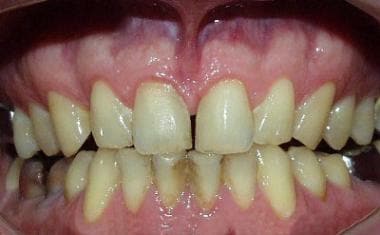 Moderate chronic gingivitis. Note that the papillae are edematous and blunted. They may bleed with brushing. Note areas of edema overlying some of the root areas. Pallor is seen in these areas. Image courtesy of Robert J. Lindberg, DMD.
Compare the image above to a healthy mouth, below.
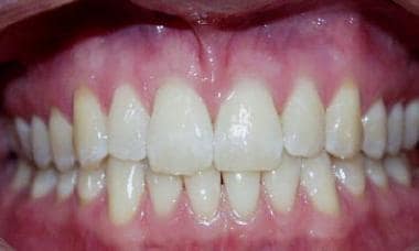 Healthy mouth and gingiva. Note the healthy light pink color of the gingiva. The interdental papillae are sharp and fill the interdental space. No local edema is present. Image courtesy of Robert J. Lindberg, DMD.
Acute necrotizing ulcerative gingivitis (ANUG, ie, trench mouth) is an acute infectious gingivitis. The term trench mouth was coined in World War I when ANUG was common among trench-bound soldiers.
Pathophysiology
The most common type of gingivitis involves the marginal gingiva and is brought on by the accumulation of microbial plaques in persons with inadequate oral hygiene. Gingivitis proceeds through an initial stage to produce early lesions, which then progress to advanced disease.
The initial stage of an acute exudative inflammatory response begins within 4 or 5 days of plaque accumulation. Both gingival fluid and transmigration of neutrophils increase. Deposition of fibrin and destruction of collagen can be noted in the initial stage. At approximately 1 week, transition to early lesions is marked by the change to predominately lymphocytic infiltrates. Monocytes and plasma cells also may be present. With time, lesions become chronic and are characterized by the presence of plasma cells and B lymphocytes. As chronic local inflammation progresses, pockets develop where the gingiva separates from the tooth. These pockets deepen and may bleed during tooth brushing, flossing, and even normal chewing. As this persistent inflammation continues, periodontal ligaments break down and destruction of the local alveolar bone occurs. Teeth loosen and eventually fall out.
ANUG is a completely different syndrome caused by acute infection of the gingiva with organisms such as Prevotella intermedia, alpha-hemolytic streptococci, Actinomyces species, or any of a number of different oral spirochetes. ANUG may result in accelerated destruction of affected tissues, as well as local or systemic spread of infection.
Noma (cancrum oris) is a syndrome in which ANUG spreads beyond the gingiva. The infection invades local tissues of the mouth and face.
Etiology
Although bacteria play a role in all forms of gingivitis, the primary cause of chronic gingivitis is inadequate oral hygiene.
Risk factors
Use of tobacco or ethanol is thought to be a risk factor.
Immune incompetence is observed more frequently in HIV-infected children. As their CD4 counts decline, incidence of gingivitis may increase. Diabetes mellitus increases the risk of gingivitis and periodontitis.
Drug-induced causes
The list of drugs that cause gingivitis and gingival bleeding is extensive.
Gingival bleeding may occur with the use of anticoagulants and fibrinolytic agents.
Phenytoin, oral contraceptive agents, and calcium channel blockers may cause gingival hyperplasia.
Gingivitis has been observed with use of protease inhibitors (eg, saquinavir, ritonavir), vitamin A and analogues, danazol, pentamidine, misoprostol, methotrexate, and gold compounds.
Gingivostomatitis has been observed in exposure to arsenic, gold, bismuth, mercury, nickel, sulfur dioxide, lead, thallium, zinc, methyl violet, and topical chlorhexidine.
Acute necrotizing ulcerative gingivitis
Acute necrotizing infection may occur as a complication of chronic gingivitis in situations in which hygiene is abandoned completely or host defenses are weakened.
ANUG is the result of soft tissue invasion by ubiquitous organisms and is not believed to be contagious. It is a risk wherever poor sanitation, diet, or oral hygiene is present. Living near livestock is an additional risk factor.
Other causes
Other causes include the following:
-
Inadequate plaque removal
-
Allergic reactions
-
Chronic debilitating disease
-
Poor nutrition
-
Lack of periodic dental examinations
Epidemiology
United States statistics
Frequency is difficult to determine because of the lack of agreement on measurement criteria. Many people believe that gingivitis begins in early childhood and that 9-17% of children aged 3-11 years have gingivitis. At puberty, prevalence rises to 70-90%. In recent years, periodontal disease, the endpoint of chronic gingivitis, slowly has decreased among adult Americans. However, chronic periodontitis is still the most prevalent chronic inflammatory condition in the elderly. [2]
ANUG may be a clinical problem in immunocompromised patients during chemotherapy. Gingivitis and resulting periodontal disease are seen more frequently in patients with either diabetes or HIV infection.
International statistics
Studies in Australia, Sweden, England, and Switzerland report gingivitis in 48-85% of children aged 3-6 years, but whether this range reflects population differences or whether it is due to different criteria used to define the disease is difficult to know. In adolescence, incidence around the world is comparable to US data (70-90%). ANUG may be found in areas where those at risk, particularly children, face poor living conditions. More recent publications show several cases in areas such as Nigeria, where ANUG and noma were observed in children younger than 14 years. [3]
Sex- and age-related demographics
Gingivitis is slightly more prevalent in males than in females because females tend to have better oral hygiene.
Adults are most commonly affected.
Children from sub-Saharan regions of Africa may be at risk for ANUG because of poor living conditions.
Prognosis
Untreated chronic gingivitis eventually results in tooth loss. After an initial cleaning and scaling in its early stages, gingivitis usually is reversible with good dental hygiene. Gingivitis generally responds well to appropriate treatment.
ANUG responds to treatment if host defenses are intact. Noma requires aggressive treatment with antibiotics and local debridement. The usual course is acute, relapsing, intermittent, and chronic.
Morbidity/mortality
Periodontal disease has been shown in some studies to be an associated factor in coronary artery disease (CAD) and cerebrovascular disease/ischemic stroke. [4, 5, 6, 7] Elevated levels of chronic inflammation (eg, C-reactive protein) have been shown to fall after treatment of periodontal disease. These elevated markers have clear association with vascular disease, so treatment of periodontal disease may theoretically have an impact on CAD and ischemic CNS disease. [8] However, a clear cause-and-effect relationship has not been demonstrated between treatment of periodontal disease and improvement of atherosclerotic diseases or outcomes. [9] A study that induced gingivitis in healthy volunteers was associated with a clear increase in inflammatory markers. [10]
A study by Sen et al that included data from 10,362 stroke-free participants, and 584 participants that had incident ischemic strokes over a 15-year period reported that periodontal disease was associated with cardioembolic (hazard ratio, 2.6; 95% confidence interval, 1.2-5.6) and thrombotic (hazard ratio, 2.2; 95% confidence interval, 1.3-3.8) stroke subtypes. The study also added that a lower stroke risk was associated with regular dental care utilization (hazard ratio, 0.77; 95% confidence interval, 0.63-0.94). [11]
A large study that enrolled 805 patients with a first myocardial infarction (MI) and 805 control patients without MI concluded that the risk of a first myocardial infarction was significantly increased in patients with periodontitis. The study found that 43% of MI patients vs 33% of matched controls had mild to severe periodontitis. After researchers controlled for smoking, diabetes, education, and marital status, individuals with periodontitis had a 28% increased risk of MI. [12, 13]
Periodontal disease in pregnancy has been associated with an increase in preterm birth and adverse pregnancy outcomes. [14] However, treatment of periodontal disease in pregnancy has not been shown to improve pregnancy outcomes. [15] Periodontitis coexisting with bacterial vaginosis is associated with higher vaginal bacterial counts. [16] Periodontitis in a person with diabetes has been associated with exacerbation of both conditions. [17, 18] Treatment of periodontitis in persons with type 2 diabetes has improved glycemic control. [19]
In the evolving picture of the global COVID-19 pandemic, it has been observed that preexisting periodontal disease is a risk factor for developing severe diease. [20]
The association of periodontal disease and rheumatoid arthritis (RA) is established. Treatment for RA often improves the periodontitis, but both require treatment concurrently. It is felt that RA predisposes to periodontal disease, but it is not clear if the inverse is true. [21]
Severe periodontal disease, as shown in the image below, may occur.
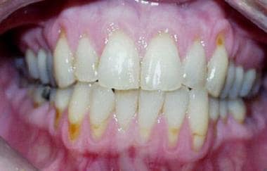 Severe periodontal disease. Loss of the gingival tissue is seen, making the teeth appear long. Even more effacement of the papillae is present. Heaped up ridges are observed in the areas overlying the roots. Image courtesy of Robert J. Lindberg, DMD.
Chronic gingivitis leads to tooth loss. ANUG may progress into the local soft tissues of the mouth, resulting in noma or cancrum oris, or may spread hematogenously to any other part of the body.
Complications
Gingivitis is not a direct significant threat to the health of a healthy individual, but it can contribute to illness and cause local and systemic complications.
ANUG that progresses to noma is associated with a mortality rate as high as 70% without proper antibiotics and debridement.
The most common complication of chronic gingivitis is progression to periodontal disease and tooth loss. Areas of chronic gingivitis may predispose the individual to the development of odontogenic abscesses by allowing a route of bacterial invasion into the periodontal space from the gingival pocket. ANUG may be locally destructive and may result in local spread of infection into the surrounding tissues (Vincent angina and noma [cancrum oris]). Potential also exists for systemic spread of infection.
Osteomyelitis of alveolar bone may arise but is uncommon.
Any dental procedures involving manipulation that causes bleeding may result in endocarditis. The presence of gingivitis increases this risk by making the gingiva more likely to bleed with simple manipulation (eg, dental scaling). Bacteria containing plaque accumulation in the gingival pockets are in direct proximity to the areas of disrupted gingiva, increasing the likelihood of bacteria escaping into the general circulation.
Patient Education
Good oral hygiene, including brushing and flossing, treats and prevents chronic gingivitis. If flossing is too much of a bother, then plaque-reducing rinses, used daily, have proven benefit.
Studies show electric toothbrushes to be more effective than manual brushes in preventing gingivitis. [22, 23]
Certain toothpastes, both herbal and non-herbal, have additional benefit.
-
Healthy mouth and gingiva. Note the healthy light pink color of the gingiva. The interdental papillae are sharp and fill the interdental space. No local edema is present. Image courtesy of Robert J. Lindberg, DMD.
-
Moderate chronic gingivitis. Note that the papillae are edematous and blunted. They may bleed with brushing. Note areas of edema overlying some of the root areas. Pallor is seen in these areas. Image courtesy of Robert J. Lindberg, DMD.
-
Severe periodontal disease. Loss of the gingival tissue is seen, making the teeth appear long. Even more effacement of the papillae is present. Heaped up ridges are observed in the areas overlying the roots. Image courtesy of Robert J. Lindberg, DMD.


