Practice Essentials
In evaluating humerus injuries, classifying the fracture and, if necessary, reducing and immobilizing the fracture are essential. Eighty percent of proximal humerus fractures are nondisplaced or minimally displaced and, therefore, can be managed nonoperatively. Associated injuries are common in patients with osteoporosis. Proximal humerus fracture accounts for 6% of all fractures and is the third most common osteoporotic fracture, after the distal radius and vertebra. Approximately 85% of proximal humerus fractures occur in individuals older than 50 years. Distal humerus fractures are associated with ipsilateral proximal forearm fractures. In adults, fractures of the distal humerus account for approximately 2% of all fractures and a third of all humerus fractures. In younger individuals, these fractures are primarily caused by high-energy traumas; in the elderly, by low-energy falls. [1] Rarely, vascular or nerve injuries are associated with humerus fractures. Radial nerve palsy associated with fractures of the shaft of the humerus is the most common nerve lesion complicating fractures of long bones. [2, 3, 4, 5, 6, 7, 8, 9, 10, 11]
Causes
Humerus fractures are caused by direct trauma to the arm or shoulder or by axial loading transmitted through the elbow. Attachments from pectoralis major, deltoid, and rotator cuff muscles influence the degree of displacement of proximal humerus fractures.
Humeral stress fractures occur with overhead throwing and occasionally with violent muscle contractions. These types of fractures are documented most commonly in baseball. As with other stress fractures, an increase in activity or stress on immature or unconditioned bone is the likely culprit. [12, 13, 14]
The most common cause of proximal humeral fractures is a fall from standing, followed by motor vehicle accident and a fall involving stairs. Additional mechanisms include violent muscle contractions from seizure activity, electrical shock, and athletic-related trauma. Proximal humeral fractures are most often closed.
Causes of humeral diaphyseal fractures include a fall from standing, a motor vehicle accident, a fall from height, and pathology related.
Distal humerus fractures are primarily caused by high-energy traumas, and in the elderly, they are most often caused by by low-energy falls. [1]
Diagnosis
Pain occurs with palpation or movement of the shoulder or elbow. Ecchymosis and edema are usually present.
Perform a careful neurovascular examination. Radial nerve injury following humerus shaft fractures is relatively common.
Humeral stress fractures are often missed. Patients have described a prodrome of milder, chronic pain with focal tenderness prior to the injury while throwing. These fractures are generally mid to distal shaft spiral fractures that are minimally displaced. [12, 13, 14]
Fractures that occur spontaneously, without apparent injury, suggest a pathologic fracture.
For the distal and diaphyseal humerus fractures, anteroposterior and lateral views of the humerus, as well as transthoracic and axillary views of the shoulder, should be adequate to visualize a fracture. CT scans are helpful if radiographs are unclear.
Treatment
Minimize the patient's movement and provide adequate analgesia to make the patient comfortable in the acute care setting. As with all fractures, provide adequate outpatient analgesia, especially during the first few days. Narcotic analgesia may be appropriate.
Operative treatment decisions are based primarily on the number of segments involved and degree of displacement. Most fractures are displaced minimally and treated conservatively. Often, 3- and 4-part fractures require surgical management because of damage of the vasculature of the humeral head. [3, 9, 10, 15, 16]
Diaphyseal fractures are classified as simple, wedge, or complex (comminuted).
Most isolated proximal and diaphyseal humerus fractures can be managed by an orthopedist in an outpatient setting. Even patients with fractures that may eventually require surgery generally may be discharged with early follow-up care if fracture is otherwise uncomplicated.
Surgical stabilization is indicated for fractures that cannot be adequately reduced or if fracture reduction cannot be controlled with functional bracing because of patient obesity, head trauma, or soft tissue injuries. [17]
The distal humerus has a triangular shape built of 2 columns and a “tie arch.” Successful management of distal humerus fractures depends on correct reduction of the fracture, reconstruction of the articular surface if needed, stability and rigidity of the fixation, and appropriate rehabilitation. [18]
Open fractures represent a surgical emergency and require extensive irrigation. Administer prophylactic antibiotics, such as cephalexin or gentamicin. Penetrating trauma requires particular neurovascular scrutiny.
Glenohumeral dislocation in conjunction with a proximal humerus fracture requires orthopedic evaluation.
Floating elbow (an ipsilateral humerus and forearm fracture) requires operative repair.
For proximal humerus fractures, complete union is expected at 6-8 weeks. Older patients often exhibit a functional decrease in shoulder range of motion (ROM). Diaphyseal fractures have a high rate of union. Residual angulation is well tolerated because of compensation by shoulder and elbow ROM.
Classification systems
Classification systems include Riseborough and Radin, which classifies distal humerus fractures according to the state of the condylar fragments; Lecestre et al, which defines supracondylar, extra-articular condylar, articular intercondylar, and comminuted fractures; Jupiter, which is based on intraoperative observations, describing high T, low T, Y, H, medial, and lateral lambda fractures; and Dubberley, which distinguishes between fracture types involving the capitellum and trochlea and comprises techniques for treatment. Internationally, the American Academy of Orthopaedic Surgeons (AO) classification is most commonly used, categorizing fractures as extra-articular, partial articular, and articular. [18]
The Neer classification system is the commonly used terminology to describe proximal humerus fractures. [4] The Neer classification for proximal humerus fractures is based on 4 fracture parts: the greater tuberosity, the lesser tuberosity, the humeral head, and the humeral shaft. If any of the 4 segments is separated by more than 1 cm from its neighbor or is angulated more than 45°, the fracture is said to be displaced. One-part fractures are nondisplaced fractures or fractures with minimal displacement. Two-part fractures are fractures in which only a single segment is displaced in relation to the other three. Three-part fractures occur when two segments are displaced in relation to the other two parts. Four-part fractures exist when all the humeral segments are displaced. [19]
Epidemiology
Humeral diaphyseal fractures account for 1.2% of all fractures. [20] Proximal humerus fractures account for 5.7% of all fractures. [20]
Proximal humerus fractures are more common in elderly persons, with the average age of 64.5 years, [21, 15] and are the third most common fracture after hip fractures and distal radius fractures. [3, 22]
Humeral diaphyseal fractures occur in a slightly younger population, with the average age being 54.8 years. [21]
In adults, fractures of the distal humerus account for approximately 2% of all fractures and a third of all humerus fractures. [1]
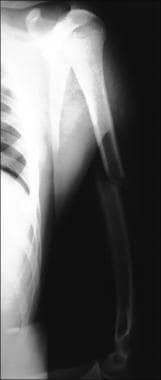
(See the image below.)
Fracture patterns are similar across all ages, though older people are more prone to fracture because of osteoporosis. A humerus fracture in a child with an inconsistent injury mechanism should raise suspicion for abuse and trigger further investigation. Young patients presenting with humeral diaphyseal fractures after high-energy injuries frequently have multiple injuries. Approximately 5% of these patients with humeral diaphyseal fractures present with spinal fractures or complex foot fractures, and about 4% present have pelvic or proximal tibial fractures. [21] Older patients tend to present with other fractures in the ipsilateral arm, usually distal radius fractures. [21]
In an epidemiologic survey of 1800 low-energy humeral fractures in an emergency department in Parma, Italy, the following were identified [16] :
-
Predominance in women: 78%.
-
Fractures of the proximal humerus represented the largest majority of humerus fractures: >85 %.
-
Incidence progressively increased with age (more than 60-fold in women and 20-fold in men).
-
Simultaneous fractures (hip in particular) were frequent, especially after 85 years of age (1 out of 8 cases).
-
Diaphyseal humerus fracture.
-
Neer classification.


