Practice Essentials
Forearm fractures are common fractures among both children and adults. These fractures are relatively more complex than other long bone fractures. The spectrum of such fractures includes isolated radius and ulna fractures, combined fractures, Galeazzi fractures, and Monteggia fractures. [1] Fractures of both the radius and the ulna together are usually the result of a fall onto an outstretched hand (FOOSH) injury. [2] These injuries can also occur as the result of a direct blow.
The forearm consists of 2 relatively parallel bones that connect 2 joints: elbow and wrist. The 2 bones themselves form joints that help in supination and pronation; therefore, forearm fractures are considered intra-articular fractures. Proper management of such fractures is necessary to restore forearm functions, including supination and pronation, elbow and wrist movements, and handgrip strength. [1]
Fractures of the forearm are classified as involving the proximal, middle, or distal shaft. Injuries to this area are intimately associated with the elbow and wrist.
In addition to diagnosis and follow-up, radiologic imaging plays a decisive role in the treatment of distal forearm fractures. Computed tomography (CT) and direct CT arthrography have become important tools in the treatment of intra-articular distal radius fractures. [3]
The upper extremity is the most commonly injured extremity; thus, it is imperative that emergency physicians are familiar with appropriate evaluation and management.
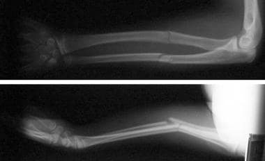
(See the image below)
The pediatric musculoskeletal system differs from that of adults. [4] The relatively greater amount of cartilage and collagen reduces the tensile strength of bone, making propagation of fractures unlikely. Forearm fractures are less identifiable on radiographs in children than in adults. Also unique to children is the growth plate, or physis (see Salter-Harris Fractures). Depending on the severity of the injury, these fractures can significantly impair further growth and functioning of the limb.
The upper extremity is involved in nearly half of all fractures seen, and wrist fractures account for about one third of these. Specifically, fractures of the forearm account for 10-45% of pediatric fractures, with most occurring distally. [5] A study looking at injuries related to skateboarding found that fracture of the radius or ulna (or both) was the most common injury (48.2%). [6]
Distal forearm fractures are prevalent among the Medicare population. Many patients who sustain these fractures have poor bone health and are at increased risk for subsequent fractures. Bone mineral density testing is underused nationwide in patients sustaining distal forearm fractures despite current guidelines. Orthopedic surgeons should ensure proper testing of patients because this may be an important time point for intervention. [7]
Signs and symptoms
Patients usually have localized pain, tenderness, and swelling at the fracture site. Any puncture or break in the skin over a fracture site should be considered evidence of an open fracture unless proven otherwise.
Tenderness or prominence of the radial head may be the only physical finding in patients with reduced Monteggia lesion or radial head fracture.
Diagnosis
Anteroposterior and lateral radiographic views of the wrist, forearm, and elbow are required when forearm fracture is suspected from clinical findings.
Forearm radiographs, which include distal joints, are inadequate for absolutely excluding associated wrist and elbow injuries, as diagnosis of radioulnar dislocation requires the x-ray beam to be centered at the joint.
One of the most common complications of these injuries is painful limitation of the range of motion, especially of pronation and supination, which is often due to underdiagnosed torsional deformity. New methods have been developed to make these torsional differences visible and quantifiable through the use of sectional imaging. [8]
Management
When a patient presents with a forearm injury, the astute emergency physician will rely on a focused history and precise examination, applied anatomic knowledge, and strong radiographic interpretative skills to avoid missed injuries and complications. [9]
The spectrum of forearm fractures includes isolated radius and ulna fractures, combined fractures, Galeazzi fractures, and Monteggia fractures. Proper management of such fractures is necessary to restore forearm functions, including supination and pronation, elbow and wrist movements, and handgrip strength. [1]
Immobilize the forearm and the upper arm, and provide effective analgesia unless the patient has other injuries with the potential for hemodynamic or respiratory instability. Specific treatment strategies include the following:
-
Nightstick fracture: Immobilize the fracture with a long-arm splint with 90° of elbow flexion and with the hand in a neutral position. Orthopedic referral is required.
-
Monteggia fracture: Immobilize with a long-arm splint (with elbow flexed 90° and forearm neutral); children may be treated by reduction and casting; adults require admission for open reduction internal fixation (ORIF).
-
Galeazzi fracture: Immobilize with a long-arm splint (with elbow flexed 90° and forearm pronated); treatment requires admission for ORIF.
-
Concomitant radius and ulna fractures: Treatment usually requires admission for an urgent ORIF, although in children younger than 10 years, these fractures may be treated by casting alone if reduced to less than 10° of angulation.
-
Torus (greenstick) fracture: Apply a long-arm cast for 4-6 weeks when angulation is less than 10°; all cases require orthopedic referral. This type of fracture occurs with only a moderate degree of trauma.
Epidemiology
Forearm fractures are among the most frequently encountered orthopedic injuries in children. Maintenance of satisfactory alignment can be problematic, and postreduction displacement with resultant malunion can occur. [10]
Because of osteoporosis, postmenopausal women have a higher rate of forearm fracture than other adults. When the mechanism of injury seems trivial, suspect a pathologic fracture associated with a cyst or a tumor. Forearm fractures in older persons are associated with increased risk of future vertebral and hip fractures. Forearm fractures are less common among black individuals because of a lower incidence of osteoporosis.
The National Electronic Injury Surveillance System database showed that among patients from birth to 19 years of age, fracture of the forearm was found to be the most common type of fracture, accounting for 17.8% of all fractures in the entire study population. Finger and wrist fractures were the second and third most common. [11]
In infants and toddlers, forearm fractures have no sex predilection. In children older than 2 years, forearm fractures are more common in boys than in girls. In older persons, fractures are more common in women than in men.
Open fractures of the forearm from gunshot wounds are serious injuries that carry high rates of nonunion and infection. Fractures with significant bone defects are at increased risk of nonunion and should be treated with stable fixation and proper soft tissue handling. Ulna fractures are at particularly high risk for deep infection and septic nonunion and should be treated aggressively. Forearm fractures from gunshots should be followed until union to identify long-term complications. [12]
Patient Education
For excellent patient education resources, visit the First Aid and Injuries Center. Also see the patient education article Broken Arm.
-
Fractures of the radius and ulna with dorsal angulation of distal fragments.
-
Torus fracture of the radius.
-
Torus fracture of the radius.


