Practice Essentials
Carpal dislocations represent a continuum of wrist injury that can lead to lunate or perilunate dislocation. The lunate cup commonly is directed in a volar direction in dislocation because of the mechanism of the injury. Perilunate dislocations result from dislocation of the distal carpal row. [1] The capitate normally rests within the lunate cup, as seen on a lateral view. With perilunate dislocations, the capitate is seen most commonly as dorsal, but it also may be volar to the lunate on lateral x-ray evaluation. As a result of the stresses involved, scaphoid fractures often accompany perilunate dislocation. [2, 3] Carpal instability may take many forms and represents a spectrum of injury including scapholunate dissociation, lunate and perilunate dislocations, scaphoid fracture, and other intercarpal instabilities. [4, 5, 6, 7, 8]
Perilunate injuries are radius-to-ulnar disruption injuries through the scaphoid and capitate bones and ligaments at some distance from the lunate. They usually occur along the greater arc of the carpus. Lunate dislocations are the least common. In lunate dislocation, the proximal articular margin and central axis of the capitate remain aligned with the distal articular surface and axis of the distal radius. The lunate is usually displaced volarly, disrupting both the capitolunate and lunatoradial joints. Both the distal and proximal sets of articular arcs are disrupted. [8]
The mechanism of injury is usually a fall onto an outstretched hand with hand rotation, which may lead to a variety of injuries. These injuries range from scapholunate strain to carpal dislocation, with scaphoid fracture at the end of the spectrum. Unfortunately, most of these injuries are not diagnosed in the ED. The injury may lead to chronic pain and instability of the wrist. [9, 10, 11]
Plain x-rays of the wrist, both anteroposterior (AP) and lateral views, are essential to diagnose wrist dislocations (as well as other carpal instabilities). CT scanning or conventional tomography is not usually needed to diagnose perilunate dislocations. [12, 13, 14, 15]
Because of the severity of pain, narcotic pain medication often is required for the first 3 days. [8]
(A lunate dislocation is shown in the radiographs below.)
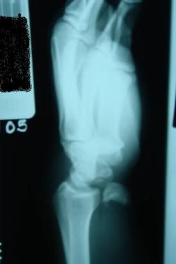 Dislocations, wrist. Lateral view of a lunate dislocation, with the classic teacup sign.
Carpal stability is based on the lunate as the central anchor for the proximal and distal carpal rows. The lunate is apposed to the radius, and the capitate rests within the lunate cup. The proximal row of carpals is connected by interosseous ligaments. Carpal stress is characterized as radial or ulnar, with some degree of axial loading. This stress is translated to all bones. Ligamentous injury results in a spectrum of injuries, including lunate and perilunate dislocations. The lunate-scaphoid ligaments may not be disrupted; if this is the case, scaphoid fracture may occur.
Patients usually present to the ED fairly soon after a fall onto an outstretched hand. The mechanism of injury is ulnar deviation of the wrist coupled with dorsiflexion. The resulting intercarpal supination places great stress on the carpals. The result can be a lunate or perilunate dislocation. [16] Often, the only symptom is wrist pain. Frequently, lunate and perilunate dislocations are not recognized at the time of the initial ED visit. [17] This emphasizes the need to consider lunate or perilunate dislocation when a patient returns to the ED a second or third time for what appears to be chronic wrist pain following an injury. The patient may have diffuse pain on palpation that is difficult to distinguish from other causes of wrist pain, including scapholunate strain, scaphoid fracture, triangular fibrocartilage complex tears, and other disorders.
Incidence of wrist injuries is estimated as 2.5% of ED visits. Wrist dislocations represent a very small portion of these visits. Because of this small proportion of wrist dislocations, they can be easily missed on initial presentation to the ED. The morbidity of wrist dislocations is tied to the frequently missed diagnosis of lunate or perilunate dislocation in the ED. [17] Often, patients are not diagnosed with these injuries until weeks following the initial injury, which can result in median nerve dysfunction, posttraumatic arthritis, and reduced functionality. [18] Many patients with undiagnosed wrist dislocation have chronic pain. Carpal instability, including radiocarpal instability, is a frequent complication. [18] Avascular necrosis of the lunate, Kienbock disease, is a potential complication of lunate dislocation.
Treatment
Prehospital care includes assessment for other injuries that may accompany the wrist injury. If no other injuries are identified, splint the wrist. Patients may be transported in their private vehicles, but the prehospital provider must emphasize the potential seriousness of the injury. Under no circumstances should a prehospital provider attempt a reduction of a suspected wrist dislocation. It may be a distal radius fracture, which requires significant care to reduce.
Patients with wrist injuries have an entire spectrum of possible injuries that represent potential disability. Although no specific fracture or dislocation may be seen on x-ray, carpal instability still may be present. Therefore, splint with plaster even if no injury is found on x-ray. Carefully splint with AP splints to the fingers until a hand specialist can evaluate the injury.
Patients in whom a wrist dislocation has been identified require referral to a hand specialist who is either an orthopedic or plastic surgeon, depending on local custom Wrist dislocations may be reduced by emergency physicians, but only after consulting with the hand specialist. The patient's own primary care physician may follow up, but it is important to stress to the primary care physician the need for hand specialist referral.
Vascular complications are unusual but may occur if an associated fracture is present, particularly of the distal radius. Soft-tissue complications include carpal ligamentous disruption, which results in carpal instability. Kienbock disease, avascular necrosis of the lunate, may occur following lunate dislocations, even if there is successful reduction in the ED.
Many patients who sustain lunate or perilunate dislocation develop chronic wrist pain or wrist instability. Remember that lunate and perilunate dislocations are part of a continuum of injury that arises from significant carpal ligamentous injury. This often results in chronic carpal instability. A study that examined the long-term impact of perilunate dislocations reported patients with these injuries experienced decreased range of motion and physical functioning, pain, and lower general health status when compared to healthy control patients. [19]
Guidelines
The American College of Radiology (ACR) Appropriateness Criteria for imaging acute hand and wrist trauma include the following key recommendations [20] :
-
For initial imaging, radiography is usually appropriate.
-
When initial imaging is negative or equivocal, repeat radiography after 10-14 days; MRI and CT scans are equivalent alternatives.
-
When initial imaging findings are acute, wrist facture and wrist tendon or ligament trauma is suspected, MRA, MRI, CTA ,and ultrasonography are equivalent alternatives for further workup.
-
When initial imaging shows malalignment but no fracture, CT of both wrists, MRI, and MRA are usually appropriate and equivalent alternatives. CTA may be appropriate in select patients.
-
Dislocations, wrist. Lateral view of a lunate dislocation, with the classic teacup sign.
-
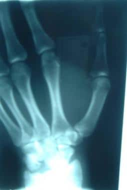
Dislocations, wrist. Anteroposterior (AP) view of a lunate dislocation.


