Practice Essentials
The elbow joint displays an elegant balance between stability and mobility. While allowing a wide range of motion, this joint has inherent stability that requires considerable force to dislocate. As a result, a significant percentage—approximately one-third—of elbow dislocations are associated with fractures of bony components of the elbow. Dislocations without associated fracture are called simple; dislocations with accompanying fracture are referred to as complex.
The elbow is among the large joints most commonly dislocated. [1] The elbow is the large joint most commonly dislocated in children. [2] Dislocations of the elbow fall in frequency just behind dislocations of the finger and shoulder. Most commonly, the elbow dislocates posteriorly. Immediate reduction is essential to reduce the risk of neurovascular or cartilaginous complications.
Elbow dislocation may be isolated, may involve damage to static supportive structures of the elbow, and may even cause fractures about the elbow. Because of this, it is important to recognize elbow dislocation and to know the appropriate management approach to avoid complications. [1]
Elbow dislocations are classified according to the direction of dislocation, with most (80%) being posterolateral. A spectrum of soft tissue injury may also be present, depending on the direction of dislocation and the energy applied. Most dislocations of the elbow can be treated nonoperatively, but recurrent instability and stiffness occur in up to 10% and 40% of patients, respectively. The aim of early surgical stabilization is to prevent these long-term complications. To avoid overtreatment, magnetic resonance imaging (MRI) is used to identify patients at greater risk of complications by determining the grade of soft tissue injury. Those with grade 3 or 4 injuries are treated with fluoroscopic examination under anesthesia. [3]
Plain radiographs are essential prior to reduction of a suspected dislocation. Postreduction films should confirm opposition of joint surfaces and should rule out previously unidentified fractures and entrapment of bony fragments within the joint space. [2]
Management of elbow dislocation should consist of immediate closed reduction and stabilization. If the patient has recurrent instability, fracture, or neurovascular compromise, operative fixation is usually required. [2]
Good long-term outcomes have been reported after nonoperative management; however, a small proportion (< 10%) of patients have a poor outcome, and some require surgical intervention. [4]
Recurrent elbow dislocations suggest chronic joint instability and may require operative fixation. [5]
Up to 10º limitation in full extension and some limitation in flexion are common, unless an intensive rehabilitation program is instituted.
Analgesics and anxiolytics are used to manage the pain associated with dislocation.
Ultrasound-guided infraclavicular block is a fast, safe, and efficient anesthesia technique that can offer an excellent alternative to sedoanalgesia and other brachial plexus blocks for management of elbow dislocation in the emergency department. [6]
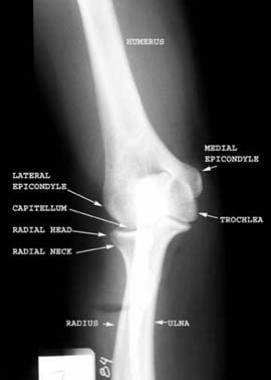
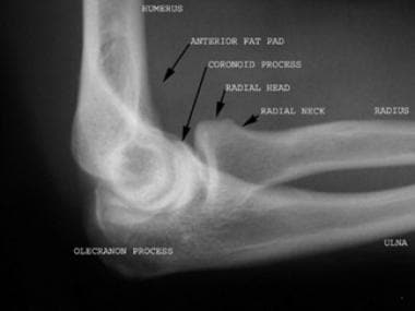
(Radiographs below show normal anatomy of the elbow.)
Pathophysiology
Simply put, the elbow is the articulation between the distal humerus and the proximal ulna and radius. The distal humerus contains the trochlea medially and the capitulum laterally. The elbow comprises 3 separate articulations. The medial aspect of the elbow includes the ulnotrochlear joint, which is primarily responsible for flexion and extension. The lateral radiocapitellar joint and the proximal radioulnar joint are mainly responsible for pronation and supination. This anatomy creates a combination of a hinge joint and a pivot joint. [1]
The elbow is generally stable due to the congruity of articular surfaces. It is further supported by static supporting structures, including the collateral ligaments on the medial and lateral side of the elbow and the joint capsule. Dynamic stabilizers of the joint are composed of the surrounding musculature. [1]
The vascular anatomy of the elbow is composed of a few structures. The brachial artery is a central component of the anterior elbow and eventually divides into the radial and ulnar arteries in the proximal forearm. The radial and ulnar arteries, along with the brachial and deep brachial arteries, create an intricate anastomosis of vessels around the elbow, including radial and ulnar collateral arteries and radial and ulnar recurrent arteries. [1]
Many nerves are present around the elbow, whose function can be compromised by an elbow dislocation. [1]
Both posterior dislocations and anterior dislocations can occur.
Posterior dislocations
A fall onto an extended abducted arm is the mechanism of injury seen in posterior dislocation of the elbow. Posterior dislocations account for most elbow dislocations. Closed posterior dislocations are not commonly associated with neurovascular injury.
These injuries frequently occur during sporting activities when a person falls on an extended elbow. In most instances, the semilunar notch of the ulna is dislocated posteriorly from the distal humerus. If no fracture is associated with the dislocation, it is described as simple and the injury is often closed with no bony protrusion through the skin. [5]
The stability of the elbow joint due to its bony structure means that significant force is required to disrupt the joint. Therefore, an associated fracture may be found along with the elbow dislocation, thus classifying the dislocation as complex. Neurovascular complications following a simple, closed, posterior dislocation are rare. [5]
(The radiograph below shows a posterior dislocation of the elbow.)
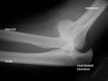 Lateral view of the elbow demonstrates a posterior dislocation of the elbow. The patient also had a nondisplaced radial head fracture.
Anterior dislocations
A strong blow to the posterior aspect of a flexed elbow may result in anterior dislocation of the elbow. This force drives the olecranon forward in relation to the humerus. Anterior dislocations and any open fractures are commonly associated with disruption of the brachial artery and/or injury to the median nerve.
The less often encountered anterior elbow dislocation requires much more force, and concern for neurovascular compromise should be greater. [5]
Epidemiology
Elbow dislocation injuries occur more often in males than in females. Dislocations occur more commonly in adults; the same force in children more often results in a supracondylar fracture of the distal humerus.
The elbow is the second most commonly dislocated major joint in adults. [4] However, anterior elbow dislocation is a rare injury in both adults and children. [2]
Outcomes and treatment satisfaction following simple elbow dislocation generally are good but are significantly worse for patients with greater levels of social deprivation and for those receiving Workers’ Compensation or Medicare insurance. Although surgeons should be aware that specific subsets of patients may benefit from early therapy, a longitudinal cohort study found that this factor did not appear to influence long-term outcomes. [7]
-
Anteroposterior radiograph of the elbow demonstrates the normal anatomy.
-
Lateral radiograph of the elbow demonstrates the normal anatomy.
-
Lateral view of the elbow demonstrates a posterior dislocation of the elbow. The patient also had a nondisplaced radial head fracture.