Background
The dramatic increase in sport diving, ecotourism, and island and coastline travel, perhaps inevitably, has returned people to the sea. Curiosity about our chondrichthyan ancestors, as well as a desire to explore that 70% of our biosphere that remains largely enigmatic, has fostered a siren call to exotic realms. Dangers exist in the sea, as with any environment for which humans are poorly adapted. Contact with hazardous marine organisms is not the least of these dangers.
Many sea creatures have improved their survival through the evolutionary development of offensive and defensive systems that are often elaborate mechanisms for delivering poison or venom to prey or predator. Most of these organisms live in temperate to tropical oceans, especially in the Indo-Pacific regions. Vast arrays of vertebrate and invertebrate creatures can envenomate humans. This article focuses on the more than 600 members of the invertebrate Conidae family of the phylum Mollusca and the class Gastropoda (ie, the cone snails). See the image below.
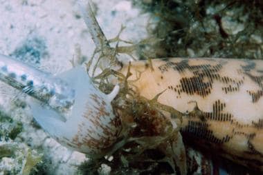 Cone snail ingesting a small fish. Cone snails incapacitate their prey by injecting a cocktail of neurotoxins, which can be dangerous to humans. Courtesy of Wikimedia Commons and David Burdick.
In the last four decades, toxinologists around the world have elucidated a wealth of information on the various classes of constituent proteins and peptides that provide each cone with its own distinctive, complex, and sophisticated bioarmamentarium. More than 2,000 toxins from an estimated more than 70,000 bioactive peptides have been identified in the Conus genus. [1] These venoms serve the cone as a primary weapon to capture prey, as defense, and possibly for other functions. [2, 3]
See Deadly Sea Envenomations, a Critical Images slideshow, to help make an accurate diagnosis.
Pathophysiology
Cone snails are carnivorous; they are divided into three groups, according to their prey: molluscivorous (hunt other gastropods; 25% genus), vermivorous (hunters of polychaete and other worms), or piscivorous (fish hunting; 10% genus). The largest group of cones are vermivores, encompassing 65% of the genus. Their habitats extend from shallow, intertidal areas to extreme deepwater areas. These marine organisms inhabit primarily tropical marine environments in the Western Atlantic, Indian, and Pacific oceans; however, a few species are found in cooler environments. Cone shells are predominantly nocturnal, burrowing in the sand and coral during the daytime.
Like all gastropods, cone snails propel themselves along the ocean floor or reefs by their muscular foot. The foot muscle, or columellar, also contracts to pull the foot in and close the aperture of the shell. To capture a much faster prey in a highly dynamic marine environment, this relatively slow-moving snail has evolved into one of the fastest known predators in the animal kingdom, with the average attack lasting only milliseconds. In an attack, the cone shells inject a cocktail of small, rapidly acting, disorienting, paralytic, and lethal oligopeptide toxins, each 15-30 residues long, into the prey.
Almost 70,000 different conotoxin peptides have been identified to date in different groups of cones. These potent peptides, which fold into small, highly structured frameworks, largely target ion channels, either voltage- or ligand-gated receptors and transporters in excitable cells. Conantokin G, exclusive to piscivore cones, subdues prey by antagonizing the NMDA receptor, causing a sleeplike state. [4] In the Gastridium clade of fish-hunting cones, including Conus geographus and Conus tulipa, insulinlike polypeptides are highly expressed in the distal duct segment. These activate insulin receptors in prey, mimicking the effects of insulin and causing prey “insulin shock” with disorientation. [5, 6] Venom mixtures are specific to each cone shell species, containing 30 - 200 conotoxin peptides and proteinaceous materials, including proteases and phospholipids. Cones are able to deploy different venom mixtures for prey capture and defense. [7]
A group of conopeptides, described as a cabal, act in a coordinated manner to produce a specific physiologic endpoint. [8] A "lightning-strike cabal" triggers an "electrical storm" by depolarizing neurons around the injection site by preventing closing of voltage-gated sodium channels and blocking potassium efflux channels. A "motor cabal" causes paralysis by blocking neuromuscular transmission through inhibition of presynaptic voltage-gated calcium channels, postsynaptic nicotinic acetylcholine receptors, acetylcholine release, or skeletal muscle voltage-gated sodium channels. Different toxic cabals in the same venom may act on the same class of target via different mechanisms. Numerous disulfide bonds determine a specific oligopeptide or polypeptide conformation for each toxin to better fit the target. These disulfide bonds also confer stability to the toxins, one result of which is their inability to be easily broken down by heat treatment. [9]
The first account of human envenomation by a cone snail was around 1670. A total of 139 cases believed to be reliable reports of cone snail envenomation have been documented worldwide. [8] Human envenomations most commonly involve piscivorous species, including C geographus (responsible for approximately 50% of all human envenomations and nearly all lethal cases reported), Conus catus, Conus aulicus, Conus gloria-maris, Conus omaria, Conus magus, Conus striatus, C tulipa, and Conus textile. Envenomation by molluscivorous species has been reported to result in serious symptoms, while envenomation by vermivorous species is associated with mild symptoms only.
Snail shell anatomy can be divided into two main portions: the body whorl and the spire. The body whorl, the lower portion of the shell, contains the soft snail body. The spire, or pointed top of the shell, can be different shapes. The whorl contains the portions of the snail essential for prey capture and movement. The cone shell detects its prey via the siphon, which is covered with chemoreceptors, although limited visual signaling may also be involved. The false mouth can be extended to engulf its prey, with a muscle contracted to retract the mouth back into the shell.
Venom, with different conotoxins formed rapidly in various portions of the venom duct due to different conotoxin gene expression profiles, [10, 11] is stored as less toxic precursors in a milky slurry in the venom bulb. When required, the precursor undergoes enzymatic cleavage of the signal peptide and the propeptide forms appropriate disulfide linkages. [12] The mature toxic solution is then delivered via a detachable radula. The radula is a dartlike, hollow, chitinous barb, formed in the radular sheath and delivered, after receiving venom in the buccal cavity, by a long, extensible proboscis. The venom sac contains approximately 20 radulae. The muscular proboscis, which may extend more than the full length to the shell spire in some species, touches a prey item and then thrusts one radula (or more, in some piscivorous cones) into the prey via circular muscles at its anterior tip. Approximately 1 to 50 microliters of venom are delivered by a radula. Venom rapidly diffuses through the poisoned prey. The radula remains attached to the cone by a cord.
Once the prey is paralyzed, the gastropod retracts the cord and engulfs the prey through the radular opening into its distensible stomach. Some cone species, such as C geographus, may distend and "net" prey with their "false mouths" before injecting venom. Digestion occurs over the ensuing several hours.
Cone shell toxins efficiently and highly selectively inhibit an extensive array of ion channels, receptors, and transporters involved in the transmission of neuromuscular signals in animals. The high target specificity of certain conotoxins toward mammalian channels is due to the fact that mammalian receptor isoforms of the specific target (eg, the nicotine receptor) are quite similar in sequence to their physiologic homologue in fish.
In the last few decades, these toxins have become the focus of some exciting molecular biological and pharmacological research. Conus venoms are remarkably diverse among species, and the large gene families that encode conotoxins show high evolutionary rates. A 2008 study suggests that this may result from either lineage-specific dietary modifications or differences in the positive impact of predator-prey interactional selection. [13, 14] To date, conotoxins have been divided into seven superfamilies, based on their disulfide bond frameworks, and they have been further divided into families based on their mechanisms of action. Several conotoxins, and their synthetic derivatives, due to their high selectivity and affinity for different ion channels, are the subjects of current clinical trials on chronic pain control, posttraumatic neuroprotection, cardioprotection, and the treatment of Parkinson disease and other neuromuscular disorders. [15]
While an extensive discussion of all discovered types of conotoxins and their specific activities is beyond the scope of this article and has served as the basis of several extensive reviews (see References), a sample of several distinct types of conotoxins and their effects are as follows:
-
ω-conotoxin - Hinders the voltage-dependent entry of calcium into the nerve terminal and inhibits acetylcholine release
-
μ-conotoxin - Modifies muscle sodium channels by occluding and thereby blocking ion conduction through the pore of voltage-gated sodium channels (VGSC), at the same site as saxitoxin and tetrodotoxin [16]
-
κ-conotoxin - Potassium channel (VGPC)-targeting peptides
-
α-conotoxin - Blocks the nicotinic acetylcholine receptor, similarly to snake alpha-neurotoxins
-
δ-conotoxin - Delays or inhibits VGSC inactivation, resulting in prolongation of the action potential; this produces a "hyperexcited state" in involved neurons and can lead to electrical hyperexcitation of the entire organism (eg, seizures in marine snails) [16]
-
S-conotoxins - Inhibit 5-HT3 channels Y-conotoxins - Competitively block muscle acetylcholine receptors
-
Conantokins - Target NMDA ( N -methyl-D-aspartate) subtype glutamate receptors
-
Conopressin - Vasopressin agonist
-
Sleeper peptide - Found primarily in C geographus, induces a deep sleep state in test animals
Ziconotide is a synthetic form of an ω-conotoxin that has been approved by the US Food and Drug Administration for intrathecal administration for severe, chronic pain patients who are intolerant or refractory to other treatments.
Cone shells are prized by shell collectors for their pleasing shape and beautiful shells, which exhibit varying, intricate, darker geometric patterns on a lighter base. A sting most commonly occurs on the hand and/or fingers of an unsuspecting handler as well as on the feet of swimmers in shallow, tropical waters. Envenomations may also occur at contact points of collection bags. Even when picked up by the spire, the cone proboscis can rapidly extend more than a shell length to envenomate the unsuspecting shell handler. Cone radulae can penetrate a 5-mm neoprene wet suit.
At the site of envenomation, local stinging is followed within minutes by numbness, paresthesias, and ischemia. The actual puncture wound may not be evident. Serious envenomations may result in nausea, cephalgia, slurred speech, drooling, ptosis, diplopia and blurred vision, generalized paralysis, coma, and respiratory failure within hours. Death is typically secondary to diaphragmatic paralysis or cardiac failure. [17] C geographus, which produces the most potent conotoxins found to date, may produce rapid cerebral edema, coma, respiratory arrest, and cardiac failure. C geographus has been given the moniker of the "cigarette snail" for the claim that an envenomated human has time to smoke a single cigarette before succumbing to the envenomation. In nonfatal envenomations, symptoms may take several weeks to resolve. Disseminated intravascular coagulation (DIC) may also be evident. The wound may be contaminated with marine organisms and can ulcerate and abscess. [18]
Etiology
The following may lead to envenomation:
-
Careless or unknowledgeable handling of a hazardous specimen
-
Unsuspecting scuba divers carrying live cone shells in a wet suit, unsecured specimen bag, or buoyancy control device
-
Accidental contact while walking, swimming, and/or diving in shallow, tropical waters
-
Increased opportunities for exposure (eg, in aquarium keepers and handlers)
Epidemiology
Frequency
United States
Conus species are not indigenous to US waters. These are more likely to be encountered while traveling abroad, by specialized aquarium staff, or by researchers studying the venom components.
International
A total of 139 human envenomations have been documented in Southern Australia, the Indo-Pacific area, the islands of the Indian Ocean, and the Brazilian coast of the Atlantic Ocean. Many unreported envenomations may have occurred.
Race, sex, and age
No relationship to age, race, or sex exists in Conus envenomation. Envenomation is more an injury of individuals engaged in either recreational or commercial shell collecting, diving, and fishing. Children are more likely to succumb to C geographus envenomation than adults. [8]
Prognosis
A high risk of death is associated with envenomation by certain species of cones, particularly C geographus, C textile, and C marmoreus. Morbidity includes mild symptoms (eg, nausea, weakness, diplopia) lasting several hours. In the 15 cases in which the time interval between envenomation and death is known, it ranged from 0.2 to 10 hours, with a mean of 2.4 hours. Thirty-three percent of deaths occurred within 1 hour of envenomation. [8] Two to 3 weeks of symptoms may be associated with more severe exposures. [8] In those who develop ulcerations at the site of envenomation, longer-term wound care may be required.
Patient Education
To assist in preventing cone shell envenomation, give patients the following instructions:
-
Properly identify cone shells.
-
Handle cone shells only with proper gloves.
-
Do not carry a live cone in a perforated or thin bag near skin, wet suits, or buoyancy control vests.
-
If a live cone must be carried, lift at the large posterior end of the shell with protective gloves. Remember, this is not always adequate protection as the proboscis can extend the entire length of the shell.
-
If the proboscis protrudes, immediately drop the cone.
-
Wear appropriate footwear when walking in intertidal areas.
-
Do not reach blindly under corals or rocks.
For patient education resources, visit the First Aid and Injuries Center. Also, see the patient education article Stingray Injury.
-
Cone snail ingesting a small fish. Cone snails incapacitate their prey by injecting a cocktail of neurotoxins, which can be dangerous to humans. Courtesy of Wikimedia Commons and David Burdick.


