Practice Essentials
In 1970, when smallpox was nearly eradicated, a previously unrecognized orthopoxvirus named monkeypox (mpox) was identified in humans. The first known human case occurred in the Equateur province of Zaire (now known as the Democratic Republic of Congo [DRC]) when a 9-year-old boy developed a smallpoxlike illness, which was eventually confirmed as human monkeypox by the World Health Organization. [1] Retrospectively, similar cases occurring in 1970-1971 from the Ivory Coast, Liberia, Nigeria, and Sierra Leone were attributed to monkeypox infection.
Monkeypox was limited to the rain forests of central and western Africa until 2003, when the first cases in the Western Hemisphere were reported. In late spring 2003, multiple persons were identified in the midwestern United States who had developed fever, rash, respiratory symptoms, and lymphadenopathy following exposure to ill pet prairie dogs (Cynomys species) infected with the monkeypox virus. [2]
In the most recent outbreak in 2022, the United Kingdom reported 9 cases of monkeypox in early May 2022, with the first identified case having recently traveled to Nigeria. From this adult index case, there were 2 confirmed transmissions within the patient's family, to another adult and a toddler. [3] On May 18, 2022, the Massachusetts Department of Public Health announced a confirmed case of monkeypox in an adult male who had recently visited Canada. [4]
As of October 19, 2022, there were over 74,700 total confirmed monkeypox cases in more than 200 different nations. Cases were present on every inhabited continent. [5] In the US, there were 27,635 total confirmed cases as of October 19, 2022. [6] By March 7, 2023, there had been 30,235 reported mpox cases in the US. [7]
Cases of monkeypox in the US peaked in early August 2022 with a 7-day moving average of 439. Owing to vaccination and avoidance in at-risk populations, the 7-day average of 48 cases have decreased as of October 19, 2022.
An overwhelming, though not exclusive, number of cases in the current outbreak are among men who have sex with men. [8] While sexual transmission has not been definitively confirmed, this mode of transmission seems likely, especially given that initial lesions are often reported at sites of sexual contact. [9, 10]
In the 2003 US outbreak, imported asymptomatic animals transmitted a nonindigenous pathogen to an indigenous susceptible animal. After an average incubation period of 12 days, the animal became ill and was capable of transmitting the pathogen to humans when in close proximity. The exact potential for human-to-human transmission and human-to-animal transmission remains unknown.
Note the image below.
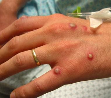 Vesicular rash on the dorsal aspect of the hand. Vesicopustules are seen; some have a central umbilication.
In July 2021, a case of monkeypox was reported in Dallas, Texas in a traveler from Nigeria. [11, 12] Later, in November of that year, a second US case was confirmed in Maryland in another traveler returning from Nigeria. [11, 13] These 2 cases represent the only reported incidents of monkeypox in the US during 2021.
Etiology
Outbreaks in western and central Africa have been linked to exposure to rats, rabbits, squirrels, monkeys, porcupines, and gazelles. Inhabitants of remote tropical rain forests may become infected from direct contact while capturing, slaughtering, and/or preparing these animals for food; ingestion has also been linked to infection. Consumption of such so-called "bush meat" is particularly hazardous because the flesh is often undercooked. Because of the diversity of animals eaten by local inhabitants, conclusions about the relative risk of meat sources are not known with certainty.
Also see Etiology.
Prognosis
Mortality rates ranging from 1-10% have been reported in Africa, but no fatalities occurred in the United States 2003 outbreak.
Also see Prognosis.
History and physical examination
Monkeypox can cause a syndrome clinically similar to smallpox but overall is less infectious and less deadly.
Transmission can occur from contact with ill animals or animal reservoirs from Western Africa (eg, prairie dogs, rabbits, rats, mice, squirrels, dormice, monkeys, porcupines, gazelles). Additionally, preparing or ingesting infected animals can transmit monkeypox infection. Finally, direct cutaneous (skin-to-skin) or respiratory contact with an animal or person who is infected can transmit the infection.
The incubation period averages 12 days, ranging from 4-20 days.
In the prodromal or preeruptive stage (lasts 1-4 days prior to the onset of rash) [14] , fever is commonly the first symptom (usually 38.5-40.5°C). The febrile illness is often accompanied by chills, drenching sweats, severe headache, backache, myalgia, malaise, anorexia, prostration, pharyngitis, shortness of breath, and cough (with or without sputum). Lymphadenopathy appears within 2-3 days after the fever. In the 2003 outbreak, 47% of patients had nodes measuring several centimeters in diameter in the cervical and submental areas.
In the exanthem (eruptive) stage, most persons develop a rash within 1-10 days after the onset of fever. The rash often starts on the face and then spreads to the rest of the body. It persists for 2-4 weeks until all lesions have shed the crusts. However, in the current outbreak, painless anogenital lesions — often without a prodrome — are being observed in persons who have had close contact with an infected person or persons, including men who have sex with men. [15]
Encephalitis with immunoglobulin M (IgM) found in the cerebrospinal fluid has been reported. [16]
The most reliable clinical sign differentiating monkeypox from smallpox and chickenpox is enlarged lymph nodes, especially the submental, submandibular, cervical, and inguinal nodes. [17]
Also see Physical Examination.
Complications
Complications include pitted scars, deforming scars, secondary bacterial infection, bronchopneumonia, respiratory distress, keratitis, corneal ulceration, blindness, septicemia, and encephalitis.
Diagnostics
On November 15, 2002, the United States Food and Drug Administration (FDA) issued an emergency use authorization (EUA) for the cobas MPXV to detect monkeypox virus DNA in swabs from human monkeypox lesions in patients with suspected monkeypox cases. [18]
Refer to the information established by the US Centers for Disease Control and Prevention (CDC) at Monkeypox: Clinical Recognition.
Also see Workup.
Treatment
The disease is usually self-limited; resolution occurs in 2-4 weeks. In the African cases, the mortality rate was 1-10%, and death was related to the patients' health status and other comorbidities. Most patients died of secondary infections. No fatalities were reported in the 2003 US outbreak.
Patients often feel poorly during the febrile stage of the illness; therefore, bedrest along with supportive care may be necessary. Hospitalization may be necessary in more severe cases; a negative pressure room is preferable.
In September 2019, the US Food and Drug Administration (FDA) approved an attenuated, live, nonreplicating smallpox and monkeypox vaccine (Jynneos) for immunization of adults at high risk for smallpox or monkeypox infection. [19, 20]
Also see Clinical management and infection prevention and control for monkeypox: Interim rapid response guidance, 10 June 2022 from the World Health Organization (WHO).
Also see Treatment and Medication.
Pathophysiology
The monkeypox (mpox) virus is a member of the genus orthopox (family Poxviridae); other members include cowpox, vaccinia, and variola (smallpox) viruses. [21] It is a zoonotic virus with primary transmission believed to occur through direct contact with infected animals or possibly by ingestion of their inadequately cooked flesh. Inoculation may be from cutaneous or mucosal lesions on the animal, especially when the skin barrier is compromised secondary to bites, scratches, or other trauma. The infection was first seen in laboratory monkeys in 1958, thus, the name monkeypox, although rodents are believed to be the major reservoir in Africa. [22, 23] A 2010 study reaffirmed that several species of forest-dwelling rodents are at risk for orthopoxvirus (including monkeypox) infection. People living in or near the forested areas may have indirect or low-level exposure, possibly leading to subclinical infection. [24]
Secondary, or human-to-human, disease transmission was found to be another possible route in an outbreak in the DRC in 1996-1997. [23] Studies of this outbreak suggested that within households, monkeypox was secondarily transmitted to 8-15% of human contacts. Prior to this, monkeypox was not identified as an important worldwide health problem because human infection rates were not known to play a significant role in the pathogenesis. Analysis of the 2003 US outbreak implicates animal-to-animal and animal-to-human transmission as the significant route of transmission. However, in the 2003 US outbreak, clear exposure to an infected animal could not be identified in 1 case, and, therefore, human-to-human transmission could not be excluded.
Human-to-human transmission has been confirmed as a major factor in the 2022 outbreak in multiple areas across the world. While many of the patients are men who have sex with men, sexual transmission of monkeypox has not been conclusively confirmed but appears likely. [8, 9, 10]
Etiology
In the DRC in 1997, animals caught from the wild were tested for the monkeypox (mpox) virus. The following animals were found to have neutralizing antibodies against the monkeypox virus, suggesting a role as natural reservoirs: domestic pig (Sus scrofa), Gambian rat (Cricetomys emini), elephant shrew (Petrodromus tetradactylus), Thomas's tree/rope squirrel (Funisciurus anerythrus), Kuhl's tree squirrel (Funisciurus congicus), and sun squirrel (Heliosciurus rufobrachium). [23]
Human-to-human transmission supplanted the prominence of animal-to-human transmission in the 1996-1997 outbreak in the DRC. Crowded living quarters, poor hygiene, discontinuation of the smallpox vaccination, and decreased herd immunity were implicated. Respiratory droplets and direct contact with mucocutaneous lesions or fomites have been postulated as routes of human-to-human transmission.
Epidemiology
Frequency
United States
In the most recent oubreak, a single confirmed case was identified May 18, 2022 in Massachusetts. [11, 13, 4] As of June 29, 2022, there were 351 confirmed cases in nearly 30 states. [6] By March 7, 2023, there had been 30,235 reported cases in the US and 38 mpox-associated deaths. [7]
During the 2003 outbreak, no cases occurred in the United States until the late spring in the Midwestern states. Between May 16 and June 20, 2003, 71 suspected cases of monkeypox (mpox) were investigated. [25] A total of 47 individuals were identified with confirmed (n = 37) or probable (n = 10) monkeypox virus infection. Monkeypox cases were confirmed on the basis of virus isolation or detection of the virus by polymerase chain reaction (PCR) from a clinical specimen (eg, skin biopsy or throat culture). Individuals who presented with fever and rash within 21 days of exposure to monkeypox and had serum positive for orthopox immunoglobulin M (IgM), but did not have culture- or PCR-positive clinical specimens, were classified as having a probable case of infection. [26, 27]
International
This condition is rare and only known to be indigenous to the rain forests of western and central Africa. [28] It was first recognized in humans in 1970 after the eradication of smallpox, possibly because of the subsequent unmasking of the infection. Surveillance reports from 1981-1986 documented 338 cases in the DRC (out of a 1982 estimated population of 5 million). In the 1996-1997 outbreak in the DRC, the attack rate was 22 cases per 1000 population.
The 2022 outbreak involved 51 locations as of June 29, 2022, and included 5,115 confirmed cases. [5]
Human infection with monkeypox has not been reported in West Africa since 1978. Monkeypox is considered endemic in northern and central DRC. Sporadic occurrences of disease are reported in neighboring countries. [29] In 2003, 11 cases and 1 death were reported from the DRC and 10 cases with no deaths were reported from Sudan in 2005. [30]
In 2009, interethnic violence in northwestern DRC lead to an influx of refugees into the Republic of the Congo (ROC). The United Nations International Children's Emergency Fund (UNICEF) sponsored a program of intensive community education in the refugee settlements that included modules on monkeypox recognition and prevention, which resulted in the indentification of 10 suspected cases of monkeypox. Seven of these 10 cases were tested and 2 were found to be positive by polymerase chain reaction assays. [31]
The results of this outreach campaign suggest that intensive community education can lead to increased capacity for detection of monkeypox in high transmission–risk settings. They also highlight the need to educate physicians in the recognition and treatment of monkeypox. [32]
Race, sex, and age
Poxvirus infections have no racial predilection, and the incidence is equal in males and females, except in the 2022 epidemic, where patients are overwhelmingly male. A survey of 528 infections confirmed from April 27, 2022 to June 24, 2022, in 16 countries found that 98% of patients were men who have sex with men, 75% were White, and 41% were HIV positive. The median patient age was 38 years. [33]
A morbidity and mortality report from the US Centers for Disease Control and Prevention revealed that, in the 2022/2023 outbreak, there were 38 mpox-related deaths (1.3 per 1000 cases). Men represented 94.7% of mortalities, with 86.8% of mortalities being Black persons. The overwhelming majority of those who succumbed were immunocompromised secondary to HIV infections. The median patient age at death was 34 years. [7]
In the African epidemics, 90% of the patients were children younger than 15 years. [34] In the 2003 US outbreak, of the confirmed cases (n = 35), 11 patients were younger than 18 years and 24 were older. Although the highest age-specific incidences and the greatest number of cases occur among persons younger than 15 years, a trend toward increasing incidence among persons aged 15-30 years has been seen in recent years. It has been hypothesized that cessation of smallpox vaccination may be a factor in the increasing incidence in this age group, but this theory fails to account for why the disease has not reemerged in countries where the disease was seen previously, such as West Africa. [30]
Prognosis
Mortality rates ranging from 1-10% have been reported in Africa, but no fatalities occurred in the United States 2003 outbreak. Death rates are disproportionately high in African children. Health status, comorbidities, vaccination status, and severity of complications influence the prognosis in the United States and Africa.
Uncomplicated cases resolve in 2-4 weeks, with only pock scars remaining.
There were 38 mpox-related death in the US during the 2022/2023 outbreak. [7]
Mortality/morbidity
The disease in the United States was generally self-limited, with resolution in 2-4 weeks, depending on the severity of the illness. However, a small subset of patients, most commonly pediatric patients, had a more severe course, with several patients requiring ICU care. [35]
Complications reported from African outbreaks include pitted scars, deforming scars, secondary bacterial infection, bronchopneumonia, respiratory distress, keratitis, corneal ulceration, blindness, septicemia, and encephalitis.
Data from the African outbreaks suggest that prior smallpox vaccination confers 85% protection from monkeypox; infection may be milder even several years after vaccination, and the incidence of complications may be reduced. [36, 37] With the 2003 US outbreak, the Centers for Disease Control and Prevention (CDC) recommended smallpox vaccination up to 2 weeks, ideally within 4 days, after a significant, unprotected exposure to a diseased animal or a confirmed human case. [38]
African cases have mortality rates of 1-10%, with the highest rates occurring in children and individuals without vaccination. In general, the prognosis is related to the amount of exposure to the virus, host immune response, comorbidities, vaccination status, and severity of complications.
Genomic sequencing of US, western African, and central African monkeypox (mpox) isolates have confirmed the existence of 2 distinct monkeypox clades. [39] The isolates from the United States were identical to the western African isolates. The disease course for individuals infected with the western African isolates is milder with less human-to-human transmission than for those infected with isolates from central Africa. [40] In 2010, a dosage comparison using a prairie dog animal model reconfirmed that the clade I (formerly known as the Congo Basin or Central African clade) of monkeypox virus is more virulent than the clade II (formerly the West African clade) of monkeypox virus. [41]
Patient Education
After the 2003 outbreak, the CDC implemented an immediate embargo on the importation of all rodents (order Rodentia) from Africa.
In addition, the Centers for Disease Control and Prevention (CDC) and the US Food and Drug Administration (FDA) prohibited the transportation or offering for transportation in interstate commerce, or the sale, offering for sale, or offering for any other type of commercial or public distribution, including release into the environment of prairie dogs and the following rodents from Africa: tree squirrels (Heliosciurus species), rope squirrels (Funisciurus species), dormice (Graphiurus species), Gambian giant pouched rats (Cricetomys species), brush-tailed porcupines (Atherurus species), and striped mice (Hybomys species).
Investigation of the exotic pet industry by state and federal authorities was triggered by the 2003 outbreak. The FDA lifted its restrictions on pet prairie dogs in 2008. The FDA consulted with the CDC and determined that the domestic restrictions placed on certain African rodents, prairie dogs, and certain other animals were no longer needed. However, the CDC restriction on the importation of all African rodents remains in effect to prevent any reintroduction of the monkeypox virus into the United States. [42]
Patients, especially those who are immunocompromised, should be counselled to avoid risky behaviors such as condomless sex and educated on monkeypox (mpox) signs and symptoms. [9]
-
Vesicular rash on the dorsal aspect of the hand. Vesicopustules are seen; some have a central umbilication.
-
Umbilicated papule on the lower part of the leg. This smaller lesion still shows the typical umbilicated morphology.
-
Lymphadenopathy in monkeypox. Large nodes in the mandibular, cervical, or inguinal region are commonly seen in monkeypox. The presence of significant lymphadenopathy helps differentiate monkeypox from smallpox and chickenpox.


