Background
Leukemia cutis is the infiltration of neoplastic leukocytes or their precursors into the epidermis, the dermis, or the subcutis, resulting in clinically identifiable cutaneous lesions. The appearance of these lesions is variable and may include flesh–colored-to-violaceous papules, plaques, or nodules. Many terms have been used to describe varying presentations of leukemia cutis. These include granulocytic sarcoma, monocytic sarcoma, primary extramedullary leukemia, and chloroma.
Leukemia cutis is a broad term used to describe any cutaneous presentation of leukemia. [1, 2, 3] Owing to the variety of hematologic malignancies or proliferative disorders that may be associated with leukemia cutis, terms such as myeloid or lymphoid leukemia cutis have been used to further classify the leukemic cells. [4]
Leukemia cutis usually occurs in the setting of a previously diagnosed systemic leukemia or lymphoproliferative disorder/myelodysplastic syndrome. In rare cases, leukemia cutis may be the first manifestation of systemic disease. [1]
The dermatologist is often instrumental in the diagnosis of leukemia cutis. Accurate diagnosis has tremendous prognostic significance and may establish a diagnosis in cases in which leukemia cutis is the harbinger of a systemic leukemic process. This is known as aleukemic leukemia cutis. Leukemia cutis can also be a manifestation of a relapse of previously treated systemic leukemia. A diagnosis of leukemia cutis in the setting of acute leukemia generally portends a poor prognosis and strongly correlates with additional sites of extramedullary involvement. In the setting of chronic leukemia, cutaneous involvement can indicate blast transformation. [1] This can significantly alter the treatment regimen for a patient. [1] Note the image below.
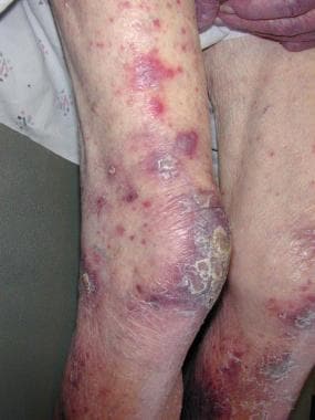 A patient with typical plum-colored lesions seen in leukemia cutis. This patient had acute myelogenous leukemia. Courtesy of Grant Anhalt, MD.
Pathophysiology
All types of leukemia result from the abnormal development of leukocytes in the bone marrow. Accumulation of genetic defects in hematopoietic precursors leads to impaired cellular differentiation, increased proliferative potential, unregulated proliferation, and defective apoptosis. Ultimately, these defects result in arrest of cell maturation and expansion of a clonal cell population. The subtype of leukemia is determined by the hematopoietic lineage of neoplastic cells and the stage at which maturational arrest occurs.
Genetic alterations predisposing to leukemia may be congenital or acquired. Advancing age, exposure to radiation, chemotherapy, and certain viral infections may promote leukemogenesis in some cases. Some antecedents are specific to a leukemia subtype, such as the association of acute T-cell leukemia-lymphoma (ATL) with human T-lymphotropic virus type I (HTLV-1). Similarly, some genetic or chromosomal abnormalities are found in specific leukemia subtypes. The association of t(15,17) with the M3 subtype of acute myeloid leukemia (acute promyelocytic leukemia) is one example.
The pathogenesis of leukemia is well-characterized in the two-hit hypothesis of acute myeloid leukemia. This model describes at least two genetic alterations required for leukemogenesis. Class II mutations cause defects in myeloid cell differentiation. These occur early and result in maturation arrest. Class I mutations promote cell survival and proliferation. These lead to progression of acute myeloid leukemia. [5]
Leukemia cutis is defined by migration of leukemic cells to the skin. Leukemia cutis has been described in patients with myeloid and lymphoid types of leukemias. Cutaneous infiltration by neoplastic lymphocytes may be seen in acute myeloid leukemia, acute lymphocytic leukemia, chronic myeloid leukemia, chronic lymphoid leukemia, hairy cell leukemia, prolymphocytic leukemia, chronic myelomonocytic leukemia, and myelodysplastic syndromes. In patients with chronic diseases, skin involvement may be associated with transformation into aggressive histology and disease progression.
Therapy-related leukemia cutis preceding a diagnosis of systemic acute leukemia has been reported among patients treated with chemotherapy for breast cancer. [4, 6, 7, 8, 9]
The pathophysiology underlying the specific migration of leukemic cells to the skin is not clear. While some associations can be made, no definitive phenotype has been demonstrated to consistently lead to leukemia cutis. A number of mechanisms have been proposed. It has been speculated that the chemokine, integrin and other adhesion (ICAM-1) molecules may play a role in skin specific homing of T and B leukemic cells. CD56 (blast neural cell adhesion molecule) expression on leukemic blasts has been associated with extramedullary disease in acute leukemia in patients with t(8;21). [10] This may be due to the role of this surface glycoprotein in cell-cell or cell-matrix adhesion. Its role is further supported by the prominent skin involvement in CD56+ cutaneous lymphomas. [11]
In the case of HTLV-I–induced leukemia, it may be from the abundant expression of the CC chemokine receptor 4 (CCR4) on the cell surface of the leukemic cells. The ligands thymus and activation-regulated chemokine (TARC/CCL17) and macrophage-derived chemokine (MDC/CCL22) are present in the skin and may explain the predilection of adult T-cell leukemia to involve the skin.
High expression of CCR4, TARC, and macrophage-derived chemokine/CCL22 in the skin has also been noted by reverse transcription–polymerase chain reaction studies. Evidence also suggests that the presence of T-cell–related antigens on the cell surface of leukemic cells in acute monocytic leukemia (AML-M5) in patients with leukemia cutis may promote selective homing to the skin. Additionally, one small study of 18 cases of myelomonocytic leukemia cutis patients showed cutaneous lymphocyte-associated antigen (CLA) staining in 14 (78%) of 18 cases. The presence of CLA may confer a specific tropism to the skin in these leukemic cells.
In addition, some authors have noted leukemic infiltrates in preexisting inflammatory lesions and skin malignancies, suggesting that chemotactic mechanisms may be involved. [12] Other factors associated with cutaneous involvement include aneuploidy of chromosome 8, acute myeloid leukemia with monocytic differentiation, and involvement of other extramedullary sites. [2]
Etiology
Both a genetic component and an environmental component appear to be involved in the pathogenesis of many types of leukemia. A variety of well-characterized chromosomal translocations result in specific leukemic syndromes.
Patients with Down syndrome have an increased risk for both megakaryoblastic leukemia and pre–B-cell leukemia.
Other genetic syndromes, including Bloom syndrome, Klinefelter syndrome, Wiskott-Aldrich syndrome, and Fanconi syndrome, have shown an increased incidence of leukemia.
CLL shows some familial tendency in approximately 20% of CLL cases.
Several genetic mutations lead to an asymmetric maturation of stem cells and result in a single clonal expansion of a severely defective stem cell. The specific mutations and phenotypic changes resulting from genetic aberration determine the subtype of leukemia. After the development of the leukemic clones, a tissue-selective homing process that leads to the infiltration of malignant cells into the epidermis, the dermis, the subcutaneous fat, and the mucosa occurs.
The molecular basis responsible for the development of leukemia cutis is not yet defined. However, initial cytogenetic studies are starting to provide insightful information that would lead to a better understanding in the pathophysiology of leukemia cutis. Prior studies have demonstrated that as many as 50% of patients with AML-M4 or AML-M5 develop leukemia cutis and other forms of EML. Karyotypic studies of leukemic cells have demonstrated the translocation of chromosomes 8 and 21 t(8;21) in these subtypes of AML. A strong association exists between aneuploidy of chromosome 8 and leukemia cutis. Other cytogenetic abnormalities noted in leukemia cutis are chromosome 3 translocations and t(6;9)(p23;q34). Chloromas, primary EMLs are associated with t(8;21), t(9;11) and inv(16) translocations. Identification of proteins coded by specific genes located in those chromosomes would assist in defining factors responsible for the development of leukemia cutis.
Environmental exposures may increase the risk of leukemia. Benzene exposure increases one's risk for AML. Ionizing radiation exposure may increase the risk of leukemia as well, particularly AML, CML, and ALL. Alkylating agents used in chemotherapy cause an increased risk of subsequent AML. The use of all trans retinoic acid to treat APL may predispose a patient to increased risk of extramedullary involvement, including leukemia cutis, which is otherwise rare in APL. Other leukemias may be caused by viral infection. These include ATLL, caused by HTLV-I and acute B-cell leukemia and large granular lymphocytic leukemia, which may be the result of Epstein-Barr virus infection.
Secondary leukemias are associated with prior hematologic disorders such as myelodysplastic syndrome or myeloproliferative disease. Therapy-related acute leukemia results from genetic damage sustained from prior radiation therapy or chemotherapy for malignancies. These are associated with the use of alkylating agents and type II topoisomerase inhibitors, and they differ from primary leukemias clinically and cytogenetically and carry a poor prognostic outcome. Therapy-related secondary leukemia with leukemia cutis as a result of chemotherapy has rarely been reported. [13]
Epidemiology
Frequency
United States
Because leukemia cutis is a relatively rare condition and because it may manifest in a variety of leukemia subtypes, the exact overall incidence of leukemia cutis is unclear. For the various subtypes, the approximate incidences are listed in Table 1 below.
Although adult T-cell leukemia/lymphoma (ATLL) is exceedingly rare in the United States, a disproportionate percentage of patients develop leukemia cutis. The rate of seroprevalence of HTLV-I in volunteer blood donors in the United States is 0.02%. Of the individuals infected with HTLV-I, only 2-4% develop ATLL. Acute myelogenous leukemia (AML) shows the second highest rates of leukemia cutis. The French-American-British (FAB) classification divides AML into 8 main subtypes M0 to M7, based on the morphology and the state of differentiation of the leukemic cells. Acute myelomonocytic leukemia (AML-M4) and AML-M5 have the highest rates of skin involvement of all the subtypes and are reported to be as high as 30-50%.
The World Health Organization (WHO) has offered an updated classification of AML, which incorporates immunophenotyping and cytogenetics. The WHO method allows more accurate classification based on factors that affect prognosis. Cases of AML that do not meet WHO criteria for a specific diagnosis fall into the category of “AML not otherwise specified.” [5] In these cases, leukemic cells are further classified by morphology, similar to the older FAB classification scheme.
More information about AML classification, including a full outline of the WHO scheme, can be found in Acute Myeloid Leukemia Staging.
The FAB classification of AML, with FAB subtype and description, is as follows:
-
M0: Undifferentiated myeloblastic
-
M1: Myeloblastic with minimal maturation
-
M2: Myeloblastic with maturation
-
M3: Promyelocytic
-
M4: Myelomonocytic
-
M4eo: Myelomonocytic with eosinophils
-
M5a: Monocytic without differentiation
-
M5b: Monocytic with differentiation
-
M6: Erythroleukemic
-
M7: Megakaryoblastic
The incidence of leukemia cutis also appears to be high among children, and cases of leukemia cutis have been documented in as many as 25-30% of infants with congenital leukemia. Most of these patients have myelogenous leukemia. In congenital leukemia, leukemia cutis does not worsen the prognosis.
In most cases of leukemia cutis, systemic disease precedes the development of skin lesions. However, in as many as 7% of patients with leukemia cutis, skin disease occurs prior to bone marrow infiltration and systemic symptoms (aleukemia cutis or primary extramedullary leukemia [EML]). Aleukemic involvement may be diffuse and papulonodular and these patients eventually develop AML. [14]
Table 1. Incidences of Types of Leukemia (Open Table in a new window)
Type of Leukemia |
Incidence in the United States |
Percentage of Patients with Leukemia Cutis (%) |
AML |
2.5 cases per 100,000 population |
13 |
Acute lymphocytic leukemia |
1.3 cases per 100,000 population |
3 |
Chronic myelogenous leukemia (CML) |
1-2 cases per 100,000 population |
2-8 |
Chronic lymphocytic leukemia (CLL) |
2.3 cases per 100,000 population |
8 |
Hairy cell leukemia |
0.6-2.9 cases per 1,000,000 population |
8 |
Adult T-cell leukemia |
Extremely low |
40-70 |
International
In general, the international incidence of leukemia cutis is thought to be similar to that in the United States. One study by Agis et al in Vienna showed a prevalence of 2.9-3.7% for AML. This is a figure similar to the rate determined by Baer et al in the United States. [15]
The exception to this rule would be the prevalence of HTLV-I–induced ATLL, which is significantly higher in the Caribbean and Japan. In Japan, 6-37% of the population is infected with HTLV-I in endemic areas. Of these, 0.5 per 1000 women and 1.5 per 1000 men will develop ATLL. In the Caribbean, 3-6% of the population is seropositive for HTLV-I. Reportedly, the rate of cutaneous involvement in ATLL ranges from 40-70%.
Race
Although specific racial, sexual, and age predilections for the subtypes of leukemia exist, no data regarding any of these factors in leukemia cutis are available.
Prognosis
The prognosis is poor, with many patients having other extramedullary disease and poor survival rates. Several studies indicate that, in the presence of leukemia cutis in AML or CML, the disease course is aggressive and the length of survival is short. Even patients with aleukemic leukemia cutis or granulocytic sarcoma progress to systemic disease and should be treated systemically from the time of diagnosis.
A study by Kaddu et al showed an average survival time in AML to be 7.5 months and in CML, 9.4 months. [16] Another study by Baer et al revealed that of 18 patients with leukemia cutis in AML, 90% had other sites of extramedullary involvement, and, in 40% of these patients, the meninges were involved. [17] In a smaller case series by Shaikh et al with only 5 patients with AML, all 5 died within 6 months of their diagnosis of leukemia cutis. [18]
The prognostic significance of leukemia cutis in CLL is less clear. In a study by Cerroni et al, CLL was associated with advanced stage and a poor prognosis. [19] One series by Su et al of 16 patients with CLL demonstrated a mean survival of 16 months, with 88% of the patients dying within 1 year. [20] Other reports suggest that finding of leukemia cutis in CLL in the absence of large cell transformation or worsening systemic disease, skin infiltration alone does not affect prognosis. [21, 22]
Patient Education
Patients should notify their physician if any fevers, skin eruptions or suspicious lumps occur.
For patient education resources, see the patient education articles Leukemia Health and Skin Biopsy.
-
Involvement of the face in a patient with acute myelogenous leukemia. Courtesy of Grant Anhalt, MD.
-
Red-brown papules can be seen in leukemia cutis. They are confluent in this patient. Courtesy of Nevena Damjanov, MD, and Elizabeth Prechtel.
-
Papules and nodules on the face of an African American patient with acute myelogenous leukemia (AML). Courtesy of Mona Mofid, MD.
-
A patient with typical plum-colored lesions seen in leukemia cutis. This patient had acute myelogenous leukemia. Courtesy of Grant Anhalt, MD.
-
This photograph shows linear areas, which are more violaceous in color, likely due to trauma to the area, such as excoriation, which results in hemorrhage into the skin. Frequent hemorrhage into the skin can make any inflammatory skin lesion appear more violaceous in patients with leukemia. Courtesy of Nevena Damjanov, MD, and Elizabeth Prechtel.
-
Low-power view of leukemia cutis acute myeloblastic leukemia (AML-M1). Note the perivascular and periadnexal infiltrate with relative epidermal sparing. Courtesy of Kim Hiatt, MD.
-
This is a higher power view of leukemia cutis acute myeloblastic leukemia (AML-M1). This photo illustrates a perivascular infiltrate of leukemic cells. The nuclei are round to oval with little cytoplasm. Courtesy of Kim Hiatt, MD.
-
Leukemia cutis of acute monocytic leukemia. Perivascular and periadnexal infiltration is also present, but the cell morphology is distinct. Many of the nuclei are folded or indented. The cytoplasm of the leukemic cells is gray-blue and more abundant than in the M1 subtype. Courtesy of Kim Hiatt, MD.
-
Low-power view of acute promyelocytic leukemia cutis with a perivascular and periadnexal but also interstitial infiltrate, with epidermal sparing but significant upper dermal edema, which could be confused with Sweet syndrome at a low-power view. Courtesy of Kim Hiatt, MD.
-
Acute promyelocytic leukemia cutis at high power. The round-to-indented nuclei with prominent cytoplasmic granules are evident. Courtesy of Kim Hiatt, MD.
-
Photo illustrates leukocyte esterase staining of the cytoplasm of the leukemic cells in acute promyelocytic leukemia. Courtesy of Kim Hiatt, MD.
-
Leukemia cutis at low power demonstrating a Grenz zone and intercalation of leukemic cells between collagen bundles. Courtesy of Keliegh Culpepper, MD.
-
Infiltration of leukemic cells between collagen bundles.
-
Infiltration of dermoepidermal junction by clonal T cells in Sézary syndrome.
-
Diffuse macules and papules on the scalp of a patient with chronic myelogenous leukemia.
-
Gingival infiltration in a patient with acute myelogenous leukemia.
-
Diffuse truncal eruption of infiltrated papules and plaques in chronic lymphocytic leukemia.
-
Close-up photo of diffuse truncal eruption of infiltrated papules and plaques in chronic lymphocytic leukemia.
Tables
Type of Leukemia |
Incidence in the United States |
Percentage of Patients with Leukemia Cutis (%) |
AML |
2.5 cases per 100,000 population |
13 |
Acute lymphocytic leukemia |
1.3 cases per 100,000 population |
3 |
Chronic myelogenous leukemia (CML) |
1-2 cases per 100,000 population |
2-8 |
Chronic lymphocytic leukemia (CLL) |
2.3 cases per 100,000 population |
8 |
Hairy cell leukemia |
0.6-2.9 cases per 1,000,000 population |
8 |
Adult T-cell leukemia |
Extremely low |
40-70 |
Cell Lineage |
CD Antigen Marker |
T cell |
CD45 (LCA) strongly positive CD45RO usually strongly positive CD3 positive but only scattered |
B cell |
CD20 strongly positive but scattered in normal B cells, weakly positive or negative in abnormal small B cells, positive in abnormal large B cells CD43 usually negative |
Granulocytes |
Lysozyme strongly positive in well and poorly differentiated granulocytes Chloroacetate esterase positive in well-differentiated granulocytes CD68 usually negative in all granulocytes CD43 positive, MPO sometimes positive |
Monocytes |
Lysozyme strongly positive in well and poorly differentiated monocytes Chloroacetate esterase usually negative CD68 positive in well-differentiated monocytes CD163 less sensitive but more specific than CD68 CD117 |


