You are in: eMedicine Specialties >
Dermatology > DISEASES OF THE VESSELS
Mondor Disease
Article Last Updated: Apr 30, 2008
AUTHOR AND EDITOR INFORMATION
Section 1 of 11
Author: Robert A Schwartz, MD, MPH, Professor and Head of Dermatology, Professor of Medicine, Professor of Pediatrics, Professor of Pathology, Professor of Preventive Medicine and Community Health, UMDNJ-New Jersey Medical School
Robert A Schwartz is a member of the following medical societies: Alpha Omega Alpha, American Academy of Dermatology, American College of Physicians, and Sigma Xi
Coauthor(s):
Matthew J Trovato, MD, Staff Physician, Department of Dermatology, UMDNJ-New Jersey Medical School
Editors: Julie C Harper, MD, Assistant Program Director, Assistant Professor, Department of Dermatology, University of Alabama at Birmingham; David F Butler, MD, Professor of Dermatology, Texas A&M University College of Medicine; Director, Division of Dermatology, Scott and White Clinic; Director Dermatology Residency Training Program, Scott and White Clinic; Christen M Mowad, MD, Associate Professor, Department of Dermatology, Geisinger Medical Center; Catherine Quirk, MD, Clinical Assistant Professor, Department of Dermatology, Brown University; Dirk M Elston, MD, Director, Department of Dermatology, Geisinger Medical Center
Author and Editor Disclosure
Synonyms and related keywords:
Mondor's disease, Mondor phlebitis, superficial thrombophlebitis of the chest wall
Background
First described in detail by Henri Mondor in 1939, this condition is a rare entity characterized by a sclerosing thrombophlebitis of the subcutaneous veins of the anterior chest wall. The sudden appearance of a subcutaneous cord, which is initially red and tender and subsequently becomes a painless, tough, fibrous band that is accompanied by tension and skin retraction, is characteristic. The condition, though benign and self-limited, has been associated with breast cancer. It requires only symptomatic therapy. However, the physician must be aware of its existence to properly diagnose it and to rule out the presence of systemic disorders, especially breast cancer.1 Subcutaneous penile vein thrombosis (penile Mondor disease) has also been described.2 Its pathogenesis is unknown. It appears suddenly as almost painless indurations on the penile dorsal surface.
A related eMedicine article is Superficial Thrombophlebitis.
Pathophysiology
The pathophysiology has been explained as pressure on the vein with stagnation of blood or as direct trauma to the vein itself. In cases that do not show such evidence, the most reasonable explanation is on the basis of repeated movement of the breast along with the contracting and relaxing pectoral muscles, which causes stretching and relaxing of the veins.3 Mondor disease may only involve 1 or more of 3 venous channels: the thoracoepigastric vein, the lateral thoracic vein, and the superior epigastric vein. The upper, inner portions of the breast are never involved.
Race
No racial or ethnic predilection is evident.
Sex
Mondor disease is 3 times more common in women than in men.4
Age
The disease can occur in persons of any age, but most patients are aged 30-60 years.4
History
- No systemic symptoms are present.
- Ask the patient about the following:
- Recent breast surgery: In one report, 7 of 15 patients had a radical mastectomy prior to the onset of Mondor thrombophlebitis on the ipsilateral side.5 Mondor disease may occur after breast reduction surgery.6
- Possibly, physical strain7
- Tight dressings and tight-fitting bras8
- Axillary shaving9
- Blood dyscrasia10
- Subcutaneous penile vein thrombosis (penile Mondor disease) has also been described.2 It is first evident as sudden and almost painless indurations on the penile dorsal surface.
Physical
- Mondor disease has a characteristic clinical picture of a sudden appearance of a linear, cordlike, thrombosed vein.
- At first, this vein is red and tender, and then, it subsequently changes into a painless, tough, fibrous band.
- The cord is accentuated by traction, elevation of the breast, or abduction of the ipsilateral arm.
- If the patient does not seek medical attention upon the initial presentation, the tenderness gradually subsides, while the thrombus organizes and recanalizes, leaving a nontender, hard, ropelike band. This band remains for varying periods up to several weeks.3
Causes
See Pathophysiology.
Cellulitis
Erythema Nodosum
Lymphangiectasia
Lymphangioma
Metastatic Carcinoma of the Skin
Other Problems to be Considered
Mondor disease may be distinguished from inflammatory cancer of the breast by the presence of sudden pain, early skin adherence, and progressive improvement. These symptoms are not commonly associated with carcinoma of the breast. The distinction from a breast abscess is made by discerning the quality of tenderness. Mondor disease is ascribed to a soreness, while an abscess presents with an exquisite tenderness.
Mondor disease may mimic a strangulated spigelian hernia.11 Mondor disease may be evident in the spigelian hernia belt and cause diagnostic confusion.
Lab Studies
- No laboratory studies are necessary for diagnosis.
- With Mondor disease of the penis, an uncommon condition usually involving the superficial dorsal veins, ultrasound and Doppler ultrasonography examination is not useful for diagnosis but may help the clinician show the patient that the disease is a benign condition.12
Imaging Studies
- Mammography results are always negative. This finding helps to differentiate Mondor disease from lesions, such as cancer, cysts, and fibroadenomata, which have positive radiography shadows.13
Histologic Findings
From histologic studies, 4 phases of the disease have been delineated: thrombus formation; thrombus organization; recanalization; and residual, thick-walled fibrosis.14
Medical Care
Treatment is entirely symptomatic.
- Hot, wet dressings and anodynes may be used for pain relief.
- The use of modalities such as enzymes, corticosteroids, antibiotics, vaccines, and anticoagulants has been studied, although none has been proven to hasten the resolution of the pathologic process.
- The major benefit that the physician can provide is reassurance.
Surgical Care
Treatment is symptomatic because, to date, the disease has proved to be benign and self-limited.
Nonsteroidal anti-inflammatory agents (NSAIDs) are used for symptomatic relief. Indomethacin is a good choice.
Drug Category: Nonsteroidal anti-inflammatory drugs
These agents have analgesic, anti-inflammatory, and antipyretic activities. Their mechanism of action is not known, but they may inhibit cyclo-oxygenase activity and prostaglandin synthesis. Other mechanisms may include inhibition of leukotriene synthesis, lysosomal enzyme release, lipoxygenase activity, neutrophil aggregation, and various cell membrane functions.
| Drug Name | Indomethacin (Indocin) |
| Description | Potent inhibitor of cyclo-oxygenase, which may decrease the local production of arachidonic acid–derived chemotactic factors for eosinophils present in sebum. |
| Adult Dose | 50 mg PO tid pc; taper as symptoms resolve |
| Pediatric Dose | <14 years: Not established >14 years: Administer as in adults |
| Contraindications | Documented hypersensitivity; GI bleeding; renal insufficiency |
| Interactions | Coadministration with aspirin increases risk of inducing serious NSAID-related adverse effects; probenecid may increase concentrations and, possibly, toxicity of NSAIDs; may decrease effect of hydralazine, captopril, and beta-blockers; may decrease diuretic effects of furosemide and thiazides; may increase PT when patient is taking anticoagulants (instruct patients to watch for signs of bleeding); may increase risk of MTX toxicity; phenytoin levels may be increased when administered concurrently |
| Pregnancy | B - Fetal risk not confirmed in studies in humans but has been shown in some studies in animals
|
| Precautions | Category D in third trimester of pregnancy (may cause closure of ductus arteriosus during the third trimester of pregnancy); may mask signs of infection; may cause fluid retention, peripheral edema, and deterioration of circulatory hemodynamics; can inhibit platelet aggregation and prolong bleeding time; liver and kidney damage may occur (rare) |
| Drug Name | Aspirin (Anacin, Ascriptin, Bayer Aspirin, Bayer Buffered Aspirin) |
| Description | Treats mild to moderate pain. Inhibits prostaglandin synthesis, which prevents formation of platelet-aggregating thromboxane A2. |
| Adult Dose | 325-650 mg PO q4-6h; not to exceed 4 g/d |
| Pediatric Dose | 10-15 mg/kg/dose PO q4-6h; not to exceed 60-80 mg/kg/d |
| Contraindications | Documented hypersensitivity; liver damage; hypoprothrombinemia; vitamin K deficiency; bleeding disorders; asthma; because of association of aspirin with Reye syndrome, do not use in children (<16 y) with flu |
| Interactions | Effects may decrease with antacids and urinary alkalinizers; corticosteroids decrease salicylate serum levels; additive hypoprothrombinemic effects and increased bleeding time may occur with coadministration of anticoagulants; may antagonize uricosuric effects of probenecid and increase toxicity of phenytoin and valproic acid; doses >2 g/d may potentiate glucose-lowering effect of sulfonylurea drugs |
| Pregnancy | C - Fetal risk revealed in studies in animals but not established or not studied in humans; may use if benefits outweigh risk to fetus
|
| Precautions | Category D in third trimester of pregnancy; may cause transient decrease in renal function and aggravate chronic kidney disease; avoid use in patients with severe anemia, in those with a history of blood coagulation defects, or in those taking anticoagulants |
Further Outpatient Care
- The natural course of Mondor disease is 3 weeks to 6 months. Reassurance during follow-up outpatient visits is most beneficial.
Complications
- Mondor phlebitis is not migratory and does not recur.
Prognosis
- Mondor disease has proven to be self-limited and benign. Its significance lies in the clinician's recognition and differentiation of it from primary, recurrent, or metastatic carcinoma, or an abscess of the breast. As are other forms of migratory thrombophlebitis, Mondor disease may be an indication of an occult carcinoma elsewhere in the body. Patients with this condition should continue to be observed.
Patient Education
- Explaining to the patient that no instance of Mondor disease has reportedly preceded or eventuated in breast cancer may be beneficial. However, patients with a history of Mondor disease should have periodic breast examinations, mammography, and additional tests searching for cancer elsewhere.
- For excellent patient education resources, visit eMedicine's Circulatory Problems Center and Cancer and Tumors Center. Also, see eMedicine's patient education articles Phlebitis, Breast Cancer, and Breast Self-Exam.
Medical/Legal Pitfalls
- Misdiagnosis of mammary carcinoma or an abscess as Mondor thrombophlebitis is a pitfall.
| Media file 1:
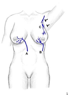
Illustration of the venous channels involved in Mondor disease. A is superior epigastric vein. B is thoracoepigastric vein. C is lateral thoracic vein. |
 | View Full Size Image | |
Media type: Image
|
- Thalhammer C, Aschwanden M. [Mondor's disease]. Dtsch Med Wochenschr. Feb 16 2007;132(7):325-6. [Medline].
- Al-Mwalad M, Loertzer H, Wicht A, Fornara P. Subcutaneous penile vein thrombosis (Penile Mondor's Disease): pathogenesis, diagnosis, and therapy. Urology. Mar 2006;67(3):586-8. [Medline].
- Hogan GF. Mondor's disease. Arch Intern Med. Jun 1964;113:881-5. [Medline].
- Weinstein EC. Mondor's disease. West J Med. Jul 1975;123(1):56-7. [Medline].
- Herrmann JB. Thrombophlebitis of breast and contiguous thoracicoabdominal wall (Mondor's disease). N Y State J Med. Dec 15 1966;66(24):3146-52. [Medline].
- Loos B, Horch RE. Mondor's disease after breast reduction surgery. Plast Reconstr Surg. Jun 2006;117(7):129e-132e. [Medline].
- Talhari C, Mang R, Megahed M, Ruzicka T, Stege H. Mondor disease associated with physical strain: report of 2 cases. Arch Dermatol. Jun 2005;141(6):800-1. [Medline].
- Oldfield MC. Mondor's disease. A superficial phlebitis of the breast. Lancet. May 12 1962;1:994-6. [Medline].
- Feller N. [Mondor's disease.]. Dapim Refuiim. Aug 1962;21:423-5. [Medline].
- Bauer-Hack K. [Contribution to Mondor's disease.]. Med Welt. Oct 13 1962;41:2152-6. [Medline].
- Losanoff JE, Basson MD, Salwen WA, Sochaki P. Mondor's disease mimicking a Spigelian hernia. Hernia. Jan 9 2008;[Medline].
- Dicuio M, Pomara G, Ales V, Fabris FM, Dahlstrand C, Morelli G. Doppler ultrasonography in a young patient with penile Mondor's disease. Arch Ital Urol Androl. Mar 2005;77(1):58-9. [Medline].
- Bircher J, Schirger A, Clagett OT, Harrison EG Jr. Mondor's disease: a vascular rarity. Mayo Clin Proc. Nov 21 1962;37:651-6. [Medline].
- Guerri G. [Histopathology and significance of cord formations on the anterolateral chest wall (Mondor's disease and syndrome).]. Arch De Vecchi Anat Patol. Oct 1960;33:829-57. [Medline].
- Holle-Robatsch S, Fink AM, Schubert C, Steiner A, Partsch H. [Mondor phlebitis associated with hepatitis C]. Vasa. Nov 2001;30(4):297-8. [Medline].
- Luis Rodríguez-Peralto J, Carrillo R, Rosales B, Rodríguez-Gil Y. Superficial thrombophlebitis. Semin Cutan Med Surg. Jun 2007;26(2):71-6. [Medline].
- Mayor M, Buron I, de Mora JC, Lazaro TE, Hernandez-Cano N, Rubio FA, et al. Mondor's disease. Int J Dermatol. Dec 2000;39(12):922-5. [Medline].
Mondor Disease excerpt Article Last Updated: Apr 30, 2008
|