Practice Essentials
Speckled lentiginous nevus is a patch of hyperpigmentation that can be seen on any area of the body. This patch contains a variable number of darkly pigmented macules and papules. Some authorities believe that speckled lentiginous nevus is a subtype of congenital melanocytic nevus. [1, 2, 3]
Patients may complain of hyperpigmented or multicolored skin lesions, with colors most often including shades of tan, brown, and black. The lesions are present at birth or noted during the individual's first years of life. Patients may be aware of an increase in the size of the lesions or changes in color over time. Skin distribution varies, with the trunk being the most common site of occurrence. It has also been reported in the oral cavity. [4]
Speckled lentiginous nevus may represent a localized defect in neural crest melanoblasts that populate a particular area of the skin. Environmental and genetic factors may also play a role. Mosaicism may be an explanation for the development of zosteriform speckled lentiginous nevus. [5]
In its earliest stage, speckled lentiginous nevus can be confused with a café au lait macule. In agminated melanocytic nevi, the background skin is not hyperpigmented, unlike that in speckled lentiginous nevus. In partial unilateral lentiginosis, multiple lentigines are grouped together on skin that has a normal color. In India, trichrome vitiligo of the face developed in at least 1 case of congenital segmental speckled lentiginous nevus in a 9-year-old girl. [6]
Electron microscopic studies show melanosomes that are fully melanized and a few premelanosomes in junctional melanocytes.
Speckled lentiginous nevus is a benign neoplasm. Malignant melanoma may develop within speckled lentiginous nevus. Predictors of the risk of malignant transformation of a speckled lentiginous nevus have yet to be determined. The surface area, the number of nevi within the speckled lentiginous nevus, and/or the presence of cytologic atypia may be factors that affect the potential for malignant transformation of a speckled lentiginous nevus. At least 20 cases of cutaneous melanoma developing within a speckled lentiginous nevus have been reported. [7, 8, 9, 10, 11, 12] In the rare instances where malignant transformation occurs within a speckled lentiginous nevus, morbidity and mortality are dependent on the stage of the secondary malignancy.
Speckled lentiginous nevi are evaluated and treated with the same level of concern as would be warranted for congenital nevi of similar size. Biopsy is necessary to rule out cytologic atypia, which may develop in a speckled lentiginous nevus.
Regular visits to a dermatologist and careful examination with the use of photography are recommended for early recognition of atypical features within a speckled lentiginous nevus. Consult a dermatologist when diagnostic and/or therapeutic uncertainty exists regarding melanocytic neoplasms in general and speckled lentiginous nevus specifically.
Pathophysiology
Speckled lentiginous nevus can be associated with different disorders. In facial features, anorexia, cachexia, and eye and skin anomalies (FACES) syndrome, cutaneous findings include zosteriform speckled lentiginous nevi. Associated findings also include ichthyosis, Ebstein syndrome (the eponym for congenital downward displacement of the tricuspid valve into the right ventricle), epidermal nevi, nevus sebaceous, scleral pigmentation, segmental neurofibromatosis type I, [13] adult-onset hearing loss, granular corneal dystrophy, and hypertrophy of the underlying pectoralis major muscle. In addition, an association with abnormality of the tongue and median nerve paresis have been described. [14, 15]
An association with phacomatosis pigmentovascularis and phacomatosis pigmentokeratotica (PPK) has also been described. [16, 17, 18, 19, 20] Loh et al present the case of a 52-year-old male with PPK and speckled lentiginous nevi on the left trunk and associated, multiple, biopsy-proven basal cell carcinomas. [21] Om et al report a case of a pediatric patient with PPK and concomitant speckled lentiginous nevus who later developed vaginal botryoid rhabdomyosarcoma. The authors attributed the PPK onset to a novel KRAS mutation. [22] Prieto-Barrios et al point out that activation of HRAS mosaic mutations can also cause PPK. [23]
Speckled lentiginous nevus syndrome has been characterized as a distinct neurocutaneous phenotype where ipsilateral neurological abnormalities are present in association with speckled lentiginous nevus. [14, 15, 16, 17, 19] This syndrome has been renamed as papular nevus spilus (PNS) syndrome to prevent confusion with macular nevus spilus. PNS syndrome is classified as a mosaic RASopathy, along with isolated nevus sebaceus, Schimmelpenning syndrome, and PKK. [24]
There has been at least a single pediatric case of PNS syndrome associated with musclar atrophy and motor neuropathy. The affected muscles were in an indentical distribution to the speckled lentiginous nevi. [25]
Epidemiology
In the general United States population, the prevalence of speckled lentiginous nevi is similar to that of congenital melanocytic nevi, with a prevalence of 0.2-2.3%, depending on age. [26] Speckled lentiginous nevus has been studied in Canadian children of Asian and white European origins, and its prevalence is the same in both ethnic groups.
No racial or sexual predilections have been noted.
Low prevalence rates of up to 0.2% have been reported in series of newborns. Approximately 80% of speckled lentiginous nevi appear at birth or during early infancy. They may present as lightly-colored café au lait macules at birth, which later develop background hyperpigmentation and darkly pigmented macules and papules over months, years, or sometimes decades.
The prevalence rate of speckled lentiginous nevus is 1.3-2.1% in school-aged children and adolescents.
In adults, the frequency of speckled lentiginous nevi larger than 1.5 cm in diameter is 2.3%.
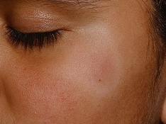
-
A large unilateral speckled lentiginous nevus that contains numerous small dark macules.
-
Extensive speckled lentiginous nevus.
-
Prominent basal layer pigmentation similar to that seen in lentigo simplex (hematoxylin-eosin stain, original magnification X200).
-
Elongated rete ridges and lentiginous proliferation of melanocytes at the dermal-epidermal junction (hematoxylin-eosin stain, original magnification X200).
-
Junctional melanocytic nevus composed of small nests of melanocytes and lentiginous melanocytic proliferation at the dermal-epidermal junction (hematoxylin-eosin stain, original magnification X100).
-
Compound melanocytic nevus with nests of uniform melanocytes in the epidermis and the dermis (hematoxylin-eosin stain, original magnification X200).