Background
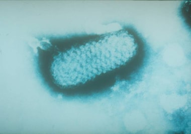
Smallpox is an acute, contagious disease caused by the variola virus, a member of the genus Orthopoxvirus, in the Poxviridae family (see the image below). Virologists have speculated that it evolved from an African rodent poxvirus 10 millennia ago. Because of the absence of an animal vector, communities had to reach a critical population (estimated at 200,000 around 3000 BCE) before endemic smallpox could be established. The name is derived from the Latin word for "spotted" and refers to the raised bumps on the face and body of the patient. (See Etiology.)
Poxviridae are linear, double-stranded deoxyribonucleic acid (DNA) viruses that replicate in the cytoplasm. Poxviridae consists of 2 subfamilies: Chordopoxvirinae, which infects vertebrates, and Entomopoxvirinae, which infects insects. Vaccinia virus, monkeypox virus, cowpox virus, and camelpox virus [1, 2] are other members of Orthopoxvirus that infect humans. (See Etiology.) Cowpox virus "scarification" by Jenner, used to induce protective immunity against smallpox, is not a single species but a group of up to 5 virus species that infects cows, humans, and other animals. [3]
The history of smallpox is remarkable not only because of the spectacular devastation it wreaked upon civilization since the dawn of humankind, but also for the astounding achievement of modern medicine, which eradicated this plague through the concerted efforts of global vaccination (see the image below). (See Treatment and Medication.)
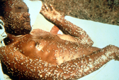 Adult with variola major with hundreds of pustular lesions centrifugally distributed. Photo from Fitzsimmons Army Medical Center slide file.
The earliest evidence of smallpox comes from ancient Egypt circa 1157 BCE, where the mummified remains of a pockmarked Ramses V were uncovered. International traders spread smallpox throughout the Old World during the 4th-15th centuries CE, while European explorers and conquerors brought the disease to the Western Hemisphere in the early 16th century.
Smallpox directly and profoundly influenced the course of human history. Its tremendous morbidity and mortality led to indiscriminate killing of kings and warlords and tipped the balance of power with regularity in Europe and elsewhere. As a result of smallpox infection, whole civilizations, including the Incas and the Aztecs, were destroyed in a single generation, and efforts to ward off the disease indelibly affected the practice of religion and medicine.
Characteristics of variola virus
The variola virus is a large, brick-shaped, double-stranded DNA virus that serologically cross-reacts with other members of the poxvirus family, including ectromelia, cowpox, monkeypox, vaccinia, and camelpox. Unlike other DNA viruses, the variola virus multiplies in the cytoplasm of parasitized host cells.
Smallpox only naturally infects humans and does not exist in a carrier state. Experimentally infected cynomolgus macaques (Macaca fascicularis) develop ordinary or hemorrhagic smallpox depending on the size of the inoculum. [4] The virus can survive in the environment for a short period, and it is most stable at low temperatures and low humidity. Variola is spread most efficiently by means of inhalation and less efficiently by means of direct contact with scabs or pustular material from skin lesions. (See the images below.) Swinepox (Suipoxvirus genus of the related Poxviridae family) may be spread by pig lice in addition to direct contact. [5]
 Characteristic skin lesion of variola viral infection on the arms and the legs of an adolescent. Photo used with the permission of the World Health Organization (WHO).
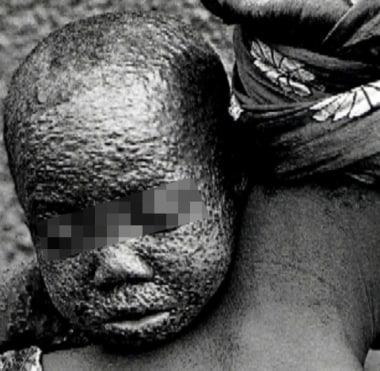
 Small child with pustular lesions due to variola viral infection. Photo used with the permission of the World Health Organization (WHO).
 Infant with advanced lesions due to variola viral infection. Photo used with the permission of the World Health Organization (WHO).
Types of smallpox
The 2 predominant variants of variola, major and minor, differ greatly in their mortality rates (30% vs 1%, respectively). Variola major was the predominant endemic strain throughout the world, and by the end of the 18th century, it was responsible for approximately 400,000 deaths a year in Europe. In patients who recovered from the disease, blindness was common, and disfiguring scars were nearly universal.
During the first half of the 20th century, all outbreaks of smallpox in Asia and most in Africa were due to variola major. Variola minor was endemic in some countries in Europe, North America, South America, and many parts of Africa.
Variola major smallpox has 4 subtypes, as follows:
-
Ordinary smallpox - The most common form, which accounts for 90% or more of smallpox cases
-
Modified smallpox - A mild form that develops in previously vaccinated persons
-
Flat smallpox (malignant smallpox) - A severe variety of smallpox in which lesions do not project above the skin surface
-
Hemorrhagic smallpox (fulminant smallpox) - A rare, very severe, highly fatal variety of smallpox in which hemorrhages develop in the skin and mucous membranes
Variola minor is less common and much less virulent; it was previously found mainly in South Africa, South America, Europe, and Australia.
Other types of smallpox include the following:
-
Variola sine eruptione (variola sine exanthemata) - Another less common form of smallpox
-
Pulmonary form of smallpox - Characterized by severe symptoms, cyanosis, and bilateral infiltrates; has been described in individuals with little or no smallpox immunity; the mortality rate of this type is undetermined
-
Pharyngeal form of smallpox - Develops in immunized individuals; this form presents with a spotty enanthem over the soft palate, uvula, and pharynx
-
Influenzalike form of smallpox - Rarely results in a rash
The pharyngeal and influenzalike forms are relatively mild, usually affect individuals who have been previously immunized, and do not cause mortality.
Immunity
Cellular immunity and humoral immunity are elicited in response to variola infection. Neutralizing antibodies can be detected during the first week of clinical illness, whereas hemagglutination-inhibition and complement-fixation antibodies are found in the second to third weeks. Neutralizing antibodies persist for many years or decades after infection, whereas levels of hemagglutination-inhibition and complement-fixation antibodies generally decrease within a year.
Cell-mediated immunity likely plays an important role in controlling disease; virus-specific cytotoxic T cells are detectable in lymphoid organs as early as 4 days after infection. These cytotoxic T cells are believed to limit viral spread by causing lysis of infected cells in the reticuloendothelial system and the skin.
The relative importance of the cellular immune response against smallpox has been demonstrated in animals. Studies show that mice with defective T cells are able to generate normal humoral responses to a viral challenge, yet they die when exposed to Orthopoxvirus concentrations that are sublethal in healthy mice. Studies in rodents and sheep have demonstrated memory in the form of virus-specific, cytotoxic lymphocyte immune responses that occur long after the initial variola infection.
Because of potential bioterrorism, interest in smallpox pathogenesis has increased. Protein analysis indicates that the variola virus G1R protein binds to cellular nuclear factor kappa-B (NF-kB), thereby inhibiting its function in cell signalling. [6] The G1R protein is highly conserved among pathogenic orthopoxviruses and is absent from the less-pathogenic vaccinia strains, thus suggesting that it may serve as a molecular therapeutic target. One report identified a novel peptide with the ability to inhibit vaccinia virus cell entry. [7]
Other studies developed a method of reliably classifying species of variola virus into major and minor species by genotype using novel real-time polymerase chain reaction (PCR) assay probes. [8] Further investigation into genetic variations between species of variola virus may reveal variable response to therapeutic targets.
History of inoculation
Intentional inoculation with subvirulent strains of variola to protect against variola major (variolation) began in India sometime before the first millennium CE. This practice spread throughout the Old World and eventually reached Europe in the early 18th century. Although variolation was capable of inducing lifelong immunity in vaccinated individuals, the practice was a risky procedure, and those inoculated had a mortality rate of approximately one tenth that of individuals with naturally occurring disease. Furthermore, treated individuals were capable of transmitting disease to untreated individuals for some time after variolation.
In one of the major accomplishments in modern medicine, Edward Jenner demonstrated in 1796 that an individual could be protected against disease. The skin could be inoculated with pustular material containing the cowpox virus, an orthopoxvirus closely related to variola. Although the heterologous immunity induced by vaccination (from the Latin word vacca, meaning cow) was not lifelong, this approach was significantly safer than variolation, and vaccination quickly spread throughout the world. In subsequent decades, the strain of virus used was sustained by means of arm-to-arm inoculation or maintained as dried material on threads.
Over time, the virus mysteriously changed from its original cowpox form to the strain of vaccinia used in current vaccines. In the latter half of the 19th century, the practice of growing virus for vaccines on the flank of calves was adopted to lessen the risk of transmitting other human diseases (eg, syphilis) during vaccination.
In the late 1940s, large-scale production of freeze-dried vaccine enabled mass vaccination campaigns and, eventually, the global eradication of smallpox. In the latter half of the 1960s, the World Health Assembly intensified its efforts to eradicate the disease by using highly potent and stable vaccine, by rapidly identifying outbreaks, and by performing ring vaccination in all contacts of a person who was infected. (See Treatment and Medication.)
The last case of endemic smallpox occurred in Somalia in 1977, and the last recorded case in humans occurred in England in 1978; this final case resulted from an accidental laboratory infection. In 1980, the World Health Organization (WHO) officially declared that smallpox had been eradicated. Currently, the only remaining known variola virus isolates are frozen in closely guarded repositories at the US Centers for Disease Control and Prevention (CDC) in the United States and at the VECTOR Institute in Russia.
After the disease was eliminated from the world, routine smallpox vaccination was stopped. The long-term consequence of eradication is that much of the world's population is now unvaccinated and at risk for smallpox infection. Currently, nearly half of the US population has not been vaccinated and has no immunity to vaccinia or variola. The remainder of the population was vaccinated 30 or more years ago and may retain partial protection from the disease. [9, 10]
Bioterrorism
Smallpox is a high-priority (category A) agent for bioterrorism, defined as follows by the CDC (see the PDF file below):
-
Easily disseminated or transmitted from person to person
-
High mortality rate and potential for significant public health effect
-
Probable instigator of panic and social disruption
-
Special actions required for public health preparedness
Bioterrorist Agents. Signs and symptoms. Chart courtesy of North Carolina Statewide Program for Infection Control and Epidemiology (SPICE), copyright University of North Carolina at Chapel Hill, www.unc.edu/depts/spice/bioterrorism.html.
Patient education
For patient education information, see Smallpox.
Etiology
Smallpox is a double-stranded, 135- to 375-kilobase (kb) DNA virus that replicates in the cytoplasm of the host cell and forms B-type inclusion bodies (Guarnieri bodies). This is in contrast to herpes viruses, which replicate in the nucleus. The orthopoxviruses are among the largest and most complex of all viruses. The virion is brick shaped, with a diameter of approximately 200 nm. (See the image below.)
Transmission
The smallpox virus is transmitted mainly through the airborne route and adheres via droplet spread of viral particles onto the mucosal surfaces of the oropharyngeal and respiratory tract. This transmission occurs through close personal contact (eg, face-to-face within 6 ft, household contact) for extended periods. Respiratory spread over long distances (eg, from one hospital floor to another) has been reported. Exposure to clothing or blankets contaminated with infected material can also result in disease. [11, 12]
Smallpox has a lower transmission rate than measles, pertussis, and influenza. Transmission through casual and limited contact has been reported in military personnel. Although rare, airborne (ie, suspended viral particles) and fomite transmission can occur. Humans are the only natural hosts of variola; nonhuman animals and insects do not carry the variola virus. Pregnant women with smallpox tend to develop hemorrhagic disease; intrauterine infection occurs in even the mildest maternal infections, resulting in premature delivery and high fetal and neonatal mortality rates.
Course of the infection
Implantation of just a few virions of smallpox into the oropharynx or respiratory tracts can cause infection. The virus infects macrophages during the first 72 hours of the incubation phase. The virus migrates and multiplies in the regional lymph nodes, resulting in asymptomatic viremia by the fourth day. The virus multiplies in the spleen, bone marrow, and lymph nodes, resulting in a symptomatic secondary viremia (ie, fever, toxemia) by the eighth day.
Finally, the virus reenters the blood in leukocytes, producing fever and toxemia, and then passes from leukocytes to adjacent cells in small blood vessels of the dermis and beneath the oropharyngeal mucosa, leading to the initial onset of the enanthem and exanthem, at which point (approximately day 14) the patient becomes infectious. (See the image below.)
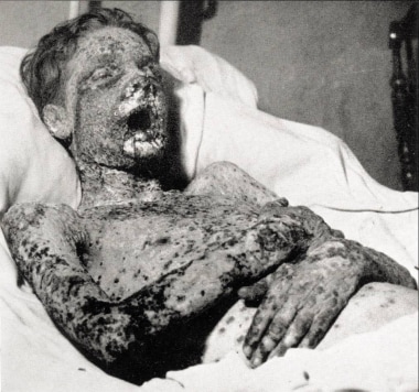 This patient with smallpox survived toxemia to succumb to secondary tissue damage days after this photo was taken. Courtesy of the US Centers for Disease Control and Prevention.
The spleen, lymph nodes, kidneys, liver, bone marrow, and other viscera may also contain large amounts of smallpox virus. Incubation periods are typically 10-12 days but can range from 7-17 days. Intrauterine infections rarely occur and usually have shorter incubation periods. Patients exposed to smallpox through routes other than the person-to-person respiratory route also have shorter incubation periods. Prior immunization, vaccinia immunoglobulin (VIG), and, possibly, antiviral chemotherapy may extend the incubation period.
Patients with smallpox are sometimes contagious upon the onset of fever (prodromal phase) but are most contagious upon rash onset. Infected persons are contagious until the last smallpox scab separates. The highest intensity of viral shedding is during the first 10 days of the rash. Infection rates among close contacts of infected persons have been reported to be between 37% and 88%. Survivors of natural smallpox infection acquire lifelong immunity.
Because of potential bioterrorism, interest in smallpox pathogenesis has increased. Protein analysis indicates that the variola virus G1R protein binds to cellular nuclear factor kappa-B (NF-kB), thereby inhibiting its function in cell signalling. [6] The G1R protein is highly conserved among pathogenic orthopoxviruses and is absent from the less-pathogenic vaccinia strains, thus suggesting that it may serve as a molecular therapeutic target. One report identified a novel peptide with the ability to inhibit vaccinia virus cell entry. [7]
Epidemiology
Occurrence in the United States
The last outbreak of smallpox in the United States was in 1947, when 12 cases were reported in New York City. In the United States, routine vaccination of the civilian population ended in 1972, and in 1990 it ended for the US military.
The most current statistics indicate that approximately 41% of the resident US population is younger than age 30 years, and that most of this group has not been vaccinated against smallpox. The immune status of those who were vaccinated 30 or more years ago has not been satisfactorily established, but some evidence shows residual immunity. Reports from the late 19th century indicate that smallpox vaccination 20-30 years previously may not protect against infection but often prevents death. No conclusive studies have shown whether people with residual immunity can transmit smallpox to nonvaccinated individuals.
International occurrence
Since 1978, no cases of smallpox have been reported in the world. The last endemic case of variola major was reported in Bangladesh in 1975; the last endemic case of variola minor was reported in Somalia in 1977. In 1978, a laboratory accident in Birmingham, England, resulted in a single case of the disease.
Smallpox is authorized to be kept for research purposes only at 2 WHO reference laboratories. One is the CDC, in Atlanta, Ga, and the other is the State Research Centre of Virology and Biotechnology, also known as the VECTOR Institute, in Koltsovo, Russia. Routine smallpox vaccinations were stopped in 1972 and smallpox was declared eradicated in 1980 after a worldwide vaccination program. In 2002, The Washington Post reported that the Central Intelligence Agency (CIA) identified possible clandestine smallpox virus stocks in 4 other nations.
Age-related demographics
The age distribution of smallpox mirrors that of the general population, although residual immunity from previous vaccination could potentially decrease disease in the older population. Historically, young or old individuals are more susceptible to severe smallpox.
Prognosis
The mortality rate in patients with untreated smallpox is 30% or higher. The more severe hemorrhagic and malignant forms of smallpox are usually fatal.
Complications
Morbidity is commonly associated with smallpox. Most patients (65-80%) recovering from infection have cutaneous scarring, which is made worse if secondary bacterial infections develop during the course of smallpox. Other complications of smallpox included dehydration and orchitis. Encephalitis occurs in 1 in 500 cases.
Dermatologic complications of smallpox include the following:
-
Formation of furuncles and/or abscesses secondary to bacterial infection
-
Sepsis
-
Pockmarks
Ophthalmologic complications of smallpox include the following:
-
Conjunctivitis
-
Corneal ulceration
-
Keratitis
-
Blindness (1% of cases)
Orthopedic complications include the following:
-
Arthritis (2% of cases)
-
Osteomyelitis variolosa
-
Symmetrical elbow joint involvement
Respiratory complications include the following:
-
Pulmonary edema
-
Pneumonitis
-
Bronchopneumonia
Mortality
Overwhelming toxemia has been the usual cause of death in smallpox. Variola major infection carries an overall fatality rate of approximately 30% (range, 15-50%) in an unvaccinated population and 3% in a vaccinated population. However, flat smallpox carries a 45.4% mortality rate in patients with discrete lesions who have been immunized. Unimmunized patients with confluent disease have a 99.3% mortality rate. Patients with hemorrhagic smallpox have a mortality rate of more than 96%, regardless of immunization status.
Variola minor infection is a less common type of smallpox and a much less severe disease, with a death rate of 1% or less.
Congenital smallpox infection results in a stillbirth rate of 35%; 50% of neonates die within their first few days of life.
-
Smallpox virion. Courtesy of US Centers for Disease Control and Prevention.
-
After exposure to the smallpox virus, a symptom-free incubation period follows. It normally lasts 10-12 days but may vary from 7-17 days. Smallpox begins with fever, headache, and severe backache. A rash appears after 2-4 days and progresses through characteristic stages of papules, vesicles, pustules, and, finally, scabs. The scabs desquamate at the end of the third or fourth week. Courtesy of the World Health Organization.
-
Smallpox rash at days 3, 5, and 7 of evolution. Lesions are denser on the face and extremities than on the trunk. They also appear on the palms of the hand and have a similar appearance. Courtesy of the World Health Organization.
-
Flat-type smallpox on day 6 of the rash. Courtesy of the US Centers for Disease Control and Prevention.
-
This patient with smallpox survived toxemia to succumb to secondary tissue damage days after this photo was taken. Courtesy of the US Centers for Disease Control and Prevention.
-
Smallpox vaccination with bifurcated needle. Reconstituted vaccine is held between the prongs of the needle and injected subcutaneously by multiple punctures; 15 rapid strokes, at right angles to the skin over the deltoid muscle, are made within a 5-mm area. Courtesy of the World Health Organization.
-
Smallpox vaccination. Evolving primary vaccination appearance. Courtesy of the US Centers for Disease Control and Prevention.
-
Typical temperature chart of a patient with smallpox infection (from Henderson, 1999).
-
Characteristic skin lesion of variola viral infection on the arms and the legs of an adolescent. Photo used with the permission of the World Health Organization (WHO).
-
Small child with pustular lesions due to variola viral infection. Photo used with the permission of the World Health Organization (WHO).
-
Infant with advanced lesions due to variola viral infection. Photo used with the permission of the World Health Organization (WHO).
-
Unvaccinated infant with the ordinary form of the variola major strain of smallpox has centrifugally distributed umbilicated pustules on day 3 in the course of the disease. Reprinted with permission from the World Health Organization (WHO).
-
Unvaccinated infant with the ordinary form of the variola major strain of smallpox has centrifugally distributed umbilicated pustules on day 5 in the course of the disease. Reprinted with permission from the World Health Organization (WHO).
-
Unvaccinated infant with the ordinary form of the variola major strain of smallpox has centrifugally distributed umbilicated pustules on day 7 in the course of the disease. Reprinted with permission from the World Health Organization (WHO).
-
The ordinary form of the variola minor strain of smallpox (alastrim) in an unvaccinated woman 12 days after the onset of skin lesions. The facial lesions are sparser and evolved more rapidly than the extremity lesions. Reprinted with permission from the World Health Organization (WHO).
-
The ordinary form of the variola minor strain of smallpox (alastrim) in an unvaccinated woman 12 days after the onset of skin lesions. The facial lesions are sparser and evolved more rapidly than the extremity lesions. Reprinted with permission from the World Health Organization (WHO).
-
The ordinary form of the variola minor strain of smallpox (alastrim) in an unvaccinated woman 12 days after the onset of skin lesions. The facial lesions are sparser and evolved more rapidly than the extremity lesions. Reprinted with permission from the World Health Organization (WHO).
-
Adult with variola major with hundreds of pustular lesions centrifugally distributed. Photo from Fitzsimmons Army Medical Center slide file.
-
Hemorrhagic-type variola major lesions. Death usually ensued before typical pustules developed. Reprinted with permission from the World Health Organization (WHO). 1988; 10-14, 35-36.


