Practice Essentials
The essential abnormality of trichorrhexis nodosa is the formation of nodes along the hair shaft through which breakage readily occurs. In 1852, Samuel Wilks of Guy's Hospital first described the condition, although the term trichorrhexis nodosa was not proposed until 1876 by M. Kaposi. Trichorrhexis nodosa is ultimately a response to physical or chemical trauma. Trichorrhexis nodosa may be acquired in patients with normal hair through exposure to a sufficient level of trauma. Trichorrhexis nodosa may also be congenital, occurring in defective, abnormally fragile hair following trivial injury. It is the most common congenital defect of the hair shaft.
It may affect the hair of the scalp, pubic area, beard, and moustache and may be particularly noticeable in persons of Afro-Caribbean descent because of hairstyling techniques. [1] Hair care practices and hairstyles often used among women of African descent may contribute to trichorrhexis nodosa and central centrifugal cicatricial alopecia. [2] The more common acquired form results from excessive or repeated trauma caused by frequent application of hair-permanent liquid, hair dyes, frequent brushing, scalp massage, and lengthy and repeated ultraviolet exposure, a reflection of the amount of trauma inflicted on the hair shafts rather than by some inherent or structural defect. [3] Trichorrhexis nodosa may also be due to malnutrition or endocrinopathy, especially iron deficiency and hypothyroidism.
See the image below.
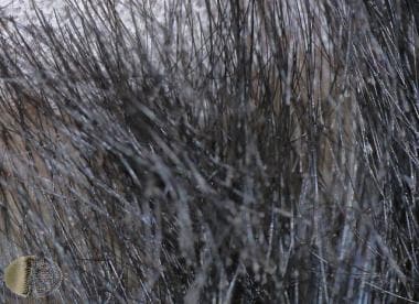 Trichorrhexis nodosa. Courtesy of DermNet New Zealand (http://www.dermnetnz.org/assets/Uploads/hair-nails-sweat/trichorrhexis-nodosa1.jpg).
Causes
See Pathophysiology and Etiology.
Prognosis
Acquired trichorrhexis nodosa is reversible with the avoidance of physical and chemical trauma. Complete resolution may take 2-4 years, depending on the growth of new anagen hairs.
Diagnostics
A CT of the brain and an EEG may aid in diagnosis if intellectual disability is present.
Tortuous intracranial vessels and bladder diverticula may be present at early stages and may suggest this diagnosis. [4]
Dermoscopy shows breakage in hair shafts at multiple levels, producing an appearance suggestive of the ends of 2 brushes aligned in opposition, resembling “thrust paint brushes.” [5, 6, 7] High-power magnification demonstrates these fibers in detail, while at low power, these structures appear as light-colored nodules or gaps located along the hair shaft. [6, 8]
Also see Laboratory Studies.
Treatment
Regardless of the presence or the absence of an underlying defect of the hair shaft, trichorrhexis nodosa is ultimately the result of trauma. Therefore, treatment is aimed at minimizing physical or chemical trauma.
Excessive brushing, hot combing, permanent waving, and other harsh hair treatments should be avoided. In acquired localized trichorrhexis nodosa, the underlying pruritic dermatosis should be treated to prevent trauma from scratching or rubbing. Underlying metabolic disorders are treated accordingly, usually through the implementation of a specifically tailored diet.
Prevention
Patients should avoid physical and chemical trauma to the hair shaft. See Pathophysiology.
Pathophysiology
Macroscopically, hair shafts affected by trichorrhexis nodosa contain small white nodes at irregular intervals throughout the length of the shaft. The number of nodes may vary from one to several, depending on the length of the hair. These nodes represent areas of cuticular cell disruption, which allows the underlying cortical fibers to separate and fray. The cortical fibers splay outwards and fracture, giving the node the microscopic appearance of 2 brooms or paintbrushes thrust together end to end by their bristles. Complete breakage often occurs at these nodes. The fractured hairs are found mainly on the scalp but they also may involve body or pubic hair.
Physical traumas that may cause sufficient damage to the hair shaft include excessive brushing, back combing, stressed hairstyles, the application of heat, and prolonged exposure to ultraviolet light. Head rolling and banging; habit tics; trichotillomania; and the scratching and pulling associated with pruritic dermatoses, such as seborrheic dermatitis and pediculosis capitis, can also result in sufficient damage. Chemical traumas include excessive exposure to salt water, shampooing, setting, perming, bleaching, and dyeing of hair.
Seasonal recurrence of trichorrhexis nodosa has been reported as the result of the cumulative effect of diverse insults to the hair. [9] The manifestations appear each summer when repeated soaking in salt water and exposure to ultraviolet light were superimposed on the traumas of shampooing and hair brushing. Trichorrhexis nodosa has also resulted from the use of chemical hair straighteners and selenium shampoo and from trauma induced by infestation with trichomycosis axillaris. Numerous studies have reproduced trichorrhexis nodosa in both normal and affected hairs by exposing them to simulated physical and chemical trauma.
Although trichorrhexis nodosa primarily occurs in normal hairs exposed to a sufficient degree of trauma, it has also been observed in patients who have an underlying weakness of the hair shaft due to a structural abnormality. Structural abnormalities associated with increased fragility include trichorrhexis invaginata (bamboo hair), pili torti, monilethrix, and pseudomonilethrix. [10] The hairs formed in these disorders demonstrate distinctive patterns of defects that result in brittle hairs. These disorders are hereditary in nature and follow autosomal dominant or recessive patterns of inheritance. Pili annulate, a hair shaft disorder without fragility, has been described with severe hair breakage linked with trichorrhexis nodosa. [11]
Trichorrhexis invaginata is an autosomal recessive disorder characterized by nodular ball-and-socket deformities resulting from intussusception of the hair shaft at the zone of keratinization. The hairs in cases of pili torti are flattened and twisted 180° around their long axis at irregular intervals along the shaft. Inheritance is autosomal dominant, although sporadic cases have been reported. Hairs affected by monilethrix show elliptical nodes separated by narrower internodes where the medulla is lacking. Hairs of pseudomonilethrix show irregular nodes, which are the protruding edges of depressions in the shaft. Both disorders are autosomal dominant in inheritance. These structural defects result in brittle hairs that are more susceptible to the effects of trauma; therefore, they are likely to demonstrate the nodes of trichorrhexis nodosa.
The underlying structural weakness of the hair shaft may result from impaired keratin formation. Hairs weakened by impaired keratin formation are seen in argininosuccinic aciduria, trichothiodystrophy, and Menkes disease.
Argininosuccinic aciduria is the autosomal recessive deficiency of argininosuccinase, the enzyme that cleaves argininosuccinic acid into arginine and fumaric acid in the urea cycle. It is comparatively rare in East Asian populations as compared with whites. [12] Hair normally contains 10.5% arginine by weight. The deficiency in arginine resulting from this disorder produces weak hair with a tendency to fracture. [13] The rare inborn deficiency of argininosuccinic acid synthetase results in low arginine levels and brittle hair. Sulfur and the sulfur-containing amino acid cystine are also important components of the hair shaft. They allow for the formation of disulfide bonds, which account in part for the strength of keratin. In cases of trichothiodystrophy, low sulfur and cystine content result in defective keratin formation and weakened hairs. The characteristic hair-shaft defect in trichothiodystrophy is a transverse fracture called trichoschisis, but trichorrhexis nodosa can also be seen.
Menkes disease is a sex-linked recessive disorder characterized by a defect in the metabolism of copper, resulting in low serum copper levels. The enzymes involved in keratin formation are copper dependent. Therefore, patients with this disorder have an inherent defect in keratinization, resulting in a twisting of hairs (pili torti) as well as weakened hairs and trichorrhexis nodosa. The hairs resulting from these metabolic and genetic defects are more susceptible to the effects of trauma; therefore, they are likely to demonstrate the nodes of trichorrhexis nodosa (see Menkes Kinky Hair Disease).
A primary congenital form of trichorrhexis nodosa is inherited as an autosomal dominant trait in some families. This form may occur as an isolated defect or with other minor ectodermal abnormalities.
Trichorrhexis nodosa has also been described in a case of hypothyroidism. [14] The accumulation of mucin in the outer root sheath is postulated to press on the inner root sheath, causing the hair-shaft defect. This accumulation may cause a defect in the differentiation and maturation of matrix cells, causing a growth defect in the hair shaft.
Trichorrhexis nodosa may also occur as part of the trichohepatoenteric syndrome (THES), an autosomal recessive syndrome also characterized by life-threatening diarrhea in infancy, immunodeficiency, liver disease, facial dysmorphism, hypopigmentation, cardiac defects, and possibly platelet abnormalities (eg, reduced platelet alpha-granules, unusual stimulated alpha granule content release, abnormal lipid inclusions, abnormal platelet canalicular system, and reduced number of microtubules). [15, 16] It has been linked to mutations in TTC37.
In another syndrome with dysmorphic features, including cleft palate, hypotrichosis with trichorrhexis nodosa, hypohidrosis, oligodontia, and ridging of nails, a heterozygous germline mutation, Ala111Thr, in the P63 gene was detected in the proband and his mother. [17]
Trichorrhexis nodosa has been described in a child with biotinidase deficiency. [18] It has also been linked with tumor necrosis factor-α inhibitor therapy. [19]
Etiology
Trichorrhexis nodosa is the result of physical or chemical trauma to the hair shaft. Incorrect use of ceramic flat irons can produce acquired trichorrhexis nodosa. [20] A primary congenital form of trichorrhexis nodosa is inherited as an autosomal dominant trait in some families. It can also occur as part of the autosomal recessive TRES (see Pathophysiology).
The cutaneous photosensitivity that is sometimes evident may be the result of defective nucleotide excision repair. Three genes, XPB, XPD, and TTDA, have been linked as causative genes for this photosensitivity. [21]
Epidemiology
Trichorrhexis nodosa is an uncommon disorder. A retrospective review of 129 hair-mount samples from 119 patients over a 10-year span found 25 cases of loose anagen hair syndrome, 6 cases of uncombable hair syndrome, and trichorrhexis nodosa in 13 patients. [22]
Acquired proximal trichorrhexis nodosa is common in Blacks and appears to occur in individuals who are genetically predisposed. Some consider it an ethnic hair disorder. [23] Acquired distal trichorrhexis nodosa primarily occurs in Asian or White persons. Acquired proximal trichorrhexis nodosa is more common in females than in males.
Congenital trichorrhexis nodosa may be present at birth, or it may appear within the first couple months of life. It can present in patients with the late form of argininosuccinic aciduria at age 2 years or older.
Acquired trichorrhexis nodosa usually presents in early to middle life.
-
Trichorrhexis nodosa. Courtesy of DermNet New Zealand (http://www.dermnetnz.org/assets/Uploads/hair-nails-sweat/trichorrhexis-nodosa1.jpg).


