Background
Tinea nigra is an uncommon superficial dermatomycosis usually caused by Hortaea werneckii (see the image below), formerly known as Phaeoannellomyces werneckii, (formerly classified as Exophiala werneckii and Cladosporium werneckii). [1]
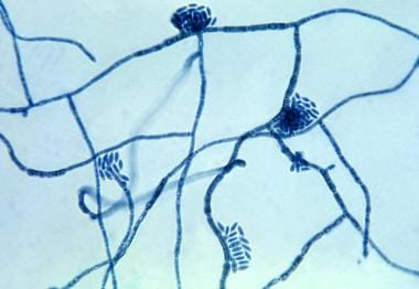 Micrograph of the Hortaea werneckii fungus, which is a causative agent of tinea nigra. Courtesy of CDC Public Health Information Library (PHIL) and Wikimedia Commons.
Tinea nigra may also be due to Stenella araguata, first described and named Cladosporium castellanii in 1973. [2] Tinea nigra appears as a hyperpigmented macule, which usually occurs on the palms. The soles and, more rarely, other areas of the body, can also be affected.
Cequeira first described tinea nigra in 1891, calling the infection keratomycosis nigricans palmaris. In 1921, Horta isolated the pathogen and gave it its original name, C werneckii.
Although H werneckii has been established as the causative fungus in most cases of tinea nigra, other species of dematiaceous fungi, such as S araguata, may produce a similar clinical picture.
A Cladophialophora strain, allegedly a new species, Cladophialophora saturnica, has been described that caused an interdigital tinea nigra – like lesion in a HIV-positive Brazilian child, successfully treated with oxiconazole. [3]
Pathophysiology
Tinea nigra is a superficial mycosis of the stratum corneum. Infection is believed to occur as a result of inoculation from a contamination source such as soil, sewage, wood, or compost subsequent to trauma in the affected area.
Typically, the incubation period for tinea nigra is 2-7 weeks, although in experimental inoculation, the incubation period was 20 years. [4] The fungus exhibits lipophilic adhesion to human skin; it is exclusively found in the stratum corneum and does not extend into the stratum lucidum.
H werneckii receives nourishment from its use of decomposed lipids. Its tolerance to an environment with a high salt concentration and a low pH allows the fungus to thrive in human skin. It has been isolated from the hypersaline waters of salterns as one of the predominant species of a group of halophilic and halotolerant melanized yeastlike fungi. [5] H werneckii has distinct mechanisms of adaptation to high-salinity environments that are not seen in salt-sensitive and only moderately salt–tolerant fungi.
A pigmentary change in the skin results in a dark-colored macule due to the accumulation of a melaninlike substance in the fungus.
Etiology
Tinea nigra is due to infection by the fungus, H werneckii, which is classified in the family Dematiaceae, class Hyphomycetes, phylum Deuteromycota. Infection occurs after inoculation subsequent to trauma. The dermatomycosis tends to occur in areas with an increased concentration of eccrine sweat glands. Hyperhidrosis appears to be a risk factor for this disease.
Epidemiology
Frequency
United States
Tinea nigra is relatively uncommon in the United States. However, numerous cases are reported in the dermatologic literature. Tinea nigra typically affects people who reside in the coastal states such as Florida, Texas, Alabama, Louisiana, Virginia, and North Carolina. Although cases of tinea nigra are also reported in patients from northern and inland regions of the United States, including New York City, Chicago, and Boston, patients often report a history of foreign travel, frequently to the Caribbean Islands. [6]
International
Tinea nigra is not uncommon in tropical regions of Central America, South America, Africa, and Asia. Epidemiologic studies of skin diseases in schoolchildren performed by direct inspection using dermatologists in Magong, Penghu, Republic of China on the island of Formosa found the prevalence of fungal infection, including tinea nigra, tinea versicolor, and tinea corporis, to be 0.24% (95% confidence interval, 0.07-0.41%). [7] Tinea nigra may present as an imported infection from endemic areas into temperate climate regions, [6] including Chile. [8]
Race
Tinea nigra appears to occur less often in the black population than in others, although this observation may reflect impaired recognition of the disease.
Sex
The female-to-male predilection for tinea nigra is 3:1.
Age
Tinea nigra most often occurs in pediatric and adolescent populations; however, individuals of any age may be affected. In a study of 12 patients in Venezuela, it was found to be more prevalent among young people with fair skin aged 3-28 years who visited beaches. [2]
Prognosis
Although the appearance of tinea nigra may be alarming because of its uncommon occurrence and its potential confusion with a more serious medical disorder (eg, malignant melanoma [9, 10] ), tinea nigra is a benign disease that is easily curable.
Patient Education
Tinea nigra may be psychologically distressing, especially because of its potential confusion with a melanoma. Therefore, the patient should be reassured of the benign nature of this condition.
-
Tinea nigra, evident as a painless cluster of brown-to-black macules. Courtesy of Dr. Peter Santalucia.
-
Tinea nigra, showing hyperkeratosis and mild acanthosis. A scant amount of perivascular lymphocytic infiltrate may be found in the papillary and subpapillary layers of the dermis (hematoxylin and eosin stain). Courtesy of Thomas N. Helm, MD.
-
Tinea nigra, with histologic section demonstrating periodic acid-Schiff–positive septate hyphae within the stratum corneum. Courtesy of Thomas N. Helm, MD.
-
Micrograph of the Hortaea werneckii fungus, which is a causative agent of tinea nigra. Courtesy of CDC Public Health Information Library (PHIL) and Wikimedia Commons.


