Background
Lipodystrophies are a heterogeneous group of diseases clinically characterized by a congenital or acquired loss of fat in circumscribed, partial, or diffuse areas of the body. As a rule, localized lipodystrophies are not associated with metabolic or systemic disorders, they tend to resolve spontaneously, and they have an excellent prognosis. The main concern is cosmetic when the lesions persist. Controversy exists in their classification, but three subsets can be identified based on the presence or the absence of preceding inflammation and the histologic findings. See the image below.
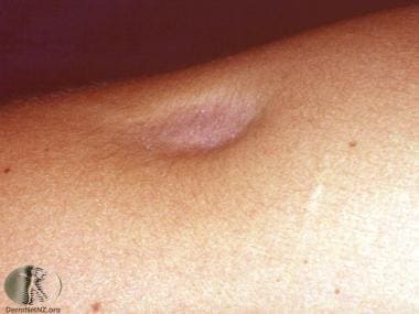 Localized lipoatrophy from a steroid injection. Courtesy of DermNet New Zealand (http://www.dermnetnz.org/assets/Uploads/dermal-infiltrative/lipoatrophy.jpg).
Centrifugal lipodystrophy or lipodystrophia centrifugalis abdominalis infantilis is the best-characterized subset. It affects children at a young age. It begins on the trunk, spreads for a few years to the neighboring abdomen or chest, and eventually resolves spontaneously in most patients.
The second group is defined by a lack of preceding inflammation and a common histologic pattern of involutional lipoatrophy. It includes such entities as lipoatrophia semicircularis; semicircular or annular dystrophy; and lipoatrophies linked to repeated trauma, pressure, or drug injections. A number of cases are idiopathic. Bleomycin, which is usually linked with flagellate erythema, may also be associated with acquired partial lipodystrophy. [1]
The third type represents a sequela from various types of panniculitis and is associated with preceding inflammation. It includes lipophagic granulomatous panniculitis and other types of panniculitis, primarily associated with connective-tissue disease.
Acquired partial lipodystrophy tends to be seen in women with late-onset lipodystrophy. [2] Some also have membranoproliferative glomerulonephritis, diabetes mellitus, and impaired autoimmune diseases such systemic lupus erythematosus, dermatomyositis, or rarely localized scleroderma.
Other Medscape articles on lipodystrophy include Dermatologic Manifestations of Generalized Lipodystrophy, Lipodystrophy in HIV, and Progressive Lipodystrophy.
Pathophysiology
The pathophysiology of localized lipodystrophy is unknown. The pathogenetic mechanism of inherited lipodystrophies at the molecular level has been linked to mutations of lamin A/C, peroxisome proliferator-activated receptor (PPAR-gamma), and other seemingly unrelated proteins. [3] In cases linked to injections of insulin, an immune response mediated by tumor necrosis factor-alpha has been hypothesized. [4, 5]
Familial partial lipodystrophy (FPLD) usually results from coding sequence mutations either in LMNA, encoding nuclear lamin A/C, or in peroxisome proliferator-activated receptor-gamma (PPARG), encoding PPAR-gamma. The LMNA form is called FPLD2 (Mendelian Inheritance in Man [MIM] 151660) and the PPARG form is called FPLD3 (MIM 604367). FPLD phenotype may be due to a single-base mutation in the DNA-binding domain of peroxisome PPAR-gamma. [6] A PPARG H449L mutation has been described in a Turkish family. [7] Impaired PPARG function through a mutation of a conserved salt bridge (R425C) producing FPLD has also been described. [8] A multicenter prospective Turkish study of 56 FPLD patients delineated pathogenic variants of the LMNA gene in nine families, with typical exon 8 codon 482 pathogenic variants in four of them. [9]
Segmental lipodystrophy may result from mosaicism, a postzygotic mutation that may be a germline or a somatic phenomenon. [10] Mosaicism of adipocytes may be challenging to classify.
Lipophagic lobular panniculitis, an entity of unknown origin, may lead to circumferential fat atrophy of the ankles and may represent an end-stage manifestation of an idiopathic lobular panniculitis of children localized to the lower part of the lower limbs. [11]
Etiology
The cause of centrifugal lipodystrophy is unknown.
In many cases, involutional lipodystrophy is idiopathic. In other cases, repeated trauma, [12] chronic pressure or compression, or local injections may be the cause. It has been reported to develop at the site of injection or in the surrounding area of injection with insulin, [13] intralesional or intra-articular steroids, antibiotics (eg, intramuscular benzathine penicillin), [14] vasopressin, and human growth hormone. [13] Localized lipoatrophy was also noted following intramuscular injection of amikacin. [15]
Localized lipoatrophy may represent the late or end stage of a preceding or concomitant panniculitis, which may be triggered by various underlying conditions. Connective-tissue disease–induced panniculitis is one of the main causes and may not be symptomatic at the time of presentation. Other causes of lipoatrophic panniculitis may include alpha-1-antitrypsin deficiency or cytophagic, factitial, and infectious panniculitides.
Epidemiology
Frequency
Few cases have been reported internationally. Centrifugal lipodystrophy and the involutional type are rare. Lipoatrophy secondary to panniculitis is more frequent.
Race
No racial predilection exists except for centrifugal lipodystrophy, which has been described mainly in Asian children.
Sex
No difference in sex has been established, except for a female predominance in the involutional type. A female predominance is also observed in connective-tissue disorder–associated lipoatrophic panniculitis.
Age
No specific age of onset exists except for centrifugal lipodystrophy. In these cases, lesions appear before age 3 years, they stop spreading by age 8 years, and they tend to resolve by age 13 years.
Connective-tissue disorder–associated lipoatrophic panniculitis may be observed at any age, but it occurs most frequently in adults aged 20-60 years. Lupus erythematosus is the most common underlying disease.
Prognosis
The prognosis is excellent in the first two groups because they are limited disorders affecting only localized areas of subcutaneous tissue. The prognosis for patients with an underlying systemic illness follows that of the primary condition. Many patients show spontaneous regression.
Lipoatrophia semicircularis responds to basic measures, which when promptly and jointly applied, resulted in 90% of the cases resolving within 6 months in one large series. [16]
Localized lipodystrophies usually have little or no associated morbidity or mortality. They are circumscribed and tend to resolve spontaneously, although panniculitis may be chronic and relapsing. Usually, no associated systemic disorder is present, except in some cases of panniculitis induced by an underlying connective-tissue disorder. Even then, symptoms tend to be mild with an excellent overall outcome.
-
Localized lipoatrophy from a steroid injection. Courtesy of DermNet New Zealand (http://www.dermnetnz.org/assets/Uploads/dermal-infiltrative/lipoatrophy.jpg).


