| Patient Education |
|
Click here for patient education.
|
|
You are in: eMedicine Specialties >
Dermatology > BACTERIAL INFECTIONS
Malakoplakia
Article Last Updated: Oct 27, 2005
AUTHOR AND EDITOR INFORMATION
Section 1 of 11
Author: Raphael J Kiel, MD, Associate Professor of Medicine, Wayne State University School of Medicine; Associate Program Director, Head of Infectious Disease Section, Department of Internal Medicine, Oakwood Hospital
Raphael J Kiel is a member of the following medical societies: American College of Physicians-American Society of Internal Medicine, American Geriatrics Society, American Medical Association, and American Medical Informatics Association
Editors: Peter Fritsch, MD, Chair, Department of Dermatology and Venereology, University of Innsbruck, Austria; David F Butler, MD, Professor of Dermatology, Texas A&M University College of Medicine; Director, Division of Dermatology, Scott and White Clinic; Director Dermatology Residency Training Program, Scott and White Clinic; Jeffrey J Miller, MD, Associate Professor, Department of Dermatology, Penn State University, Milton S Hershey Medical Center; Glen H Crawford, MD, Assistant Clinical Professor, Department of Dermatology, University of Pennsylvania School of Medicine; Chief, Division of Dermatology, The Pennsylvania Hospital; William D James, MD, Paul R Gross Professor of Dermatology, University of Pennsylvania School of Medicine; Vice-Chair, Program Director, Department of Dermatology, University of Pennsylvania Health System
Author and Editor Disclosure
Synonyms and related keywords:
malacoplakia soft plaque, genitourinary tract plaque, genitourinary tract nodule, GU tract nodule, GU tract plaque, malacoplakia, cutaneous malakoplakia, cutaneous malacoplakia
Background
Malakoplakia is an inflammatory condition presenting as a plaque or a nodule that usually affects the genitourinary tract but may rarely involve the skin. Malakoplakia was first described in the early 1900s as yellow soft plaques that were seen on the mucosa of the urinary bladder. Microscopically, malakoplakia is characterized by the presence of foamy histiocytes with distinctive basophilic inclusions, which are known as Michaelis-Gutmann bodies.
Cutaneous malakoplakia is rare but presents in patients who are immunocompromised and have defects in macrophage function. Lesions are yellow-to-pink papules, but they can present as nodules or ulcerations and are often diagnosed only after biopsy. Cultures of the lesions can yield bacteria, most commonly Escherichia coli.
Pathophysiology
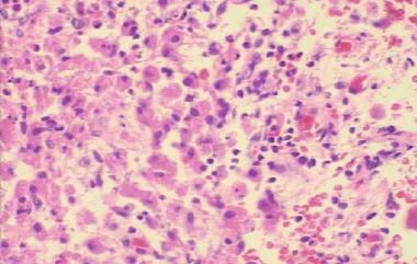
Malakoplakia is believed to result from the inadequate killing of bacteria by macrophages or monocytes that exhibit defective phagolysosomal activity. Partially digested bacteria accumulate in monocytes or macrophages and lead to the deposition of calcium and iron on residual bacterial glycolipid. The presence of the resulting basophilic inclusion structure, the Michaelis-Gutmann body, is considered pathognomonic for malakoplakia.
Studies have suggested that a decreased intracellular cyclic guanosine monophosphate (cGMP) level may interfere with adequate microtubular function and lysosomal activity, leading to an incomplete elimination of bacteria from macrophages and monocytes.
Frequency
United States
The total number of patients with malakoplakia is fewer than 500. Most patients have genitourinary tract disease, although involvement of the gastrointestinal tract and other visceral organs has been described. Cutaneous malakoplakia is rare, with fewer than 50 cases reported in the literature. Because this disease is often diagnosed after biopsy of nondescript lesions, the true incidence is most likely higher.
International
International frequency of malakoplakia is the same as in the United States.
Mortality/Morbidity
Malakoplakia most often occurs in patients who are immunocompromised. Often, malakoplakia is resistant to treatment, with a mean duration of the skin lesions of 3-6 months.
- Mortality in patients with malakoplakia is most often due to an underlying condition.
- Significant morbidity relates to the chronicity of the condition, which can resist local and systemic therapy. Draining sinuses, persistence of disfiguring skin lesions, and involvement of visceral organs constitute significant morbidity in patients with malakoplakia.
Race
Cutaneous malakoplakia is more commonly reported in whites (39%) than in African Americans (19%) or Asians (12%), although the patient's ethnic group is often not specified in case reports.
Sex
Cutaneous malakoplakia occurs with a male-to-female ratio of 2.3:1.
Age
Cutaneous malakoplakia presents in patients with a broad age range. The median age at the time of presentation is 53 years. Occurrence of the disease is associated more with the presence of an immunocompromised state affecting monocyte and macrophage function than with age.
History
- Patients typically present with a history of immunosuppression due to renal transplantation, diabetes mellitus, or lymphoma or a history of receiving long-term therapy with systemic corticosteroids.
- Close to one quarter of patients present with internal organ involvement, most commonly in the retroperitoneal area, the kidney, the bladder, or the colon. Symptoms reflect the organ system involved, but patients can often present with draining sinuses originating from deeper organ involvement.
- Malakoplakia rarely presents in patients with HIV infection and AIDS, which may be because of the preservation of monocytic antimicrobial function in these patients.
- Malakoplakia can mimic colonic, pancreatic, oropharyngeal, or genital tract carcinoma. Involvement of the bone and the lungs has also caused diagnostic difficulty.
Physical
- Lesions present as a papule, a plaque, or an ulceration that is yellow to pink or skin colored. Lesions may have associated drainage.
- Common locations include the perianal or inguinal areas, the buttocks, and the abdominal wall.
- Draining abscesses/sinuses have been reported to occur near the urethra, the thigh, the vulva, and the perianal area. One patient with malakoplakia who presented with an axillary abscess has been described.
- Lesions may be clinically misdiagnosed as an abscess, a malignancy, a lymphoma, or a benign skin tag.
- The area may appear fluctuant, or it may present as a solitary tender nodule or a group of papules.
- Often, lesions are chronic but are not debilitating to the patient.
Causes
Defective macrophage killing of bacteria, most commonly E coli, leads to malakoplakia. Defective macrophage killing results in an accumulation of bacterial degradation products and a granulomatous reaction, which clinically manifests as the formation of a papule, a plaque, or an ulceration. Although E coli is the most common gram-negative bacteria isolated, other enteric bacteria may be found on culture. Staphylococcus aureus and Pseudomonas aeruginosa may be cultured.
Risk factors for the development of malakoplakia include the following:
- Prolonged therapy with systemic corticosteroids
- Organ transplantation
- Diabetes mellitus
- Lymphoma
- Rheumatoid arthritis
Actinomycosis
Sarcoidosis
Squamous Cell Carcinoma
Other Problems to be Considered
Histiocytosis X
Malignant lymphoma
Histiocytoma
Granular cell tumor
Skin abscess
Botryomycosis
Lab Studies
- Culture the draining sinuses for aerobic, anaerobic, fungal, and mycobacterial pathogens.
Imaging Studies
- Positron emission tomography scanning has been used to diagnose malakoplakia in 2 cases of renal disease. Intense uptake of fluorodeoxyglucose was noted in the kidney.
- MRI of a patient with renal malakoplakia has shown multiple nodules with low signal on T1-weighted, T2-weighted, and early and late postgadolinium images.
Procedures
- Perform a biopsy of the skin lesion. Tissue processing follows by using routine hematoxylin and eosin stain, periodic acid–Schiff stain, von Kossa stain for calcium, and Perls Prussian blue stain for iron.
Histologic Findings
Sheets of ovoid histiocytes with fine eosinophilic cytoplasmic granules (von Hansemann cells) are seen on routine staining. Histiocytes with 5- to 15-mm basophilic inclusions with concentric laminations (Michaelis-Gutmann bodies) are diagnostic. Michaelis-Gutmann bodies demonstrate positive results using periodic acid–Schiff stain and are diastase resistant. They stain with von Kossa stain for calcium and Perls Prussian blue stain for iron.
Electron microscopy results show that Michaelis-Gutmann bodies consist of lysosomes filled with partially digested bacteria. Gram stain may demonstrate gram-negative bacteria. Immunohistochemical studies demonstrate positive results for CD68 antibodies, lysosomes, and a-chymotrypsin. Immunostaining with polyclonal anti–Mycobacterium bovis antibody may demonstrate organisms in patients with malakoplakia.
Medical Care
- Therapy with antibiotics that concentrate in macrophages (eg, quinolone, trimethoprim-sulfamethoxazole) is associated with a high cure rate. Antibiotic therapy directed against E coli in combination with surgery provides the best chance of cure.
- Bethanechol, a choline agonist, has been used in combination with antibiotics and surgery. Bethanechol may correct the decreased cGMP levels that are believed to interfere with complete bacterial killing.
- Ascorbic acid has been used to increase the cGMP and cyclic adenosine monophosphate (cAMP) levels in monocytes, which may represent an effective strategy for therapy. An insufficient number of patients have been treated with ascorbic acid to determine its overall effect.
- Discontinuation of immunosuppressive drug therapy is usually needed to effectively treat malakoplakia.
Surgical Care
- Excision of skin lesions and drainage of abscesses are fundamental to diagnosis and treatment.
- Surgery combined with antibiotic therapy should be directed against E coli or other organisms recovered on culture.
Consultations
- Dermatologist
- Infectious disease specialist
The goals of pharmacotherapy are to reduce morbidity and to prevent complications.
Drug Category: Antibiotics
Antimicrobials directed against gram-negative bacteria, especially E coli, are used to treat patients with malakoplakia. In addition, antibiotics that concentrate in macrophages are used. Quinolone antibiotics (eg, ciprofloxacin) and sulfonamides (eg, trimethoprim-sulfamethoxazole) are indicated. Bethanechol and ascorbic acid have also been used in the treatment of patients with malakoplakia.
| Drug Name | Ciprofloxacin (Cipro) |
| Description | Fluoroquinolone that has activity against Pseudomonas and Streptococcus species, MRSA, Staphylococcus epidermidis, and most gram-negative organisms but has no activity against anaerobes. Inhibits bacterial DNA synthesis and, consequently, growth. Continue treatment for at least 2 d (7-14 d typical) after signs and symptoms have disappeared. |
| Adult Dose | 250-500 mg PO bid for 7-14 d |
| Pediatric Dose | <18 years: Not recommended >18 years: Administer as in adults |
| Contraindications | Documented hypersensitivity |
| Interactions | Antacids, iron salts, and zinc salts may reduce serum levels; administer antacids 2-4 h before or after taking fluoroquinolones; cimetidine may interfere with metabolism of fluoroquinolones; reduces therapeutic effects of phenytoin; probenecid may increase serum concentrations; may increase toxicity of theophylline, caffeine, cyclosporine, and digoxin (monitor digoxin levels); may increase effects of anticoagulants (monitor PT) |
| Pregnancy | C - Safety for use during pregnancy has not been established.
|
| Precautions | In prolonged therapy, perform periodic evaluations of organ system functions (eg, renal, hepatic, hematopoietic); adjust dose in renal function impairment; superinfections may occur with prolonged or repeated antibiotic therapy |
| Drug Name | Trimethoprim and sulfamethoxazole (Bactrim, Bactrim DS, Septra, Septra DS) |
| Description | Inhibits bacterial growth by inhibiting synthesis of dihydrofolic acid. Antibacterial activity of TMP-SMZ includes common urinary tract pathogens except P aeruginosa. |
| Adult Dose | 160 mg TMP/800 mg SMZ PO q12h for 10-14 d |
| Pediatric Dose | <2 months: Do not administer >2 months: 10-20 mg TMP/kg/d PO/IV divided tid/qid for 14 d |
| Contraindications | Documented hypersensitivity; megaloblastic anemia due to folate deficiency |
| Interactions | May increase PT when used with warfarin (perform coagulation tests and adjust dose accordingly); coadministration with dapsone may increase blood levels of both drugs; coadministration with diuretics increases incidence of thrombocytopenia purpura in elderly patients; phenytoin levels may increase with coadministration; may potentiate effects of methotrexate in bone marrow depression; hypoglycemic response to sulfonylureas may increase with coadministration; may increase levels of zidovudine |
| Pregnancy | C - Safety for use during pregnancy has not been established.
|
| Precautions | Discontinue at first appearance of skin rash or sign of adverse reaction; obtain CBC counts frequently; discontinue therapy if significant hematologic changes occur; goiter, diuresis, and hypoglycemia may occur with sulfonamides; prolonged IV infusions or high doses may cause bone marrow depression (if signs occur, administer 5-15 mg/d leucovorin); caution in folate deficiency (eg, patients with chronic alcoholism, elderly patients, patients receiving anticonvulsant therapy, patients with malabsorption syndrome); hemolysis may occur in patients with G-6-PD deficiency; patients with AIDS may not tolerate or respond to TMP-SMZ; caution in renal or hepatic impairment (perform urinalyses and renal function tests during therapy); administer fluids to prevent crystalluria and stone formation |
Drug Category: Cholinergic agents
These agents increase the contractility of the bladder.
| Drug Name | Bethanechol hydrochloride (Duvoid, Myotonachol, Urabeth, Urecholine) |
| Description | Used for selective stimulation of the bladder to produce contractions to initiate micturition and to empty the bladder. Found to be most useful in patients who have bladder hypocontractility, provided they have functional and coordinated sphincters. Rarely used because of difficulty in timing the effect and because of gastrointestinal tract stimulation. |
| Adult Dose | 10-50 mg PO tid/qid |
| Pediatric Dose | Not established |
| Contraindications | Documented hypersensitivity; peptic ulcer disease; obstructive pulmonary disease; bradycardia; vasomotor instability; hypotension; atrioventricular conduction defects; hyperthyroidism; epilepsy; mechanically obstructed GI or GU tract |
| Interactions | Concurrent administration with ganglion-blocking compounds may cause decrease of blood pressure to critical levels |
| Pregnancy | C - Safety for use during pregnancy has not been established.
|
| Precautions | Urinary retention secondary to possible reflux of urine into kidneys may occur |
Drug Category: Vitamins
Vitamins with the ability to induce collagen fibril synthesis are used.
| Drug Name | Ascorbic acid (C-Crystals, Ascorbicap, Cevi-Bid, Vita-C) |
| Description | For collagen synthesis and tissue repair. |
| Adult Dose | 100-250 mg PO qd/bid for a minimum of 2 wk |
| Pediatric Dose | 100-300 mg PO in divided doses for a minimum of 2 wk |
| Contraindications | Documented hypersensitivity; pregnancy if large dose used |
| Interactions | Decreases effects of warfarin and fluphenazine; increases aspirin levels |
| Pregnancy | A - Safe in pregnancy
|
| Precautions | Pregnancy category C when doses higher than RDA used; prolonged high doses may cause renal calculi, especially in patients with diabetes |
Further Inpatient Care
- Generally, cutaneous malakoplakia is treated in an outpatient setting.
Further Outpatient Care
- Patients should receive follow-up observation until the skin lesions resolve clinically.
- Patients may require a prolonged course of antibiotic therapy.
Deterrence/Prevention
- Patients should notify the physician if they develop a chronic skin lesion.
- Immunosuppressive agents should be used with caution in patients with a prior history of malakoplakia.
Complications
- Complications include local disfigurement and organ dysfunction with visceral involvement.
Prognosis
- Malakoplakia usually has a benign clinical course.
Patient Education
- Instruct patients who are immunosuppressed, who have undergone organ transplantation, or who are receiving systemic corticosteroids to notify the physician if they notice a fleshy mass that is growing slowly and ulcerating.
Medical/Legal Pitfalls
- Malakoplakia may be confused with cutaneous malignancy. Analysis of a biopsy specimen is often the only way to determine if malignancy exists.
| Media file 2:
Michaelis-Gutmann body in a patient with malakoplakia. |
 | View Full Size Image | |
Media type: Photo
|
- Almagro UA, Choi H, Caya JG, Norback DH. Cutaneous malakoplakia. Report of a case and review of the literature. Am J Dermatopathol. Fall 1981;3(3):295-301. [Medline].
- Andrés L, Etxegarai L, López JI, et al. Malakoplakia associated with colorectal adenocarcinoma. Ann Saudi Med. Jan-Feb 2005;25(1):50-2. [Medline].
- Barnard M, Chalvardjian A. Cutaneous malakoplakia in a patient with acquired immunodeficiency syndrome (AIDS). Am J Dermatopathol. Apr 1998;20(2):185-8. [Medline].
- Biggar WD, Keating A, Bear RA. Malakoplakia: evidence for an acquired disease secondary to immunosuppression. Transplantation. Feb 1981;31(2):109-12. [Medline].
- Chevallier A, Battaglione V, Brunner C, et al. [Cutaneous malacoplakia. An immunohistochemical and ultrastructural case study]. Arch Anat Cytol Pathol. 1998;46(3):193-8. [Medline].
- Choudhury M, Bajaj P, Jain R, et al. Malakoplakia of bone. A case report. Acta Cytol. May-Jun 2001;45(3):404-6. [Medline].
- Douglas-Jones AG, Rodd C, James EM, Mills RG. Prediagnostic malakoplakia presenting as a chronic inflammatory mass in the soft tissues of the neck. J Laryngol Otol. Feb 1992;106(2):173-7. [Medline].
- Feldmann R, Breier F, Duschet P, et al. Cutaneous malakoplakia on the forehead. Dermatology. 1997;194(4):358-60. [Medline].
- Font RL, Bersani TA, Eagle RC Jr. Malakoplakia of the eyelid. Clinical, histopathologic, and ultrastructural characteristics. Ophthalmology. Jan 1988;95(1):61-8. [Medline].
- Kogulan PK, Smith M, Seidman J, et al. Malakoplakia involving the abdominal wall, urinary bladder, vagina, and vulva: case report and discussion of malakoplakia-associated bacteria. Int J Gynecol Pathol. Oct 2001;20(4):403-6. [Medline].
- Lewin KJ, Fair WR, Steigbigel RT, et al. Clinical and laboratory studies into the pathogenesis of malacoplakia. J Clin Pathol. Apr 1976;29(4):354-363. [Medline].
- Lowitt MH, Kariniemi AL, Niemi KM, Kao GF. Cutaneous malacoplakia: a report of two cases and review of the literature. J Am Acad Dermatol. Feb 1996;34(2 Pt 2):325-32. [Medline].
- Mehregan DR, Mehregan AH, Mehregan DA. Cutaneous malakoplakia: a report of two cases with the use of anti-BCG for the detection for micro-organisms. J Am Acad Dermatol. Aug 2000;43(2 Pt 2):351-4. [Medline].
- Moore WM 3rd, Stokes TL, Cabanas VY. Malakoplakia of the skin: report of a case. Am J Clin Pathol. Aug 1973;60(2):218-21. [Medline].
- Nieland ML, Silverman AR, Borochovitz D, Saferstein HL. Cutaneous malakoplakia. Am J Dermatopathol. Fall 1981;3(3):287-94. [Medline].
- Nuciforo PG, Moneghini L, Braidotti P, et al. Malakoplakia of the pancreas with diffuse lymph-node involvement. Virchows Arch. Jan 2003;442(1):82-5. [Medline].
- Palou J, Torras H, Baradad M, et al. Cutaneous malakoplakia. Report of a case. Dermatologica. 1988;176(6):288-92. [Medline].
- Pang LC. Pulmonary malakoplakia coexistent with tuberculosis of the hilar lymph node mimicking malignancy. Respiration. Jan-Feb 2005;72(1):95-100. [Medline].
- Perdiki M, Lazaris AC, Theodoropoulos GE, et al. Cecum malakoplakia: a tumor-like lesion with coexistent adenocarcinoma. Int J Gastrointest Cancer. 2002;32(2-3):157-60. [Medline].
- Remond B, Dompmartin A, Moreau A, et al. Cutaneous malacoplakia. Int J Dermatol. Aug 1994;33(8):538-42. [Medline].
- Rizzo E, Sandmeier D, Hack I, et al. Malakoplakia and colonic adenoma: a rare association. Ann Diagn Pathol. Dec 2004;8(6):364-6. [Medline].
- Sarkell B, Dannenberg M, Blaylock WK, Patterson JW. Cutaneous malacoplakia. J Am Acad Dermatol. May 1994;30(5 Pt 2):834-6. [Medline].
- Sencer O. Malakoplakia of the skin. Arch Pathol Lab Med. Jul 1980;104(7):392. [Medline].
- Sencer O, Sencer H, Uluoglu O, et al. Malakoplakia of the skin. Ultrastructure and quantitative x-ray microanalysis of Michaelis-Gutmann bodies. Arch Pathol Lab Med. Aug 1979;103(9):446-50. [Medline].
- Sheerin NS, Bhatacharia KF, Webb MC. Positron emission tomography in a patient with renal malacoplakia. Am J Kidney Dis. Sep 2003;42(3):E36-9. [Medline].
- Thomas DA, Sharp HR, Lawson CW, Mitchell DB. Malakoplakia of the oropharynx. J Laryngol Otol. Jul 2001;115(7):578-9. [Medline].
- Vanbrabant P, Drieskens O, Blockmans D. 18-fluoro-deoxyglucose positron emission tomography may contribute to the diagnosis and follow-up of malakoplakia. Acta Clin Belg. May-Jun 2004;59(3):138-42. [Medline].
- Wittenberg PG, Douglass MC, Azam M, et al. Cutaneous malacoplakia in a patient with the acquired immunodeficiency syndrome. Arch Dermatol. Feb 1998;134(2):244-5. [Medline].
- Zimina OG, Rezun S, Armao D, et al. Renal malacoplakia: demonstration by MR imaging. Magn Reson Imaging. Oct 2002;20(8):611-4. [Medline].
- van der Voort HJ, ten Velden JA, Wassenaar RP, Silberbusch J. Malacoplakia. Two case reports and a comparison of treatment modalities based on a literature review. Arch Intern Med. Mar 11 1996;156(5):577-83. [Medline].
Malakoplakia excerpt Article Last Updated: Oct 27, 2005
|