Sinnya S, Wheller L, Carroll M, Williamson R, De'Ambrosis B. Verruciform xanthoma of the penis: a rare Australian case. Australas J Dermatol. 2015 Nov. 56 (4):e99-101. [QxMD MEDLINE Link].
Mohsin SK, Lee MW, Amin MB, Stoler MH, Eyzaguirre E, Ma CK, et al. Cutaneous verruciform xanthoma: a report of five cases investigating the etiology and nature of xanthomatous cells. Am J Surg Pathol. 1998 Apr. 22 (4):479-87. [QxMD MEDLINE Link].
Snider RL. Verruciform xanthomas and lymphedema. J Am Acad Dermatol. 1992 Dec. 27 (6 Pt 1):1021-3. [QxMD MEDLINE Link].
Grosshans E, Laplanche G. Verruciform xanthoma or xanthomatous transformation of inflammatory epidermal nevus?. J Cutan Pathol. 1981 Oct. 8 (5):382-4. [QxMD MEDLINE Link].
Haustein UF. [Xanthomatous cells in inflammatory linear verrucous epidermal nevus or nevoid verruciform xanthoma?]. Dermatol Monatsschr. 1984. 170 (7):475-8. [QxMD MEDLINE Link].
Getz GI, Parag-Sharma K, Reside J, Padilla RJ, Amelio AL. Identification of NSDHL mutations associated with CHILD syndrome in oral verruciform xanthoma. Oral Surg Oral Med Oral Pathol Oral Radiol. 2019 Jul. 128 (1):60-69. [QxMD MEDLINE Link].
Rawal SY, Kalmar JR, Tatakis DN. Verruciform xanthoma: immunohistochemical characterization of xanthoma cell phenotypes. J Periodontol. 2007 Mar. 78 (3):504-9. [QxMD MEDLINE Link].
Khaskhely NM, Uezato H, Kamiyama T, Maruno M, Kariya KI, Oshiro M, et al. Association of human papillomavirus type 6 with a verruciform xanthoma. Am J Dermatopathol. 2000 Oct. 22 (5):447-52. [QxMD MEDLINE Link].
Hegde U, Doddawad VG, Sreeshyla H, Patil R. Verruciform xanthoma: A view on the concepts of its etiopathogenesis. J Oral Maxillofac Pathol. 2013 Sep. 17 (3):392-6. [QxMD MEDLINE Link].
Ide F, Obara K, Yamada H, Mishima K, Saito I, Kusama K. Cellular basis of verruciform xanthoma: immunohistochemical and ultrastructural characterization. Oral Dis. 2008 Mar. 14 (2):150-7. [QxMD MEDLINE Link].
Zegarelli DJ, Aegarelli-Schmidt EC, Zegarelli EV. Verruciform xanthoma. A clinical, light microscopic, and electron microscopic study of two cases. Oral Surg Oral Med Oral Pathol. 1974 Nov. 38 (5):725-34. [QxMD MEDLINE Link].
Aggarwal S, Aggarwal A, Gill S, Bakshi Y, Singh HP. Verruciform xanthoma of oral cavity- a case report. J Clin Diagn Res. 2014 Jul. 8 (7):FD11-2. [QxMD MEDLINE Link].
Shahrabi Farahani S, Treister NS, Khan Z, Woo SB. Oral verruciform xanthoma associated with chronic graft-versus-host disease: a report of five cases and a review of the literature. Head Neck Pathol. 2011 Jun. 5 (2):193-8. [QxMD MEDLINE Link].
Qi Y, Sun Q, Yang P, Song A. A case of multiple verruciform xanthoma in gingiva. Br J Oral Maxillofac Surg. 2014 Jan. 52 (1):e1-3. [QxMD MEDLINE Link].
Kanitakis J, Euvrard S, Butnaru AC, Claudy A. Verruciform xanthoma of the scrotum in a renal transplant patient. Br J Dermatol. 2004 Jan. 150 (1):161-3. [QxMD MEDLINE Link].
Hsia LL, Kuppalli S, Erdag GE, Wick MR, Patterson JW, Wilson BB. What is your diagnosis? verruciform xanthoma. Cutis. 2014 Sep. 94 (3):115, 140-1. [QxMD MEDLINE Link].
Marques YM, de Andrade CR, Machado de Sousa SC, Navarro CM. Oral verruciform xanthoma: a case report and literature review. Case Rep Pathol. 2014. 2014:641015. [QxMD MEDLINE Link].
Shetty A, Nakhaei K, Lakkashetty Y, Mohseni M, Mohebatzadeh I. Oral verruciform xanthoma: a case report and literature review. Case Rep Dent. 2013. 2013:528967. [QxMD MEDLINE Link].
Buchner A, Hansen LS, Merrell PW. Verruciform xanthoma of the oral mucosa. Report of five cases and review of the literature. Arch Dermatol. 1981 Sep. 117 (9):563-5. [QxMD MEDLINE Link].
Hu JA, Li YN, Li SY, Ying H. [Study of the clinicopathology on verruciform xanthoma of the oral cavity]. Shanghai Kou Qiang Yi Xue. 2005 Aug. 14 (4):370-3. [QxMD MEDLINE Link].
Mostafa KA, Takata T, Ogawa I, Ijuhin N, Nikai H. Verruciform xanthoma of the oral mucosa: a clinicopathological study with immunohistochemical findings relating to pathogenesis. Virchows Arch A Pathol Anat Histopathol. 1993. 423 (4):243-8. [QxMD MEDLINE Link].
Oliveira PT, Jaeger RG, Cabral LA, Carvalho YR, Costa AL, Jaeger MM. Verruciform xanthoma of the oral mucosa. Report of four cases and a review of the literature. Oral Oncol. 2001 Apr. 37 (3):326-31. [QxMD MEDLINE Link].
Platkajs MA, Scofield HH. Verruciform xanthoma of the oral mucosa. Report of seven cases and review of the literature. J Can Dent Assoc. 1981 May. 47 (5):309-12. [QxMD MEDLINE Link].
Rowden D, Lovas G, Shafer W, Sheikh K. Langerhans cells in verruciform xanthomas: an immunoperoxidase study of 10 oral cases. J Oral Pathol. 1986 Jan. 15 (1):48-53. [QxMD MEDLINE Link].
Shin HI, Choi KS, Nagatsuka H, Murata M, Nagai N. Verruciform xanthoma of the oral mucosa: an immunohistochemical and ultrastructural study of two cases. Oral Oncol. 1997 Jul. 33 (4):279-83. [QxMD MEDLINE Link].
Sklavounou A, Laskaris G, Angelopoulos A. Verruciform xanthoma of the oral mucosa. Dermatologica. 1982 Jan. 164 (1):41-6. [QxMD MEDLINE Link].
Yu CH, Tsai TC, Wang JT, Liu BY, Wang YP, Sun A, et al. Oral verruciform xanthoma: a clinicopathologic study of 15 cases. J Formos Med Assoc. 2007 Feb. 106 (2):141-7. [QxMD MEDLINE Link].
de Andrade BA, Agostini M, Pires FR, Rumayor A, Carlos R, de Almeida OP, et al. Oral verruciform xanthoma: a clinicopathologic and immunohistochemical study of 20 cases. J Cutan Pathol. 2015 Jul. 42 (7):489-95. [QxMD MEDLINE Link].
Philipsen HP, Reichart PA, Takata T, Ogawa I. Verruciform xanthoma--biological profile of 282 oral lesions based on a literature survey with nine new cases from Japan. Oral Oncol. 2003 Jun. 39 (4):325-36. [QxMD MEDLINE Link].
Blanco C, Miranda C, Fernández F, Val-Bernal JF, Garijo F, Saiz-Bustillo R. Verruciform xanthoma of the lip: two lesions in a woman. Am J Dermatopathol. 1988 Apr. 10 (2):176-8. [QxMD MEDLINE Link].
Colonna TM, Fair KP, Patterson JW. A persistent lower lip lesion. Verruciform xanthoma. Arch Dermatol. 2000 May. 136 (5):665-6, 669. [QxMD MEDLINE Link].
Toida M, Koizumi H. Verruciform xanthoma involving the lip: a case report. J Oral Maxillofac Surg. 1993 Apr. 51 (4):432-4. [QxMD MEDLINE Link].
Amantea A, Gaudio E, Catricalà C, Donati P, Balus L. [Verruciform xanthoma of the penis]. G Ital Dermatol Venereol. 1989 Jan-Feb. 124 (1-2):37-40. [QxMD MEDLINE Link].
George WM, Azadeh B. Verruciform xanthoma of the penis. Cutis. 1989 Aug. 44 (2):167-70. [QxMD MEDLINE Link].
Kraemer BB, Schmidt WA, Foucar E, Rosen T. Verruciform xanthoma of the penis. Arch Dermatol. 1981 Aug. 117 (8):516-8. [QxMD MEDLINE Link].
Laguna Urraca G, Concha López A, Tudela Patón MP. [Verruciform xanthoma of the penis]. Actas Urol Esp. 1990 May-Jun. 14 (3):210-3. [QxMD MEDLINE Link].
Lonsdale RN. Verruciform xanthoma of the penis. Br J Urol. 1992 Nov. 70 (5):574-5. [QxMD MEDLINE Link].
Pellice C Jr, Sole M, Pellice C, Carretero P. [Verruciform xanthoma of penis]. J Urol (Paris). 1987. 93 (1):41-2. [QxMD MEDLINE Link].
Torrecilla Ortíz C, Marco Pérez LM, Dinares Prat J, Autonell J. [Verruciform xanthoma of the penis]. Actas Urol Esp. 1997 Sep. 21 (8):797-9. [QxMD MEDLINE Link].
Blankenship DW, Zech L, Mirzabeigi M, Venna S. Verruciform xanthoma of the upper-extremity in the absence of chronic skin disease or syndrome: a case report and review of the literature. J Cutan Pathol. 2013 Aug. 40 (8):745-52. [QxMD MEDLINE Link].
Cumberland L, Dana A, Resh B, Fitzpatrick J, Goldenberg G. Verruciform xanthoma in the setting of cutaneous trauma and chronic inflammation: report of a patient and a brief review of the literature. J Cutan Pathol. 2010 Aug. 37 (8):895-900. [QxMD MEDLINE Link].
Erşahin C, Szpaderska AM, Foreman K, Yong S. Verucciform xanthoma of the penis not associated with human papillomavirus infection. Arch Pathol Lab Med. 2005 Mar. 129 (3):e62-4. [QxMD MEDLINE Link].
Zhou H. [Verruciform xanthoma of glans penis: report of a case]. Zhonghua Bing Li Xue Za Zhi. 2012 Feb. 41 (2):127. [QxMD MEDLINE Link].
Kukreja M, Kamal M, Ray R, Mannan AA. Verruciform xanthoma of the penis in a young male masquerading as squamous cell carcinoma: case report. Gulf J Oncolog. 2011 Jul. 12(5):65-8. [QxMD MEDLINE Link].
Xia TL, Li GZ, Na YQ, Guo YL. Verruciform xanthoma of the penis: report of a case. Chin Med J (Engl). 2004 Jan. 117 (1):150-2. [QxMD MEDLINE Link].
Takiwaki H, Yokota M, Ahsan K, Yokota K, Kurokawa Y, Ogawa I. Squamous cell carcinoma associated with verruciform xanthoma of the penis. Am J Dermatopathol. 1996 Oct. 18 (5):551-4. [QxMD MEDLINE Link].
Requena L, Sarasa JL, Martin L, Pique E, Farina MC, Olivares M, et al. Verruciform xanthoma of the penis with acantholytic cells. Clin Exp Dermatol. 1995 Nov. 20 (6):504-8. [QxMD MEDLINE Link].
Cuozzo DW, Vachher P, Sau P, Frishberg DP, James WD. Verruciform xanthoma: a benign penile growth. J Urol. 1995 May. 153 (5):1625-7. [QxMD MEDLINE Link].
Canillot S, Stamm C, Balme B, Perrot H. [Verruciform xanthoma of the penis]. Ann Dermatol Venereol. 1994. 121 (5):404-7. [QxMD MEDLINE Link].
Geiss DF, Del Rosso JQ, Murphy J. Verruciform xanthoma of the glans penis: a benign clinical simulant of genital malignancy. Cutis. 1993 May. 51 (5):369-72. [QxMD MEDLINE Link].
Balus S, Breathnach AS, O'Grady AJ. Ultrastructural observations on 'foam cells' and the source of their lipid in verruciform xanthoma. J Am Acad Dermatol. 1991 May. 24 (5 Pt 1):760-4. [QxMD MEDLINE Link].
Ronan SG, Bolano J, Manaligod JR. Verruciform xanthoma of penis. Light and electron-microscopic study. Urology. 1984 Jun. 23 (6):600-3. [QxMD MEDLINE Link].
Sette CS, Wachholz PA, Brandão LS, Marques GF, Casafus FS, Soares CT. Verruciform xanthoma on the penis: an unusual location. Clin Exp Dermatol. 2015 Oct. 40 (7):807-8. [QxMD MEDLINE Link].
Arzberger E, Oliveira A, Hofmann-Wellenhof R, Zalaudek I, Cerroni L, Komericki P. Dermoscopy and reflectance confocal microscopy in verruciform xanthoma of the glans penis. J Am Acad Dermatol. 2015 Jun. 72 (6):e147-9. [QxMD MEDLINE Link].
Leong FJ, Meredith DJ. Verruciform xanthoma of the vulva. A case report. Pathol Res Pract. 1998. 194 (9):661-5; discussion 666-7. [QxMD MEDLINE Link].
Santa Cruz DJ, Martin SA. Verruciform xanthoma of the vulva. Report of two cases. Am J Clin Pathol. 1979 Feb. 71 (2):224-8. [QxMD MEDLINE Link].
de Rosa G, Barra E, Gentile R, Boscaino A, Di Prisco B, Ayala F. Verruciform xanthoma of the vulva: case report. Genitourin Med. 1989 Aug. 65 (4):252-4. [QxMD MEDLINE Link].
Reich O, Regauer S. Recurrent verruciform xanthoma of the vulva. Int J Gynecol Pathol. 2004 Jan. 23 (1):75-7. [QxMD MEDLINE Link].
Fite C, Plantier F, Dupin N, Avril MF, Moyal-Barracco M. Vulvar verruciform xanthoma: ten cases associated with lichen sclerosus, lichen planus, or other conditions. Arch Dermatol. 2011 Sep. 147 (9):1087-92. [QxMD MEDLINE Link].
Mehra S, Li L, Fan CY, Smoller B, Morgan M, Somach S. A novel somatic mutation of the 3beta-hydroxysteroid dehydrogenase gene in sporadic cutaneous verruciform xanthoma. Arch Dermatol. 2005 Oct. 141 (10):1263-7. [QxMD MEDLINE Link].
Orchard GE, Wilson Jones E, Russell Jones R. Verruciform xanthoma: an immunocytochemical study. Br J Biomed Sci. 1994 Mar. 51 (1):28-34. [QxMD MEDLINE Link].
Frankel MA, Rhodes HE, Euscher ED. Verruciform xanthoma in an adolescent: a case report. J Low Genit Tract Dis. 2012 Jan. 16 (1):70-4. [QxMD MEDLINE Link].
Kishimoto S, Takenaka H, Shibagaki R, Nagata M, Yasuno H. Verruciform xanthoma in association with a vulval fibroepithelial polyp. Br J Dermatol. 1997 Nov. 137 (5):816-20. [QxMD MEDLINE Link]. [Full Text].
Griffel B, Cordoba M. Verruciform xanthoma in the anal region. Am J Proctol Gastroenterol Colon Rectal Surg. 1980 Apr. 31 (4):24-5. [QxMD MEDLINE Link].
Duray PH, Johnston YE. Verruciform xanthoma of the nose in an elderly male. Am J Dermatopathol. 1986 Jun. 8 (3):237-40. [QxMD MEDLINE Link].
Than T, Birch PJ, Dawes PJ. Verruciform xanthoma of the nose. J Laryngol Otol. 1999 Jan. 113 (1):79-81. [QxMD MEDLINE Link].
Jensen JL, Liao SY, Jeffes EW 3rd. Verruciform xanthoma of the ear with coexisting epidermal dysplasia. Am J Dermatopathol. 1992 Oct. 14 (5):426-30. [QxMD MEDLINE Link].
Chyu J, Medenica M, Whitney DH. Verruciform xanthoma of the lower extremity--report of a case and review of literature. J Am Acad Dermatol. 1987 Oct. 17 (4):695-8. [QxMD MEDLINE Link].
Kimura S. Verruciform xanthoma of the scrotum. Arch Dermatol. 1984 Oct. 120 (10):1378-9. [QxMD MEDLINE Link].
Nakamura S, Kanamori S, Nakayama K, Aoki M. Verruciform xanthoma on the scrotum. J Dermatol. 1989 Oct. 16 (5):397-401. [QxMD MEDLINE Link].
Shindo Y, Mikoshiba H, Okamoto K, Morohashi M. Verruciform xanthoma of the scrotum. J Dermatol. 1985 Oct. 12 (5):443-8. [QxMD MEDLINE Link].
Salamanca J, Alemany I, Sosa G, Pinedo F, Hernando S, Martín-Acosta P. Esophageal verruciform xanthoma following radiotherapy. Gastroenterol Hepatol. 2012 May. 35 (5):317-20. [QxMD MEDLINE Link].
Licci S, Campo SM, Ventura P. Verruciform xanthoma of the esophagus: an uncommon entity in an unusual site. Endoscopy. 2010. 42 Suppl 2:E330. [QxMD MEDLINE Link].
Herrera-Goepfert R, Lizano-Soberón M, García-Perales M. Verruciform xanthoma of the esophagus. Hum Pathol. 2003 Aug. 34 (8):814-5. [QxMD MEDLINE Link].
Travis WD, Davis GE, Tsokos M, Lebovics R, Merrick HF, Miller SP, et al. Multifocal verruciform xanthoma of the upper aerodigestive tract in a child with a systemic lipid storage disease. Am J Surg Pathol. 1989 Apr. 13 (4):309-16. [QxMD MEDLINE Link].
Graff SG, Burk JL Jr, McKean TW. Verruciform xanthoma. First case reported in a black person. Oral Surg Oral Med Oral Pathol. 1978 May. 45 (5):762-7. [QxMD MEDLINE Link].
Ryu da J, Lee SH, Yuk JI, Kim HJ, Huh JK, Park KH. Verruciform xanthoma of the palatal gingiva: a report of two cases. J Korean Assoc Oral Maxillofac Surg. 2013 Dec. 39 (6):292-6. [QxMD MEDLINE Link].
Agarwal-Antal N, Zimmermann J, Scholz T, Noyes RD, Leachman SA. A giant verruciform xanthoma. J Cutan Pathol. 2002 Feb. 29 (2):119-24. [QxMD MEDLINE Link].
Kimura M, Ohto H, Shibata A, Enomoto A, Umemura M. Clinicopathological and Immunohistochemical Characteristics of Verruciform Xanthoma of the Lower Gingiva: A Case Report. J Clin Diagn Res. 2016 Jun. 10 (6):ZD05-6. [QxMD MEDLINE Link]. [Full Text].
Nowparast B, Howell FV, Rick GM. Verruciform xanthoma. A clinicopathologic review and report of fifty-four cases. Oral Surg Oral Med Oral Pathol. 1981 Jun. 51 (6):619-25. [QxMD MEDLINE Link].
Hashimoto K, Prada S, Lopez AP, Hoyos JG, Escobar M. CHILD syndrome with linear eruptions, hypopigmented bands, and verruciform xanthoma. Pediatr Dermatol. 1998 Sep-Oct. 15 (5):360-6. [QxMD MEDLINE Link].
Zamora-Martinez E, Martin-Moreno L, Barat-Cascante A, Castro-Torres A. Another CHILD syndrome with xanthomatous pattern. Dermatologica. 1990. 180 (4):263-6. [QxMD MEDLINE Link].
Gantner S, Rütten A, Requena L, Gassenmaier G, Landthaler M, Hafner C. CHILD syndrome with mild skin lesions: histopathologic clues for the diagnosis. J Cutan Pathol. 2014 Oct. 41 (10):787-90. [QxMD MEDLINE Link].
Cooper TW, Santa Cruz DJ, Bauer EA. Verruciform xanthoma. Occurrence in eroded skin in a patient with recessive dystrophic epidermolysis bullosa. J Am Acad Dermatol. 1983 Apr. 8 (4):463-7. [QxMD MEDLINE Link].
Meyers DC, Woosley JT, Reddick RL. Verruciform xanthoma in association with discoid lupus erythematosus. J Cutan Pathol. 1992 Apr. 19 (2):156-8. [QxMD MEDLINE Link].
Miyamoto Y, Nagayama M, Hayashi Y. Verruciform xanthoma occurring within oral lichen planus. J Oral Pathol Med. 1996 Apr. 25 (4):188-91. [QxMD MEDLINE Link].
Gehrig RD, Baughman RA, Collins JF. Verruciform xanthoma in a young male patient with a past history of pemphigus vulgaris. Oral Surg Oral Med Oral Pathol. 1983 Jan. 55 (1):58-61. [QxMD MEDLINE Link].
Drummond JF, White DK, Damm DD, Cramer JR. Verruciform xanthoma within carcinoma in situ. J Oral Maxillofac Surg. 1989 Apr. 47 (4):398-400. [QxMD MEDLINE Link].
Mannes KD, Dekle CL, Requena L, Sangueza OP. Verruciform xanthoma associated with squamous cell carcinoma. Am J Dermatopathol. 1999 Feb. 21 (1):66-9. [QxMD MEDLINE Link].
Allen CM, Kapoor N. Verruciform xanthoma in a bone marrow transplant recipient. Oral Surg Oral Med Oral Pathol. 1993 May. 75 (5):591-4. [QxMD MEDLINE Link].
Stiff KM, Cohen PR. Vegas (Verruciform Genital-Associated) Xanthoma: A Comprehensive Literature Review. Dermatol Ther (Heidelb). 2017 Mar. 7 (1):65-79. [QxMD MEDLINE Link]. [Full Text].
Zegarelli DJ, Zegarelli-Schmidt EC, Zegarelli EV. Verruciform xanthoma. Further light and electron microscopic studies, with the addition of a third case. Oral Surg Oral Med Oral Pathol. 1975 Aug. 40 (2):246-56. [QxMD MEDLINE Link].
Cobb CM, Holt R, Denys FR. Ultrastructural features of the verruciform xanthoma. J Oral Pathol. 1976 Jan. 5 (1):42-51. [QxMD MEDLINE Link].
Ohnishi T, Shiraishi H, Fukaya S, Tanaka T, Watanabe S. Verruciform xanthoma: report of three patients with comparative dermoscopic study. Clin Exp Dermatol. 2015 Mar. 40 (2):156-9. [QxMD MEDLINE Link].
Guo Y, Dang Y, Toyohara JP, Geng S. Successful treatment of verruciform xanthoma with imiquimod. J Am Acad Dermatol. 2013 Oct. 69 (4):e184-6. [QxMD MEDLINE Link].
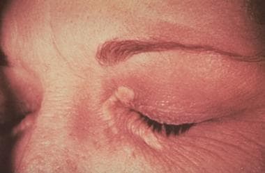 Xanthelasma. Courtesy of Duke University Medical Center.


