Practice Essentials
A dilated pore of Winer is a hair structure anomaly that appears as an enlarged solitary comedo. [1] Most commonly, it appears on the face of a middle-aged person. This condition is not associated with acne vulgaris. See the images below.
Pathophysiology and etiology
A dilated pore of Winer is a tumor of the intraepidermal follicle and infundibulum of a pilosebaceous apparatus. [2] An immunohistochemical study using monoclonal antibodies against cytokeratins and involucrin confirmed differentiation toward the infundibulum and partly toward the isthmus. [3]
The cause of a dilated pore of Winer is unknown. Winer suggested that an infection or an obstruction of the follicle ostium is the stimulus for the development of a dilated pore of Winer in a process similar to those of inflammatory cystic acne or other cystic conditions.
Signs and symptoms
Most patients with dilated pore of Winer consult a dermatologist after many years of dealing with an unsightly, enlarged pore on the face. [4]
Patients with dilated pore of Winer usually have a past or present history of severe acne.
Patients report needing to repeatedly express a keratotic plug from the center of the dilated pore of Winer. The expression of this plug allows the further removal of caseous, white, soggy keratin from the deeper portion of the pore. Once the content of the dilated pore is expressed, a keratotic material similar to the original plug refills the pore within several weeks.
A dilated pore of Winer usually appears as a solitary large comedo on the face, predominantly on the upper lip, cheek, or forehead. The lesion can also be found on the trunk, most commonly the back. A rare incidence involving external ear canal has also been reported. [5]
The skin surrounding the pore appears to be unchanged, with no inflammation or induration.
Manipulation of a dilated pore of Winer may lead to infection and scarring.
Diagnostics
Also see Histologic Findings.
Dermoscopic findings
Dermoscopic examination shows a pinkish nodule with peripheral vessels in a regular pattern. The individual vessels at the periphery of the lesion extend towards the center. The size of the vessels decreases with every progressive branching. The center of the lesion reveals a dilated ostium filled with terminal hairs. [6]
Biopsy
Histologic examination of a biopsy specimen from the lesion is the only way to make a definitive diagnosis.
Management
No medical treatment is available for a dilated pore of Winer. Excision of the entire lesion and closure of the resulting surgical defect are curative.
Superficial treatments, such as electrodesiccation, cauterization, coagulation, dermabrasion, or carbon dioxide laser surgery, are less effective because of the deeply invaginated base of a dilated pore of Winer.
A dermatopathologist may be consulted.
Epidemiology
Most cases of dilated pore of Winer are reported in older adults in both the American and European literature.
Most cases of dilated pore of Winer have been reported in white males.
Although dilated pores are found in both sexes, they appear to occur in men more often than in women.
Most cases of dilated pore of Winer are diagnosed in individuals older than 40 years; however, many individuals report that they have had the lesions for many decades, usually starting when they are aged 20-60 years. [7]
Prognosis
Incomplete excision results in regeneration of the dilated pore from the residual infundibular lining. Prognosis is excellent with no known report of death. One case of trichoid basal cell carcinoma in a dilated pore has been reported. [8] More commonly, chronic manipulation and expression of the keratotic plug from inside the pore may lead to inflammation and infection of the surrounding tissue.
In addition to basal cell carcinoma and squamous cell carcinoma, there is at least 1 reported case of dilated pore of Winer in a collision tumor with melanoma in situ. [9]
-
Hematoxylin and eosin stain. Original magnification X40. Courtesy of Lawrence Machtinger, MD.
-
Image shows an epidermal lining that is atrophic near the ostium but progressively hypertrophic and proliferative, with numerous rete ridges, in the deeper part of the cavity (hematoxylin and eosin, original magnification X100).
-
The cavity is filled with laminated keratin (hematoxylin and eosin, original magnification X100).
-
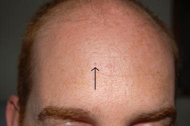
Dilated pore of Winer on forehead.
-
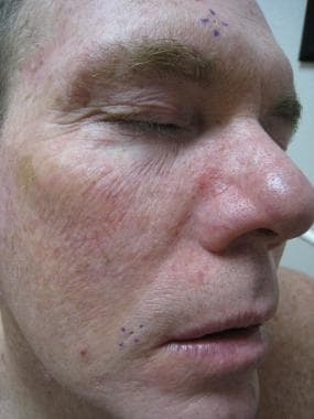
Dilated pores of Winer on forehead and lateral upper labial region.
-
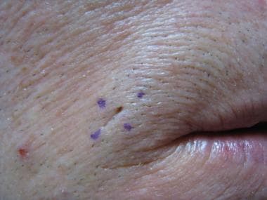
Dilated pore of Winer on lateral upper labial region.


