Practice Essentials
Since Hippocrates first described digital clubbing in patients with empyema, digital clubbing has been associated with various underlying pulmonary, cardiovascular, neoplastic, infectious, hepatobiliary, mediastinal, endocrine, and gastrointestinal diseases. Finger clubbing also may occur, without evident underlying disease, as an idiopathic form or as a Mendelian dominant trait. Clubbing is a clinically descriptive term, referring to the bulbous uniform swelling of the soft tissue of the terminal phalanx of a digit with subsequent loss of the normal angle between the nail and the nail bed.
Digital clubbing is classified into primary (ie, idiopathic, hereditary) and secondary forms. Digital clubbing may be symmetric bilaterally, or it may be unilateral or involve a single digit. Anatomic considerations, such as the classic measurement of the Lovibond angle or the more recently derived index of nail curvature by Goyal et al [1] , usually can be identified on simple physical examination and can be used to identify digital clubbing and to monitor this dynamic process objectively. Various imaging modalities have been used not only to evaluate clubbing but also to help identify possible clues to its development.
Signs and symptoms
Also see History.
Clubbing is a clinical finding characterized by bulbous fusiform enlargement of the distal portion of a digit (see the image below).
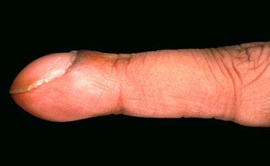 Clubbed fingernail.
When the profile of the distal digit is viewed, the angle made by the proximal nail fold and nail plate (Lovibond angle) typically is less than or equal to 160°. In clubbing, the angle flattens out and increases as the severity of the clubbing increases. If the angle is greater than 180°, definitive clubbing exists. An angle between 160-180° falls in a gray area and may indicate early stages of clubbing or a pseudoclubbing phenomenon.
Individuals without clubbing display a diamond-shaped window at the base of the nail beds when the dorsum of 2 fingers from the opposite hands are opposed. The distal angle between the 2 opposed nails should be minimal. In individuals with digital clubbing, the diamond window is obliterated and the distal angle between the nails increases with increasing severity of clubbing.
The nail moves more freely in patients with clubbing; therefore, the examiner may note a spongy sensation as the nail is pressed toward the nail plate. The sponginess results from increased fibrovascular tissue between the nail and the phalanx. The skin at the base of the nail may be smooth and shiny.
Obliteration in clubbed fingers of the diamond-shaped window normally produced when the dorsal surfaces of the corresponding finger of each hand are opposed (Schamroth sign) may useful for the identification of clubbing. [2]
Diagnostics
Also see Imaging Studies.
Because clubbing typically is secondary to an underlying pathological process, perform pertinent laboratory studies for primary medical disorders that are suggested clinically.
Microscopically, the collagen fibrils and cells are separated by a distance greater than that seen in histologically normal specimens. This increased separation results in a less dense nail bed matrix. Primitive fibroblasts are seen with large nuclei, basophilic cytoplasm, and long reticular processes. Increased and scattered extravascular lymphocytes and, less often, a moderately increased number of tissue eosinophils also are noted in the nail beds of some specimens. The periosteum of the nail bed may be thickened with increased vascular penetration.
Eventually, increased collagen is laid down in all types of chronic clubbing. The mat of collagen fibers may be abnormally thick and dense. The walls of the vascular components increase in thickness and are encased in a thick fibrous sheet. At this stage of clubbing, the histologic and morphologic changes probably are irreversible.
Management
No specific treatment for clubbing is available. Treatment of the underlying pathological condition may decrease the clubbing or, potentially, reverse it if performed early enough. Once substantial chronic tissue changes, including increased collagen deposition, have occurred, reversal is unlikely. Treatment for related problems, such as pain, is symptomatic. One patient with pachydermoperiostosis responded to etoricoxib. [3]
No specific surgical procedures are performed for clubbing. Appropriate surgical treatment of underlying disease, such as tumor removal in patients with lung cancer, may improve or reverse clubbing, provided that permanent morphologic changes have not occurred.
Further outpatient care should be guided by the specific primary disease, which is the cause of the clubbing. This should be evaluated on a case-by-case basis.
Consultations
Clubbing is a clinical sign of many pathological processes; therefore, consultation with specialists may be necessary to diagnose the underlying disease. Patients with primary hypertrophic osteoarthropathy should be evaluated for associated findings, possibly including myelofibrosis. [4, 5]
Background
Clubbing is a feature of pachydermoperiostosis (PDP), a rare genodermatosis characterized by pachydermia, digital clubbing, periostosis, and an excess of affected males. [6] Although usually an autosomal dominant model with incomplete penetrance and variable expression, both autosomal recessive and X-linked inheritance have been suggested in some PDP families.
Primary hypertrophic osteoarthropathy (PHO), a rare hereditary disorder with digital clubbing, subperiosteal new bone formation, and arthropathy, has been linked mutations in the 15-hydroxy-prostaglandin dehydrogenase (15-PGDH) encoding gene HPGD, which causes PHO. [7, 8, 9] A mutation was identified in a large Pakistani family with isolated congenital nail clubbing. [10] In another study of homozygous mutations, an intragenic deletion that results in frameshift and a missense mutation was associated with a severe PHO phenotype. A heterozygous carrier of a stop mutation had isolated digital clubbing. PHO with severe arthralgia may be due to a gene mutation in SLCO2A1, which encodes the prostaglandin transport protein. [11]
Pathophysiology
The specific pathophysiologic mechanism of digital clubbing remains unknown. Many theories have been proposed, yet none have received widespread acceptance as a comprehensive explanation for the phenomenon of digital clubbing. As stated best by Samuel West in 1897, "Clubbing is one of those phenomena with which we are all so familiar that we appear to know more about it than we really do."
Alterations in size and configuration of the clubbed digit result from changes in the nail bed, beginning with increased interstitial edema early in the process. As clubbing progresses, the volume of the terminal portion of the digit may increase because of an increase in the vascular connective tissue and change in quality of the vascular connective tissue, although some cases have been associated with spurs of bone on the terminal phalanx.
Although clubbing is a common physical finding in many underlying pathological processes, surprisingly, the mechanism of clubbing remains unclear. Different pathological processes may follow different pathways to a common end. Many studies have shown increased blood flow in the clubbed portion of the finger.
High-resolution magnetic resonance imaging uisng a contrast agent in 4 patients with finger clubbing and 4 healthy volunteers documented nail bed hypervascularization as linked with clubbed nails. [12] Most researchers agree that this results from an increase in distal digital vasodilation, the cause of which is unknown. Also unknown is the exact mechanism by which increased blood flow results in changes in the vascular connective tissue under the nail bed.
Many researchers agree that the common factor in most types of clubbing is distal digital vasodilation, which results in increased blood flow to the distal portion of the digits. Whether the vasodilation results from a circulating or local vasodilator, neural mechanism, response to hypoxemia, genetic predisposition, or a combination of these or other mediators is not agreed on currently.
Evidence that favors the presence of a circulating vasodilator derives from the association of clubbing with cyanotic congenital heart disease. Many potential vasodilators, which usually are inactivated as blood passes through the lungs, bypass the inactivation process in patients with right-to-left shunts. Patients with tetralogy of Fallot with substantial shunting have a high incidence of clubbing. After surgical correction diminishes the shunt, the clubbing improves. Also previously observed is clubbing confined to the feet in patients with late untreated patent ductus arteriosus in whom blood from the pulmonary artery bypasses the lungs and is shunted into the descending aorta. In the absence of a shunt, the circulating vasodilator may be produced by the lung tissue, or, possibly, it passes through the pulmonary circulation without becoming inactivated. Proposed vasodilatory factors include ferritin, prostaglandins, bradykinin, adenine nucleotides, and 5-hydroxytryptamine.
A neural mechanism has been proposed with particular consideration of the vagal system. An increased incidence of digital clubbing has been associated with the pathology and disease of vagally innervated organs. Furthermore, regression of clubbing after vagotomy has been reported. Although some factor related to the vagal system is a possible contributor to the development of clubbing, especially clubbing occurring with hypertrophic osteoarthropathy, the hypothesis of a neural mechanism has decreased in popularity because of the lack of evidence of clubbing in neurologic disorders and the presence of clubbing in diseases of organs not innervated by the vagal system.
Hypoxia has been proposed as an alternative explanation for clubbing in cyanotic heart disease and pulmonary diseases. An increase in hypoxia may activate local vasodilators, consequently increasing blood flow to the distal portion of the digits; however, in most cases, hypoxia is absent in the presence of clubbing, and many diseases with noted hypoxia are not associated with clubbing.
Genetic inheritance and predisposition also may play a role in digital clubbing. Hereditary clubbing is observed in 2 forms, including idiopathic hereditary clubbing and clubbing associated with pachydermoperiostosis. The 2 forms are believed to be separate entities. Both demonstrate autosomal dominant inheritance with incomplete penetrance.
More recently, platelet-derived growth factor released from fragments of platelet clumps or megakaryocytes has been proposed as the mechanism by which digital clubbing occurs. [13] The fragments are large enough to lodge in the vascular beds of the fingertips, and, subsequently, they release platelet-derived growth factor. This factor has been shown to have general growth-promoting activity and causes increased capillary permeability and connective tissue hypertrophy.
Etiology
Clubbing can be idiopathic or secondary to many underlying pathologies in various organ systems.
Causes of idiopathic or primary clubbing include pachydermoperiostosis, familial clubbing, and hypertrophic osteoarthropathy. [14, 15]
Causes of secondary clubbing include the following [16] :
-
Pulmonary disease - Lung cancer, [17] cystic fibrosis, interstitial lung disease, [18] idiopathic pulmonary fibrosis, [19] sarcoidosis, [20] lipoid pneumonia, empyema, pleural mesothelioma, pulmonary artery sarcoma, [21] lung hydatid cysts, [22] Eisenmenger syndrome (a severe form of pulmonary arterial hypertension), [23] and pulmonary metastases (see Dermatologic Manifestations of Pulmonary Disease)
-
Cardiac disease - Cyanotic congenital heart disease, [24] other causes of right-to-left shunting, and bacterial endocarditis (see Dermatologic Manifestations of Cardiac Disease); a scoring system for empiric treatment of infective endocarditis includes clubbing [25]
-
Gastrointestinal disease - Ulcerative colitis, Crohn disease, primary biliary cirrhosis, cirrhosis of the liver, hepatopulmonary syndrome, [26] leiomyoma of the esophagus, achalasia, and peptic ulceration of the esophagus (see Dermatologic Manifestations of Gastrointestinal Disease) [27]
-
Skin disease - Pachydermoperiostosis, Bureau-Barrière-Thomas syndrome, Fischer syndrome, palmoplantar keratoderma, [28] and Volavsek syndrome
-
Malignancies - Thyroid cancer, thymus cancer, Hodgkin disease, [29] and disseminated chronic myeloid leukemia (POEMS [polyneuropathy, organomegaly, endocrinopathy, monoclonal gammopathy, and skin changes] syndrome is a rare paraneoplastic syndrome secondary to a plasma cell dyscrasia in which clubbing may be seen. [30, 31] Other findings including peripheral neuropathy, organomegaly, endocrinopathy, monoclonal plasma proliferative disorder, skin changes, sclerotic bone lesions, Castleman disease, thrombocytosis, papilledema, peripheral edema, pleural effusions, ascites, and white nails.)
Hypertrophic osteoarthropathy in adults is often a sign of internal cancer. [35] Other stigmata may also be evident, such as acquired keratoderma taking on a yellow velvety appearance with accentuation of dermatoglyphic lines.
As a paraneoplastic syndrome, it is most commonly associated with non–small-cell lung cancer, [36] although it may occur with metastatic melanoma and other cancers. [37, 38] Hypertrophic pulmonary osteoarthropathy is evident in 1- 5 % of all patients with non–small-cell lung cancer. [39] The full spectrum of hypertrophic pulmonary osteoarthropathy has been observed mostly in adults with liver disease and cancers and in children and adolescents with cystic fibrosis. [40]
Among hemodialysis patients with end-stage renal disease, the finding of clubbing was linked with the calcium-phosphorus product. [41]
Nail changes in 100 chronic renal failure patients undergoing hemodialysis and 100 matched controls were assessed. [42] Nail disorders were more prevalent in the renal failure patients (76%) than in the control group (30%). The half-and-half nail was the most common finding (20%), followed by absent lunula, onycholysis, brittle nail, Beau lines, clubbing, longitudinal ridging, onychomycosis, subungual hyperkeratosis, koilonychias, total leukonychia, splinter hemorrhage, pitting, and pincer nail deformity.
Epidemiology
Frequency
Idiopathic or primary clubbing is rare, while the occurrence of secondary clubbing depends on the underlying disease.
Primary digital clubbing has been reported to occur in 89% of patients diagnosed with pachydermoperiostosis. This syndrome most often occurs in young males.
Of patients with idiopathic pulmonary fibrosis, 65% have clinical digital clubbing. In these patients, an increased occurrence has been shown in patients with higher grades of smooth muscle proliferation in the lungs.
Clubbing has been reported in 29% of patients with lung cancer and is observed more commonly in patients with non–small cell lung carcinoma (35%) than in patients with small cell lung carcinoma (4%).
Digital clubbing was reported in 38% of patients with Crohn disease, 15% of patients with ulcerative colitis, and 8% of patients with proctitis. Clubbing was observed in up to one third of Ugandan patients with pulmonary tuberculosis. [43] It was not associated with stage of HIV infection, extensive disease, or hypoalbuminemia.
Race
Racial predilection of secondary clubbing depends on the racial prevalence of the underlying disease.
Sex
Sex predilection of secondary clubbing of the fingers depends on the prevalence of the underlying disease.
Prognosis
Since clubbing of the fingers is a clinical finding, it is not an independent cause of mortality; however, mortality associated with underlying pathology in patients with secondary clubbing widely varies. [44]
Morbidity is minimal and typically is associated with the cosmetic appearance of clubbed digits.
Treatment of the underlying pathological condition may decrease the clubbing or, potentially, reverse it if performed early enough. Once substantial chronic tissue changes, including increased collagen deposition, have occurred, reversal is unlikely. Prognosis of the underlying disease should be determined on an individual basis.
-
Clubbed fingernail.