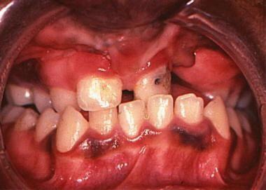
Continually Updated Clinical Reference
 
 
  All Sources     eMedicine     Medscape     Drug Reference     MEDLINE
 
You are in: eMedicine Specialties > Dermatology > Diseases Of The Oral Mucosa

Oral Melanoacanthoma

Last Updated: December 4, 2006
Email to a Colleague
Synonyms and related keywords: cutaneous melanoacanthoma, mucosal melanoacanthoma, mucosal lesions, cutaneous lesions, pigmented seborrheic keratosis, melanocyte and keratinocyte proliferation, pigmented macular lesions, benign nonovoid melanoepithelioma

  AUTHOR INFORMATION Section 1 of 10    Click here to go to the next section in this topic
Author Information Introduction Clinical Differentials Workup Treatment Follow-up Miscellaneous Pictures Bibliography

Author: Talib Najjar, DMD, MDS, PhD, Professor, Department of Oral and Maxillofacial Surgery, University of Medicine and Dentistry of New Jersey

Coauthor(s): Thomas A Chiodo, DDS, Staff Dentist, Department of Oral and Maxillofacial Surgery, University of Medicine and Dentistry of New Jersey

Editor(s): Neil Shear, MD, Professor and Chief of Dermatology, Professor of Medicine, Pediatrics and Pharmacology, University of Toronto Medical School; Head of Dermatology, Sunnybrook Women's College Health Sciences Center, Canada; Michael J Wells, MD, Associate Professor, Department of Dermatology, Texas Tech University Health Sciences Center; M Joyce Rico, MD, Consulting Staff, Department of Dermatology, Fujisawa Healthcare, Inc; Glen H Crawford, MD, Assistant Clinical Professor, Department of Dermatology, University of Pennsylvania School of Medicine; Chief, Division of Dermatology, The Pennsylvania Hospital; and Dirk M Elston, MD, Director, Department of Dermatology, Geisinger Medical Center

Disclosure


  INTRODUCTION Section 2 of 10   Click here to go to the previous section in this topic Click here to go to the top of this page Click here to go to the next section in this topic
Author Information Introduction Clinical Differentials Workup Treatment Follow-up Miscellaneous Pictures Bibliography

Background: Melanoacanthoma is a rare condition of the cutaneous and mucosal epithelia that has been reported only in the last century. The lesion is characterized by a proliferation of both melanocytes and keratinocytes that results in pigmented macular or plaquelike lesions (see Image 1).

Initially, the cutaneous lesions were suspected to be malignant, and subsequently, the lesions were linked to a transformation to low-grade malignancy. Malignant transformation is reported in at least 2 cases; however, most lesions are generally considered benign and should be treated conservatively. No cases of recurrence or metastasis are reported in either the mucosal or cutaneous variants, although when examined clinically, the 2 types seem to have substantial differences. Cutaneous and mucosal melanoacanthoma differ in terms of patient age, patient race, and site.

In 1927, Bloch provided the earliest known description of melanoacanthoma, which was reported in the German literature (Schneider, 1981). Bloch described several skin lesions, which he originally called benign nonovoid melanoepithelioma of the skin. He further subdivided his cases into 2 types: In the histologic samples studied, type 1 had both dendritic melanocytes and keratinocytes, whereas the histologic appearance of type 2 was similar to that of type 1, but it lacked the dendritic melanocytes. Other authors have since redefined this type.

In 1960, Mishima and Pinkus reexamined the condition and further refined the diagnostic terminology (Wright, 1983). The condition that Bloch designated as type 1 is currently called melanoacanthoma, and type 2 is currently called pigmented seborrheic keratosis.

Although cutaneous lesions of melanoacanthoma were reported as early as 1927, Tomich et al at the American Academy of Oral Pathology first reported oral mucosal lesions in 1978 (Schneider, 1981). Although many authors subsequently published case reports, Goode et al published the first case report in 1983 (Goode, 1983). This was a retrospective review of 10 cases of oral melanoacanthoma reported in the literature.

To date, oral melanoacanthoma remains a rare condition, with approximately 25 reported cases. The cutaneous variant is also rare; however, it is more prevalent than the mucosal variant.

Pathophysiology: The cutaneous and mucosal variants of melanoacanthoma have similar histologic appearances. The lesion consists of proliferating melanocytes and keratinocytes, which result in large pigment-containing dendritic cells. The dendritic cells are present throughout the middle and upper layers of the epithelium.

The presence of inflammation in the mucosal variant is the factor that histologically differentiates the cutaneous and mucosal forms. Inflammation in the skin lesions is believed to be a secondary reactive phenomenon, and it is not related to the neoplastic process. Inflammation occurs almost universally in patients with mucosal lesions. The presence of inflammation and the spontaneous resolution of oral lesions are suggestive of a reactive process rather than a neoplastic process. The observation of trauma and inflammation associated with oral lesions has led to the conclusion that the mucosal variant is likely the result of a reactive process rather than a neoplastic process (Horlick, 1988).

Frequency:

  • In the US: Oral melanoacanthoma is rare, with only approximately 25 cases observed since 1978, when the lesions were first reported. The cutaneous variant is more common, but it is still relatively rare among skin lesions. Among all the cases reported, specific patterns are described with respect to the race, sex, and age of patients with cutaneous melanoacanthomas and those with mucosal melanoacanthomas.

Race:

  • Cutaneous lesions of melanoacanthoma are reported almost exclusively in white patients.
  • The mucosal variant is reported almost exclusively in black patients.
  • Some sporadic mucosal cases are reported in Asian individuals.

Sex: The prevalence for both variants of melanoacanthoma is fairly equal in both sexes, with a slight female predominance. The female-to-male ratio is approximately 3:2.

Age: The age distributions of the 2 types of melanoacanthoma differ (Chandler, 1997).

  • Cutaneous lesions are found in patients with a mean age of approximately 60 years.
  • Mucosal lesions appear in patients with a mean age of approximately 25 years.


  CLINICAL Section 3 of 10   Click here to go to the previous section in this topic Click here to go to the top of this page Click here to go to the next section in this topic
Author Information Introduction Clinical Differentials Workup Treatment Follow-up Miscellaneous Pictures Bibliography

History: Because the lesions of oral melanoacanthoma are not painful, physicians usually discover them on routine oral examination.

Physical: The clinical features of cutaneous and mucosal lesions may mimic those of other pigmented lesions. For example, the clinical and histologic features of oral melanoacanthoma lesions can resemble those of melanoma in situ.

  • Cutaneous lesions
    • Cutaneous lesions are reported in the scalp, eyelid, ear, nose, neck, thorax, and abdomen.
    • Cutaneous lesions may be found on any area of the head and neck, as well as on the chest, abdomen, back, or legs.
    • The lesions are generally asymptomatic, flat or slightly raised hyperpigmented areas.
    • The color of the lesions ranges from brown to black to blue.
    • Lesions are usually isolated, with no apparent precipitating factors.
    • Lesions are slow growing and are usually present for months before treatment is sought.
  • Mucosal lesions
    • Although the appearance of mucosal lesions is similar to that of cutaneous lesions, mucosal lesions have a more rapid onset and rate of growth.
    • Mucosal lesions occur mostly on the lip, buccal mucosa, or palate, and they are precipitated by a traumatic event.
    • The diameter of the mucosal lesions can range from a few millimeters to several centimeters.

Causes: The role of trauma in the development of the lesion remains controversial, but any irritant must be removed.
  DIFFERENTIALS Section 4 of 10   Click here to go to the previous section in this topic Click here to go to the top of this page Click here to go to the next section in this topic
Author Information Introduction Clinical Differentials Workup Treatment Follow-up Miscellaneous Pictures Bibliography

Seborrheic Keratosis


Other Problems to be Considered:

Melanoma in situ
Melanosis
Superficial melanoma
Junctional melanocytic nevus
Melanotic macule
Pigmented basal cell epithelioma

Quick Find
Author Information
Introduction
Clinical
Differentials
Workup
Treatment
Follow-up
Miscellaneous
Pictures
Bibliography

Click for related images.

Related Articles
Seborrheic Keratosis


Patient Education
Click here for patient education.


  WORKUP Section 5 of 10   Click here to go to the previous section in this topic Click here to go to the top of this page Click here to go to the next section in this topic
Author Information Introduction Clinical Differentials Workup Treatment Follow-up Miscellaneous Pictures Bibliography

Procedures:

  • The clinical features of cutaneous and mucosal lesions may mimic those of other pigmented lesions; therefore, definitive diagnosis is based on histologic examination.
  • Incisional or excisional biopsy may be performed based on the size and the anatomic location of the lesion.
Histologic Findings: Microscopic examination reveals a hyperplastic edematous stratified squamous epithelium with acanthosis and elongated widened rete ridges. Increased melanin pigmentation is present in the basal layer (see
Image 3). In addition, many proliferating dendritic melanocytes or clear cells extend upward into the prickle-cell layers (see Image 4).

The clinical and histologic features of oral melanoacanthoma lesions can resemble those of melanoma in situ. Usually, no evidence of cellular pleomorphism or abnormal mitotic activity is seen in oral melanoacanthomas, and this feature excludes malignancy.

In mucosal lesions, mixed chronic inflammatory cells and numerous melanin-laden macrophages densely infiltrate the stratum corneum. This inflammatory process may indicate the reactive rather than the neoplastic nature of the lesion.

  TREATMENT Section 6 of 10   Click here to go to the previous section in this topic Click here to go to the top of this page Click here to go to the next section in this topic
Author Information Introduction Clinical Differentials Workup Treatment Follow-up Miscellaneous Pictures Bibliography

Medical Care: No medical treatment for mucosal or cutaneous lesions is known. A low risk of recurrence exists.

Surgical Care: Surgical excision is the treatment of choice for both mucosal and cutaneous lesions.

  FOLLOW-UP Section 7 of 10   Click here to go to the previous section in this topic Click here to go to the top of this page Click here to go to the next section in this topic
Author Information Introduction Clinical Differentials Workup Treatment Follow-up Miscellaneous Pictures Bibliography

Complications:

  • No evidence of malignant transformation has been reported.
  • No complications arise from incisional or excisional biopsy.

Prognosis:

  • Initially, cutaneous lesions were suspected to be malignant, and subsequently, the lesions were linked to a transformation to low-grade malignancy.
    • Malignant transformation has been reported in at least 2 cases.
    • However, most lesions are considered benign and should be treated conservatively.
  • No cases of recurrence or metastasis have been reported in either the mucosal variant or cutaneous variant.
  • A low risk of recurrence exists.
  MISCELLANEOUS Section 8 of 10   Click here to go to the previous section in this topic Click here to go to the top of this page Click here to go to the next section in this topic
Author Information Introduction Clinical Differentials Workup Treatment Follow-up Miscellaneous Pictures Bibliography

Medical/Legal Pitfalls:

  • Melanoacanthoma is a benign lesion with little legal impact, however; the clinical and morphologic differential diagnosis is essential to rule out malignant melanoma. Melanoma is a fatal lesion with clear medical and legal consequences.
  PICTURES Section 9 of 10   Click here to go to the previous section in this topic Click here to go to the top of this page Click here to go to the next section in this topic
Author Information Introduction Clinical Differentials Workup Treatment Follow-up Miscellaneous Pictures Bibliography

Caption: Picture 1. 
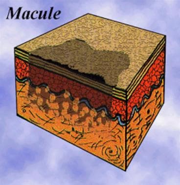
Diagram of a pigmented epithelial macule.
Click to see larger pictureClick to see detailView Full Size Image
Picture Type: Image
Caption: Picture 2. Intraoral melanoacanthoma lesion on the mandibular gingiva.
Click to see larger pictureClick to see detailView Full Size Image
Picture Type: Photo
Caption: Picture 3. Increased melanin pigmentation in the basal layer of a melanoacanthoma (hematoxylin and eosin, original magnification X10).
Click to see larger pictureClick to see detailView Full Size Image
Picture Type: Photo
Caption: Picture 4. Proliferating dendritic melanocytes in the prickle-cell layers of a melanoacanthoma (hematoxylin and eosin, original magnification X40).
Click to see larger pictureClick to see detailView Full Size Image
Picture Type: Photo
  BIBLIOGRAPHY Section 10 of 10   Click here to go to the previous section in this topic Click here to go to the top of this page
Author Information Introduction Clinical Differentials Workup Treatment Follow-up Miscellaneous Pictures Bibliography

  • Andrews BT, Trask DK: Oral melanoacanthoma: a case report, a review of the literature, and a new treatment option. Ann Otol Rhinol Laryngol 2005 Sep; 114(9): 677-80[Medline].
  • Buchner A, Merrell PW, Carpenter WM: Relative frequency of solitary melanocytic lesions of the oral mucosa. J Oral Pathol Med 2004 Oct; 33(9): 550-7[Medline].
  • Chandler K, Chaudhry Z, Kumar N, et al: Melanocanthoma: a rare cause of oral hyperpigmentation. Oral Surg Oral Med Oral Pathol Oral Radiol Endod 1997 Nov; 84(5): 492-4[Medline].
  • Contreras E, Carlos R: Oral melanoacanthosis (melanoachantoma): report of a case and review of the literature. Med Oral Patol Oral Cir Bucal 2005 Jan-Feb; 10(1): 11-2; 9-11[Medline].
  • Frey VM, Lambert WC, Seldin RD, et al: Intraoral melanoacanthoma. J Surg Oncol 1984 Oct; 27(2): 93-6[Medline].
  • Goode RK, Crawford BE, Callihan MD, Neville BW: Oral melanoacanthoma. Review of the literature and report of ten cases. Oral Surg Oral Med Oral Pathol 1983 Dec; 56(6): 622-8[Medline].
  • Horlick HP, Walther RR, Zegarelli DJ, et al: Mucosal melanotic macule, reactive type: a simulation of melanoma. J Am Acad Dermatol 1988 Nov; 19(5 Pt 1): 786-91[Medline].
  • Matsuoka LY, Glasser S, Barsky S: Melanoacanthoma of the lip. Arch Dermatol 1979 Sep; 115(9): 1116-7[Medline].
  • Schneider LC, Mesa ML, Haber SM: Melanoacanthoma of the oral mucosa. Oral Surg Oral Med Oral Pathol 1981 Sep; 52(3): 284-7[Medline].
  • Whitt JC, Jennings DR, Arendt DM, Vinton JR: Rapidly expanding pigmented lesion of the buccal mucosa. J Am Dent Assoc 1988 Oct; 117(5): 620-2[Medline].
  • Wright JM, Binnie WH, Byrd DL, Dunsworth AR: Intraoral melanoacanthoma. J Periodontol 1983 Feb; 54(2): 107-11[Medline].

Oral Melanoacanthoma excerpt